ECG criteria
- The P wave precedes the QRS complexes; it is usually positive in leads I and II, and generally positive in aVF — a combination of signs and the P axis should be evaluated, rather than aVF in isolation.
- The P wave is negative in aVR; often biphasic (+/−) in V1, and usually positive in chest leads, though the shape is variable.
- P wave morphology is identical across all cycles on a single recording (distinguishing it from wandering atrial pacemaker and ectopic rhythms).
- The PQ interval is usually constant, within the typical range of 0.12–0.20 s (120–200 ms); deviations (prolongation, variability) are described separately as AV conduction disturbances.
- PP intervals are regular (accounting for physiological respiratory variability); RR regularity is determined by AV conduction and ventricular rhythm and is not an obligatory sign of sinus origin for atrial activity.
- Heart rate is typically 60–100 bpm in a resting adult — this is the normal heart rate range, not a criterion of sinus origin as such (outside this range is sinus bradycardia or tachycardia).
- Usually, each P wave is followed by a single QRS complex (1:1 conduction) in the absence of AV block; if AV block is present, some P waves may not be conducted — atrial activity remains sinus, but the rhythm is described accounting for the AV conduction disturbance.
What it looks like on ECG
The key diagnostic element is the P wave. It reflects atrial depolarization starting in the region of the sinus node (upper part of the right atrium near the opening of the superior vena cava), so the wavefront spreads from top to bottom and from right to left — the vector is directed toward leads I, II, aVF and away from lead aVR.
Signs typical of sinus origin for atrial activity:
| Parameter | Typical picture in sinus rhythm |
|---|---|
| P wave in II | positive, rounded, height ≤ 2.5 mm |
| P wave in aVR | negative |
| P wave in V1 | often biphasic (+/−) |
| P duration | usually < 0.12 s (120 ms) in adults |
| PQ interval | usually 0.12–0.20 s, constant |
| P to QRS ratio | usually 1:1, in the absence of AV block |
Evaluation is performed in steps: first, the P wave is identified, then its positivity in I and II and negativity in aVR are checked (positivity in aVF is a typical, but not strictly mandatory sign: with a borderline P-axis direction, an isoelectic or occasionally weakly negative P in the inferior lead is possible), then the shape of P across different cycles is compared — it must be identical (this distinguishes stable sinus rhythm from a wandering pacemaker or ectopic atrial rhythms). Next, the PQ interval and its constancy are evaluated separately as a characteristic of AV conduction, rather than atrial activity itself. Finally, PP regularity is assessed — the distance between adjacent P waves should be virtually identical; minor physiological variability within the respiratory cycle is permissible and termed sinus arrhythmia.
Sinus atrial activity can be combined with an irregular ventricular rhythm in AV block or extrasystole — these conditions must be explicitly noted in the conclusion and are not described simply as "sinus rhythm". In first- or second-degree AV block, it is termed sinus rhythm (atrial activity of sinus origin) with the specified degree of AV conduction disturbance. In complete (third-degree) AV block, the atria are activated by the sinus node independently of the ventricles — this is a sinus atrial rhythm with AV dissociation and a subsidiary escape ventricular rhythm, rather than a unified "sinus rhythm" in the previous sense. In extrasystole, the rhythm is described as sinus rhythm complicated by (supraventricular or ventricular) extrasystoles, rather than an unreserved sinus rhythm.
Real ECGs: 8 examples
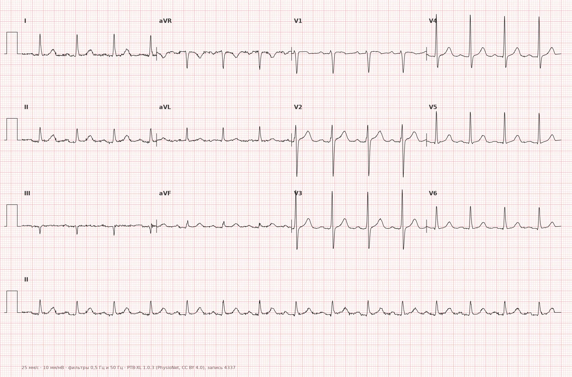
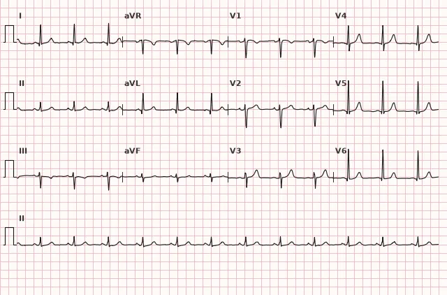
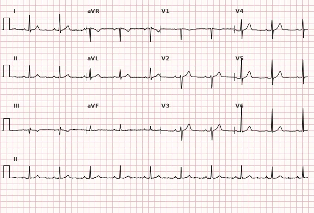
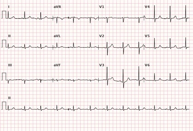
ECG from the open PTB-XL dataset (Wagner P. et al., Scientific Data, 2020; PhysioNet), CC BY 4.0 license. Rendering by WhiteCoat Pro.
Mechanism
The sinus node (sinoatrial node) is a cluster of pacemaker cells in the wall of the right atrium near the opening of the superior vena cava. These cells possess the highest automaticity among all parts of the conduction system — they spontaneously depolarize faster than atrial, AV nodal, and ventricular cells, which is why the sinus node normally sets the rhythm for the entire heart.
Excitement spreads from the sinus node across the atria (forming the P wave), reaches the AV node where physiological conduction delay normally occurs (PQ interval), and then via the bundle of His and its branches reaches the ventricular myocardium (QRS complex). The ECG picture typical of a sinus origin of the rhythm describes only the first stage — impulse formation and atrial activation; the state of the AV node and His-Purkinje system may be normal or impaired, which is evaluated separately. The rate of sinus rhythm is regulated by the autonomic nervous system: sympathetic stimulation accelerates node cell depolarization (speeding up the rhythm), while parasympathetic (vagal) stimulation slows it down.
Normal
Sinus rhythm with an HR of 60–100 bpm in a resting adult is a normal physiological variant requiring no action (some sources use a narrower reference of 60–90 bpm, but this is not a universally accepted strict boundary). In children, normal sinus rate is significantly higher and depends on age; in trained adults, a rate below 60 bpm can be physiological. Minor rate fluctuations associated with respiration (acceleration on inspiration, deceleration on expiration), especially in children and young adults, are also within normal limits and are described as sinus arrhythmia.
Differential diagnosis
— Ectopic atrial rhythm: a P wave of different morphology (may be negative in II, aVF), indicating an impulse source outside the sinus node; occasionally, a low ectopic atrial focus yields a P morphology close to sinus, so when in doubt, the combination of leads and clinical context are considered.
— Wandering atrial pacemaker: P wave morphology gradually changes from cycle to cycle, and the PQ interval varies — the rhythm source "wanders" between the sinus node and atrial foci.
— AV nodal rhythm: the P wave is absent before the QRS, or is negative in II and located close to or within the QRS complex (retrograde conduction).
— Sinus tachycardia/bradycardia: identical P morphology, but the HR falls outside 60–100 bpm in a resting adult — these are variants of the same sinus rhythm origin with an altered rate, not separate rhythms; thresholds of 60 and 100 apply to adults, whereas age-adjusted norms are used for children.
— Atrial fibrillation: the P wave is absent, replaced by f waves, and RR intervals are absolutely irregular.
Clinical significance and management
The ECG picture of sinus rhythm is a descriptive characteristic confirming the sequence of atrial activation typical of the sinus node. It does not confirm normal function of the entire conduction system and does not rule out AV block, bundle branch block, ventricular pre-excitation, or other conduction disturbances — these conditions are assessed separately and indicated in the conclusion alongside the sinus rhythm. Sinus atrial activity by itself is not a pathology and requires no treatment.
It is important for the physician to: 1) ensure that the rhythm is truly sinus (rather than a mimicking ectopic atrial or junctional rhythm with retrograde P waves) by verifying P-wave morphology across multiple leads; 2) assess the heart rate — differentiating a normal sinus rhythm from sinus tachycardia or bradycardia; 3) evaluate PP regularity independently of RR regularity, ruling out sinus arrhythmia, pauses, AV block, or extrasystoles, and explicitly stating them with a distinct term if present (and in complete AV block, separately describing the sinus atrial rhythm and the independent escape ventricular rhythm); 4) include the description "sinus rhythm, HR N bpm" in the standard ECG report as a baseline against which intervals, axis, complex morphology, and signs of pathology are subsequently evaluated.
Common interpretation errors
— Mistaking an artifact or a U wave from the preceding cycle for a P wave, especially during tachycardia when the P wave is superimposed on the T wave.
— Failing to verify P-wave morphology in different leads (limiting evaluation to lead II alone), which leads to missing ectopic rhythms with coincidentally similar P waves.
— Confusing normal respiratory sinus arrhythmia in young individuals with pathological rhythm irregularity.
— Writing "sinus rhythm" in complete AV block or pronounced extrasystole without clarification — it is more accurate to separately indicate the sinus atrial activity and the nature of the conduction disturbance or escape rhythm.
— Writing "sinus rhythm" without specifying the heart rate: it is recommended to state the heart rate in the standard rhythm description.
1. Which ECG sign supports the sinus origin of the rhythm?
A positive P wave in II and a negative P wave in aVR reflect the atrial depolarization vector typical of an impulse originating from the sinus node.
2. According to classical criteria, which heart rate corresponds to a normal sinus rhythm in a resting adult?
The range of 60–100 bpm is the classical reference point for a normal resting heart rate in adults.
3. How should an ECG be correctly described in complete AV block if P waves of sinus morphology are unrelated to QRS complexes?
In complete AV block, the atria and ventricles function independently: the atria are depolarized by the sinus node, while the ventricles are driven by an escape rhythm; this requires a separate description rather than a single umbrella term "sinus rhythm".
Frequently asked questions
How to distinguish sinus rhythm from an ectopic atrial rhythm?
The primary supporting sign is P-wave morphology: in sinus rhythm, the P wave is typically positive in leads I and II, generally positive in aVF, and negative in aVR, with a consistent contour across all cycles. In an ectopic atrial rhythm, the P-wave axis differs from the typical one, and P is frequently negative in inferior leads. This sign supports the diagnosis but is not always sufficient in isolation — it requires evaluating the ensemble of leads and the clinical context, as some low ectopic atrial foci yield a similar P-wave axis.
Is sinus rhythm dangerous?
No, a typical ECG presentation of sinus-origin impulses represents a normal physiological variant. Risk arises only with rate abnormalities (tachycardia, bradycardia), regularity disturbances (pauses, blocks), or concomitant AV and intraventricular conduction disturbances, which are described using distinct terms.
What heart rate is considered normal for sinus rhythm?
Classically, 60–100 bpm in resting adults. This is a range of normal heart rates rather than a mandatory criterion for sinus origin — sinus rhythm can exist outside of these limits, in which case it is termed sinus bradycardia or sinus tachycardia.
Can sinus rhythm be irregular?
Yes, minor physiological variability of PP intervals related to respiration is termed sinus arrhythmia and is considered a normal variant, especially in children and young adults. RR intervals may also be irregular due to other causes — for instance, AV block or extrasystoles; in such cases, atrial activity remains sinus, but the conduction disturbance or premature beats are specified separately.
How is rhythm described in complete AV block if the atria are driven by the sinus node?
In this case, they are described separately: sinus atrial rhythm (with the P-wave rate indicated) and independent ventricular escape rhythm (indicating its rate and source), alongside noting the AV dissociation. The unqualified term "sinus rhythm" for the entire ECG is not used in this scenario because the ventricles are not driven by the sinus node.
ECG Course
Interpretation from basics to myocardial infarction — learn to read any ECG using a systematic algorithm.
- Video lessons and theory for every topic
- Simulator case studies under every lesson
- Unlimited ECG simulator practice
- Study guide: "All ECGs in 25 minutes"
ECG Strip Simulator
ECG, radiology, histology, pathomorphology, pharmacology — thousands of flashcards with detailed explanations.
- Real ECG strips, not textbook drawings
- Instant answers and explanations
- Streaks and progress tracking in your dashboard
Related terms
More in the section «Basics: waves, intervals, axes»
Sources
- Orlov, V.N. «Electrocardiography Guide»
- Hampton J. The ECG Made Easy
- Surawicz B., Knilans T. Chou's Electrocardiography in Clinical Practice
- AHA/ACCF/HRS Recommendations for the Standardization of ECG, 2009
Educational material for medical students and physicians. Does not replace clinical guidelines and physical examination; the ECG must always be interpreted in conjunction with the clinical picture.