ECG criteria
- Measure the QRS amplitude (maximum absolute deviation from the isoelectric line — the tallest R wave or deepest Q/S wave; for a QS complex, from the isoelectric line to the nadir of the QS) in each of the 6 limb leads (I, II, III, aVL, aVR, aVF)
- If the amplitude is < 5 mm (0.5 mV) in all 6 limb leads, it indicates low limb lead voltage (the rounded formulation "no more than 5 mm" is also used in educational practice)
- Measure the QRS amplitude using the same method in each chest lead (V1–V6)
- If the amplitude is < 10 mm (1.0 mV) in all 6 chest leads, it indicates low chest lead voltage (rounded as "no more than 10 mm")
- Generalized low voltage is classified separately — the criterion is met in both limb and chest leads
- Check the recording calibration (1 mV = 10 mm) — false low voltage occurs with incorrect gain settings
- Exclude technical causes: poor electrode contact, artifact, incorrect gain
What it looks like on ECG
QRS complexes are reduced in amplitude uniformly across all leads of a single group — distinguishing it from localized voltage reduction (e.g., only in inferior leads following an infarction).
| Lead group | Low voltage threshold |
|---|---|
| Limb leads (I, II, III, aVL, aVR, aVF) | < 5 mm in all 6 |
| Chest leads (V1–V6) | < 10 mm in all 6 |
| Generalized | both criteria simultaneously |
Amplitude is measured as the maximum absolute deviation of the QRS complex from the isoelectric line — either to the peak of the highest R wave or to the nadir of the deepest Q or S wave, depending on which of these deviations is greater; the peak-to-peak span between positive and negative peaks is not used for this criterion because it can falsely mask low voltage or distort evaluation. Measurements are taken in millimeters at standard calibration of 10 mm/mV and a speed of 25 mm/s. The morphology of the complexes is usually preserved — narrow, without pathological morphology, simply small. It is often combined with a low-amplitude P wave and a flattened or low T wave, particularly in pericardial effusion. The electrical axis is harder to determine due to low amplitude, and sometimes appears indeterminate simply due to technical measurement difficulties.
Real ECGs: 8 examples
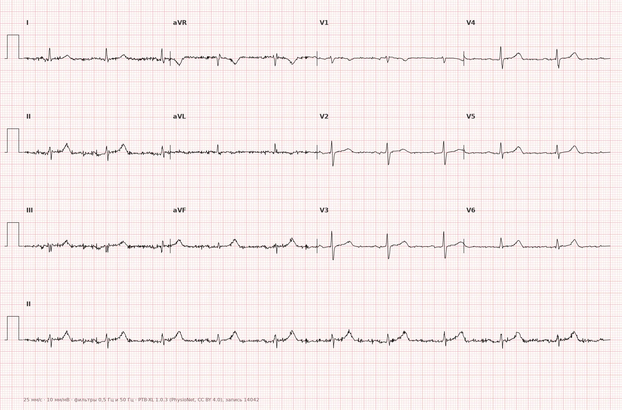
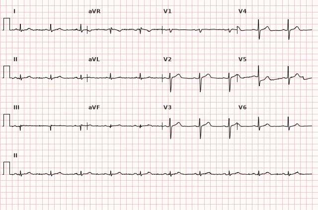
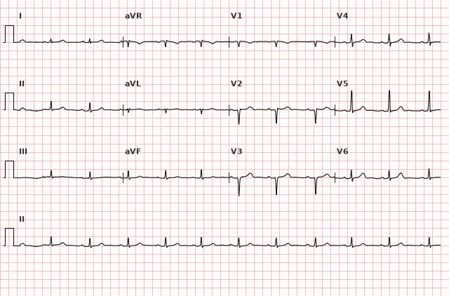
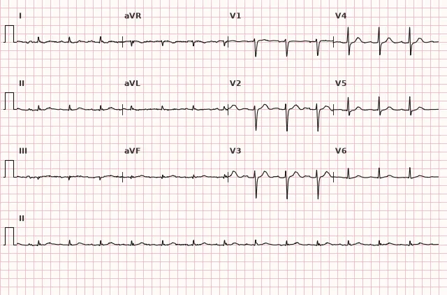
ECG from the open PTB-XL dataset (Wagner P. et al., Scientific Data, 2020; PhysioNet), CC BY 4.0 license. Rendering by WhiteCoat Pro.
Mechanism
The surface QRS amplitude depends on the magnitude of the ventricular depolarization electrical vector and the extent to which this vector is attenuated by the medium between the heart and the electrode. Voltage reduction occurs in two ways: reduction of the electrical signal itself (loss, replacement, or infiltration of the myocardium decreases the net vector) or attenuation of the surface electrical field on its way to the skin. In pericardial effusion, fluid increases the distance between the heart and electrodes and alters the conditions of electrical field propagation, causing it to scatter and attenuate by the time it reaches the skin. In COPD and emphysema, the mechanism is different: hyperinflation increases the distance between the heart and electrodes, alters the position of the heart in the thoracic cavity, and weakens the transmission of potentials through poorly conductive aerated lung tissue.
Causes
- Pericardial effusion and cardiac tamponade — one of the main causes of significant low voltage; isolated pericarditis without effusion does not usually reduce voltage
- Constrictive pericarditis
- Hypothyroidism (myxedema, often with effusion)
- COPD and pulmonary emphysema (air cushion between the heart and chest wall)
- Obesity, pronounced subcutaneous edema
- Diffuse fibrosis or infiltrative cardiomyopathies (amyloidosis — especially with discrepancy between low voltage and thickened walls on echocardiography)
- Extensive prior myocardial infarction with scar replacement of the myocardium
- Diffuse myocarditis with myocardial edema or necrosis
- Pneumothorax, pleural effusion
- End-stage heart failure with pronounced remodeling
- Technical cause: incorrect amplifier calibration (pseudo-low voltage)
Differential diagnosis
- Normal variant in healthy adults — also occurs in healthy individuals; the diagnosis is purely descriptive, not a syndrome
- Incorrect apparatus calibration — check the calibration pulse (should be 10 mm at 1 mV), recalculate for other settings
- Technical artifacts (poor contact, dried gel) — usually asymmetric, affecting one or two leads rather than all
- Localized amplitude reduction after infarction (e.g., only in leads with pathological Q waves) — this is not true low voltage, but loss of myocardial mass in the necrotic zone
- Low voltage due to obesity or COPD — more often an isolated finding, but the absence of electrical alternans, tachycardia, or pulsus paradoxus (a clinical sign, rather than ECG) does not rule out tamponade; when the clinical picture is incomplete, decisions are made based on the totality of data and echocardiography
Clinical significance and management
Low QRS voltage on its own is neither a diagnosis nor a reason for urgent action; it is a descriptive ECG sign requiring clinical interpretation. The physician should first assess the patient's condition and rule out an emergency cause rather than immediately undertaking technical checks. When tamponade is suspected (hypotension, jugular venous distension, muffled heart sounds, tachycardia, pulsus paradoxus), the combination of low voltage and electrical alternans increases the probability of the diagnosis. Urgent echocardiography is performed in a stable patient with such suspicion. In an unstable patient, evaluation must not delay urgent resuscitation assessment, emergency bedside echocardiography, and — in confirmed or highly probable tamponade — urgent drainage of the effusion (pericardiocentesis or surgical intervention as indicated). In a stable patient without signs of tamponade, the next step is to check calibration and electrode contact quality, and compare with prior ECGs. If the cause is known (COPD, obesity, hypothyroidism), the patient is stable, and the voltage is unchanged from prior recordings, immediate additional evaluation may not be required. However, for newly identified or progressive low voltage, discrepancy with the clinical picture, or suspicion of a new condition, targeted investigations including echocardiography are necessary. If infiltrative cardiomyopathy is suspected (discrepancy between low voltage and thickened myocardium on echocardiography), further evaluation for amyloidosis is indicated.
Common interpretation errors
- Failing to check the calibration pulse and confusing incorrect amplifier settings with true low voltage
- Diagnosing low voltage based on one or two leads rather than the entire lead group (limb or chest)
- Failing to correlate the finding with the clinical picture — missing tamponade in the presence of clear signs of cardiac compression, delaying urgent drainage with technical checks or waiting for elective echocardiography
- Forgetting to rule out pleural effusion and pneumothorax as a cause in an ICU patient
- Interpreting low amplitude in an extensive post-infarction scar as an independent syndrome rather than a consequence of myocardial mass loss
1. How should QRS amplitude be measured to assess the low voltage criterion?
The low voltage criterion utilizes the maximum absolute deviation from the isoelectric line rather than the peak-to-peak span between positive and negative peaks, which can distort the evaluation.
2. Which cause of low QRS voltage is associated with increased distance and attenuation of the electrical field due to fluid surrounding the heart?
Pericardial effusion increases the distance between the myocardium and electrodes and alters the propagation of the electric field, attenuating the signal—a classic mechanism in cardiac tamponade.
3. What should be done in the presence of low QRS voltage and clinical signs of tamponade in an unstable patient?
In an unstable patient with clinical tamponade, workup must not delay emergency resuscitation assessment, bedside echocardiography, and urgent fluid drainage.
Frequently asked questions
What is the amplitude threshold for low QRS voltage?
In limb leads: QRS amplitude less than 5 mm in all six leads. In precordial leads: less than 10 mm in all six leads V1–V6. When both criteria are met simultaneously, it is referred to as generalized low voltage. Some textbooks use the rounded phrasing "no more than 5/10 mm".
Is low QRS voltage dangerous?
The sign itself is not dangerous, but it may indicate cardiac tamponade, constrictive pericarditis, infiltrative cardiomyopathy, or severe COPD. The risk is determined by the underlying disease, not the ECG finding itself.
How to distinguish true low voltage from calibration error?
It is necessary to check the calibration pulse on the tracing: at standard settings, 1 mV should produce a 10 mm deflection. If the calibration is correct and the amplitude is still low across all leads in the group, the voltage is genuinely low.
Why does cardiac tamponade reduce QRS voltage?
Pericardial effusion increases the distance and alters the propagation conditions of the electrical field between the myocardium and surface electrodes; therefore, the QRS amplitude on the skin may decrease. Low voltage is specifically associated with significant effusion or tamponade, rather than pericarditis per se.
Does low QRS voltage occur in healthy individuals?
Yes, in some healthy individuals, especially those with overweight or prominent subcutaneous fat, low QRS voltage can be an isolated finding with no underlying heart disease.
ECG Course
Interpretation from basics to myocardial infarction — learn to read any ECG using a systematic algorithm.
- Video lessons and theory for every topic
- Simulator case studies under every lesson
- Unlimited ECG simulator practice
- Study guide: "All ECGs in 25 minutes"
ECG Strip Simulator
ECG, radiology, histology, pathomorphology, pharmacology — thousands of flashcards with detailed explanations.
- Real ECG strips, not textbook drawings
- Instant answers and explanations
- Streaks and progress tracking in your dashboard
Related terms
More in the section «Basics: waves, intervals, axes»
Sources
- Goldberger's Clinical Electrocardiography, latest edition
- Hampton. The ECG in Practice
- Surawicz, Knilans. Chou's Electrocardiography in Clinical Practice
- AHA/ACCF/HRS Recommendations for the Standardization and Interpretation of the ECG, 2009
Educational material for medical students and physicians. Does not replace clinical guidelines and physical examination; the ECG must always be interpreted in conjunction with the clinical picture.