ECG criteria
- Sokolow-Lyon index: S(V1) + R(V5 or V6) > 35 mm (in adults)
- R(aVL) > 11 mm
- Gubner-Ungerleider criterion: R(I) + S(III) > 25 mm
- Cornell voltage criterion: R(aVL) + S(V3) > 28 mm in men, > 20 mm in women
- Romhilt-Estes criteria: point score based on voltage and non-voltage features (axis, ST-T, left atrial enlargement, QRS duration)
- Evaluation in context: QRS width, axis, ST-T segment, patient age and body habitus
What it looks like on ECG
QRS wave amplitudes exceed normal values in one or multiple leads while maintaining a normal complex width (narrow QRS unless a concomitant bundle branch block is present). Precordial leads are assessed most frequently: deep S in V1–V2 and tall R in V5–V6. In limb leads — tall R in I and aVL, deep S in III.
| Criterion | Leads | Cut-off |
|---|---|---|
| Sokolow-Lyon | S(V1) + R(V5/V6) | > 35 mm |
| Gubner-Ungerleider | R(I) + S(III) | > 25 mm |
| Cornell | R(aVL) + S(V3) | > 28 mm (M), > 20 mm (F) |
| aVL isolated | R(aVL) | > 11 mm |
In physiological or isolated high voltage, the ST segment and T wave are frequently unremarkable. The appearance of downsloping ST depression and asymmetric T wave inversion in leads with tall R waves (V5–V6, I, aVL) represents a strain pattern, which supports the diagnosis of left ventricular hypertrophy with systolic overload; however, this pattern is non-specific and requires clinical evaluation, as similar ST-T changes occur in ischemia, drug effects, and other conditions.
Real ECGs: 8 examples
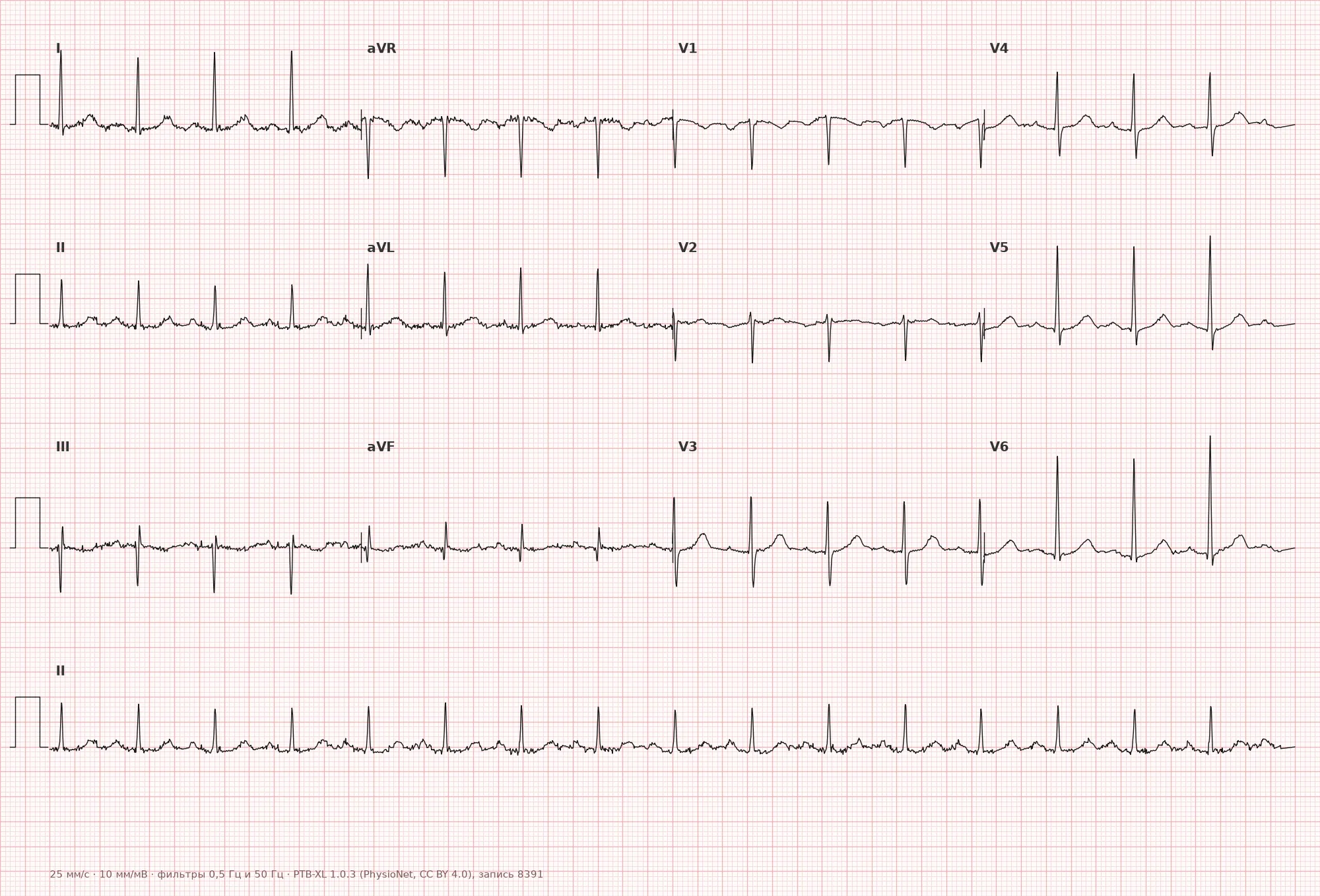
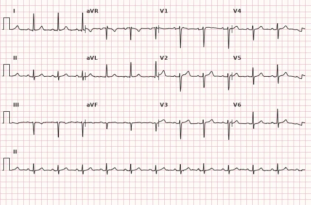
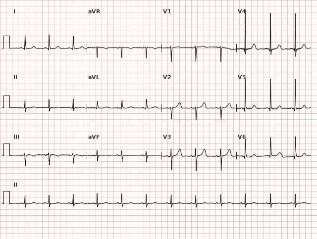
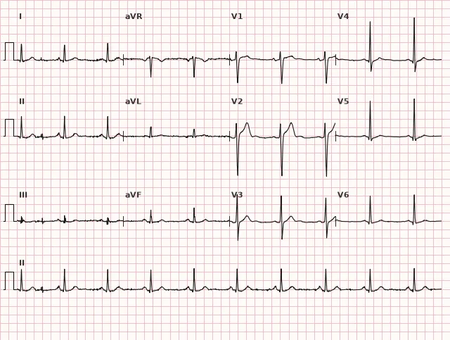
ECG from the open PTB-XL dataset (Wagner P. et al., Scientific Data, 2020; PhysioNet), CC BY 4.0 license. Rendering by WhiteCoat Pro.
Mechanism
QRS amplitude reflects the net electrical activity of the ventricular myocardium recorded by the electrode. It depends on myocardial mass and orientation (hypertrophy increases the depolarization vector), the distance between the heart and the electrode (lean body habitus, thin chest wall, children and young adults result in higher amplitude), and signal attenuation in intervening tissues (adipose tissue, lung air, fluid attenuate the signal). Technical factors—such as poor electrode contact or incorrect calibration—can also alter the measured amplitude without any changes in the myocardium itself.
Causes
- Normal variant in young individuals, thin subjects, and athletes with a thin chest wall
- Left ventricular hypertrophy (arterial hypertension, aortic stenosis)
- Right ventricular hypertrophy
- Hypertrophic cardiomyopathy
- Athlete's heart (physiological hypertrophy in trained athletes)
- Technical causes: poor electrode contact, machine calibration error
Differential diagnosis
Left ventricular hypertrophy — high voltage is only one of the ECG criteria; scoring systems (Sokolow-Lyon, Cornell, Romhilt-Estes) incorporate voltage and, in some cases, non-voltage features, but the absence of some criteria does not exclude LVH. QRS widening is not a typical feature of LVH itself and requires separate evaluation for bundle branch block or other intraventricular conduction delays. Anatomic hypertrophy is confirmed by echocardiography or other imaging modalities. Wolff-Parkinson-White (WPW) syndrome is recognized by a shortened PR interval, delta wave, and widened QRS; high voltage is neither a typical nor mandatory feature of WPW, and LVH voltage criteria are unreliable in pre-excitation. Left bundle branch block — amplitude may be high, QRS is wide (≥120 ms) with characteristic morphology; in complete LBBB, standard LVH voltage criteria have low reliability, and interpretation is guided by block morphology and clinical data. Technical artifact — verify the calibration pulse (must be 10 mm) and correct electrode placement.
Clinical significance and management
Isolated high QRS voltage in a young, healthy individual without risk factors typically requires no further workup and is regarded as a normal variant. When combined with other criteria for hypertrophy (ST-T changes, axis deviation, left atrial enlargement), the physician should suspect structural heart disease and refer the patient for echocardiography to confirm hypertrophy and determine its etiology. LVH voltage criteria (Sokolow-Lyon, Cornell, Romhilt-Estes) exhibit high specificity but low sensitivity, particularly in obese individuals, in whom voltage may remain low despite true hypertrophy.
Common interpretation errors
- Diagnosing LVH based solely on voltage without assessing the ST-T segment, axis, and clinical picture
- Failing to check the calibration signal prior to measuring amplitudes
- Confusing high voltage in thin, young patients with pathologic hypertrophy
- Ignoring QRS widening that alters interpretation (blockade, WPW) in favor of another diagnosis
- Applying adult thresholds for the Cornell criterion without sex adjustment
1. Which of the following criteria belongs to the Sokolow-Lyon index?
The classical precordial variant of the Sokolow-Lyon index uses the sum of S(V1) and the maximal R wave in V5 or V6.
2. What distinguishes high QRS voltage from LVH with systolic overload?
Systolic overload adds characteristic ST-T changes to the voltage criteria, whereas isolated high voltage does not produce these changes.
3. In which patient group is high QRS voltage most commonly a normal variant?
A thin chest wall in young and thin individuals decreases the distance from the heart to the electrode, which increases amplitude in the absence of structural pathology.
Frequently asked questions
Does high QRS voltage always mean left ventricular hypertrophy?
No. It is only one criterion that must be correlated with QRS width, cardiac axis, ST-T changes, and the clinical presentation. In young, thin individuals, high voltage is frequently a normal variant.
How to distinguish high QRS voltage from LVH with overload?
In simple high voltage, the ST segment and T wave are typically unremarkable. The appearance of downsloping ST depression and asymmetric T wave inversion in leads with tall R waves indicates hypertrophy with systolic overload, although this pattern is not strictly specific.
Is high QRS voltage dangerous on its own?
The sign itself is not dangerous and does not require immediate intervention. The risk is determined by the underlying condition, if present, such as arterial hypertension or hypertrophic cardiomyopathy.
Which criterion is most commonly used to diagnose LVH on an ECG?
The Sokolow-Lyon index and the Cornell voltage criterion are used most frequently, sometimes combined with Romhilt-Estes point scoring to increase diagnostic accuracy.
Is additional investigation necessary when high voltage is found incidentally?
If there are no other ECG signs of overload and no clinical risk factors, additional workup is not mandatory. When combined with ST-T changes or risk factors, echocardiography is indicated.
ECG Course
Interpretation from basics to myocardial infarction — learn to read any ECG using a systematic algorithm.
- Video lessons and theory for every topic
- Simulator case studies under every lesson
- Unlimited ECG simulator practice
- Study guide: "All ECGs in 25 minutes"
ECG Strip Simulator
ECG, radiology, histology, pathomorphology, pharmacology — thousands of flashcards with detailed explanations.
- Real ECG strips, not textbook drawings
- Instant answers and explanations
- Streaks and progress tracking in your dashboard
Related terms
More in the section «Basics: waves, intervals, axes»
Sources
- Goldberger A.L. Clinical Electrocardiography: A Simplified Approach, 2017
- Surawicz B., Knilans T.K. Chou's Electrocardiography in Clinical Practice, 2008
- Orlov V.N. Guide to Electrocardiography
- AHA/ACCF/HRS Recommendations for the Standardization of the Electrocardiogram, 2009
Educational material for medical students and physicians. Does not replace clinical guidelines and physical examination; the ECG must always be interpreted in conjunction with the clinical picture.