ECG criteria
- Measured from the beginning of the P wave to the beginning of the QRS complex (the first deflection of Q or R)
- Measurement is performed in the lead where the onset of P and the onset of QRS are clearly visible, often lead II; chest leads are used if visualization is better
- Normal in adults: 120–200 ms (0.12–0.20 s)
- Shortening: less than 120 ms
- Prolongation: more than 200 ms — the criterion for first-degree AV block
- Usually relatively constant within a single rhythm; minor changes are possible with changes in heart rate and autonomic tone, without dropped QRS complexes and without progressive lengthening
- The PR interval typically shortens during tachycardia and prolongs during bradycardia, but fixed adult thresholds are applied taking into account the clinical context; in children, age- and rate-dependent reference values are used
What it looks like on ECG
The PQ (PR) interval is measured from the beginning of the P wave to the beginning of the QRS complex, meaning it includes the P wave itself and the isoelectric PQ (PR) segment following it. Measurement is performed in the lead where the onset of the P wave and the onset of the QRS complex are clearly visible — often lead II, but if P visualization is poor in lead II, chest leads can be used if the onsets of P and QRS are clearer there.
| Parameter | Significance |
|---|---|
| Normal in adults | 120–200 ms |
| Shortening | < 120 ms |
| Prolongation (first-degree AV block) | > 200 ms |
| 1 mm at 25 mm/s | 40 ms |
During a stable sinus rhythm, the PQ (PR) interval is relatively constant from cycle to cycle, although minor fluctuations can occur due to respiratory sinus arrhythmia and changes in vagal tone. If it progressively increases before a dropped QRS complex, this is a sign of second-degree AV block Mobitz type I (Wenckebach periodicity). In second-degree AV block Mobitz type II, conducted complexes have a stable PQ (PR) interval, but there is a sudden blocking of a regular P wave — a dropped QRS without prior interval lengthening. With 2:1 conduction (every second P wave blocked), the type of block usually cannot be determined from a single ECG. In complete (third-degree) AV block, there is no fixed relationship between P and QRS: P waves and QRS complexes exist independently (AV dissociation), and a stable PQ (PR) interval does not exist.
In children, assessment is performed using age- and heart rate-dependent reference ranges rather than a single range; in adolescents, the upper limit of normal may approach adult values.
Real ECGs: 8 examples
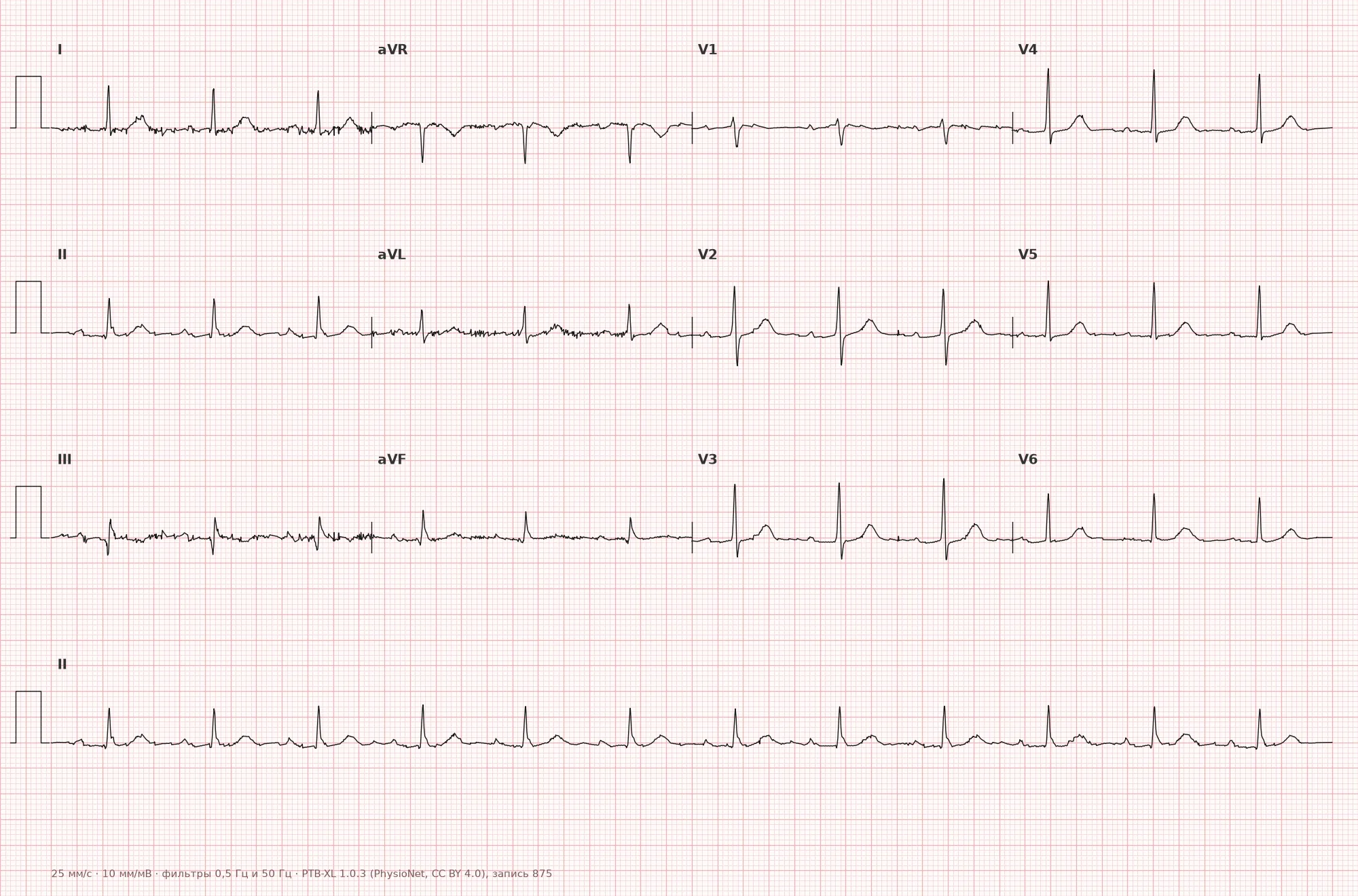
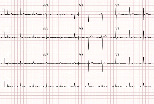
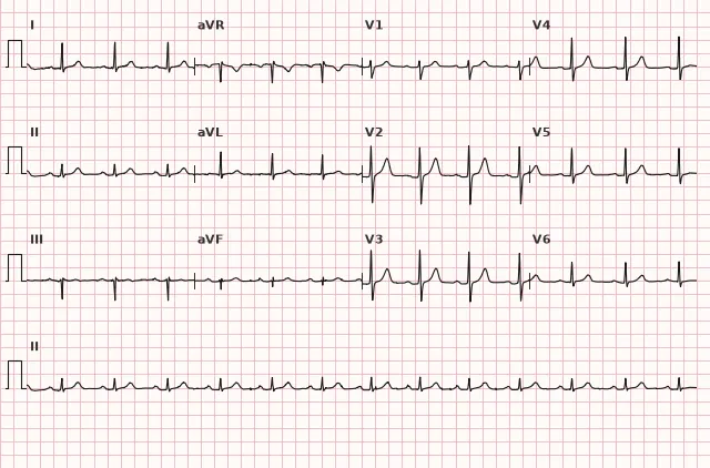
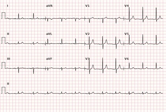
ECG from the open PTB-XL dataset (Wagner P. et al., Scientific Data, 2020; PhysioNet), CC BY 4.0 license. Rendering by WhiteCoat Pro.
Mechanism
The PQ (PR) interval reflects the total time of atrial depolarization and impulse propagation through the AV node, the bundle of His, and its branches prior to the onset of ventricular depolarization. The AV node accounts for the majority of this time (physiological delay), where conduction is slow due to the properties of nodal cells — this ensures mechanical synchronization: the atria have time to contract and fill the ventricles with blood before ventricular systole. Prolongation of the interval indicates slowed conduction at any of these levels — most commonly in the AV node, less frequently in the atria or the His-Purkinje system. Shortening indicates either accelerated AV conduction, bypass of the AV node via an accessory pathway (as in WPW syndrome), or a rhythm originating closer to the ventricles (e.g., from the AV junction).
Normal Variants
A short PR interval near the lower limit of normal (close to 120 ms) can be found in young individuals without signs of pre-excitation. A PR interval greater than 200 ms by definition corresponds to first-degree AV block: in elderly individuals, during sleep, and with high vagal tone, such prolongation may be physiological and clinically benign, but terminologically it remains first-degree AV block rather than a normal variant in the strict sense. A typical, though not absolute, physiological tendency is PR shortening during tachycardia and prolongation during bradycardia due to the rate dependency of AV conduction; fixed normal thresholds should not be mechanically adjusted based solely on heart rate, and interpretation must always be correlated with the clinical situation.
Pathological changes
Prolongation of the PQ (PR) interval greater than 200 ms:
- First-degree AV block (isolated delay in the AV node or His system)
- ischemia or myocardial infarction involving the AV node (most commonly inferior infarction involving the right coronary artery)
- myocarditis
- use of drugs that slow AV conduction (beta-blockers, calcium channel blockers, digoxin, class IA, IC, III antiarrhythmics)
- hyperkalemia
- increased vagal tone, hypothyroidism
- degenerative diseases of the conduction system (Lenègre-Lev disease)
Shortening of the PQ (PR) interval < 120 ms:
- ventricular pre-excitation pattern (WPW phenomenon) with a delta wave and widened QRS
- short PR without signs of pre-excitation (narrow QRS, no delta wave) — may be a normal variant or reflect accelerated AV conduction; the cause is assessed based on the clinical context and rhythm
- junctional rhythm or accelerated junctional rhythm
- sometimes a normal variant during tachycardia
Differential diagnosis
Prolonged PR in first-degree AV block should be differentiated from a concealed blocked atrial premature beat — in the latter case, the PQ (PR) interval preceding the dropped complex does not fit the regular sinus rhythm, and the P wave is deformed or superimposed on the preceding T wave.
Progressive PQ (PR) prolongation with periodic QRS dropping is second-degree AV block Mobitz type I, not true first-degree AV block (where drops do not occur at all).
A short PR with a delta wave (ventricular pre-excitation pattern, WPW pattern) is distinguished from a short PR without signs of pre-excitation by the presence of the delta wave itself and QRS widening — in the absence of pre-excitation, the QRS complex remains narrow. The diagnosis of WPW syndrome (rather than simply a pattern) is made only when pre-excitation is combined with symptomatic tachyarrhythmia.
In complete (third-degree) AV block, there is no fixed P-QRS relationship and no stable PQ (PR) interval; individual distances between P and the subsequent QRS complexes can be measured technically, but they vary and do not reflect AV conduction, so they are not used to assess AV conduction — this is sometimes confused with an extremely prolonged PR, although the correct interpretation is AV dissociation.
Clinical significance and management
Isolated first-degree AV block (PQ 200–220 ms) is usually benign and does not require treatment in the absence of symptoms and progression. PR prolongation can be a marker of conduction disturbance; the risk of progression to higher-degree block is assessed not by the PR value alone, but by the clinical context, QRS width, presence of symptoms, and dynamics on repeat ECGs. Marked prolongation (> 300 ms) can impair hemodynamics due to the loss of atrial contribution to ventricular filling and requires investigating the cause — ischemia, medications, electrolyte disturbances.
The physician should: compare with previous ECGs (new prolongation requires attention), evaluate medications that slow AV conduction, check potassium levels, and in the presence of symptoms (syncope, weakness) or progressive prolongation — perform Holter monitoring and consult a specialist to assess the risk of progression to higher-degree block.
An asymptomatic ventricular pre-excitation pattern (WPW pattern) requires risk assessment, particularly in young individuals and during physical exertion. In the presence of tachycardia paroxysms related to an accessory pathway (WPW syndrome), referral to a rhythm specialist is indicated to assess the possibility of accessory pathway catheter ablation.
Common interpretation errors
- Measuring the PQ (PR) interval in a lead where the onset of the P wave merges with the baseline and is poorly visible — choose the lead with the most distinct P and QRS onset, often lead II
- Confusing the PQ (PR) segment with the PQ (PR) interval — the segment does not include the P wave, while the interval does
- Failing to recognize progressive PQ (PR) prolongation prior to a dropped complex — a sign of Mobitz type I rather than simple prolongation
- Attempting to use individual P-QRS intervals during complete AV block as an indicator of AV conduction, even though there is no relationship between P and QRS
- Ignoring rate dependency — during tachycardia, physiological shortening is not always pathology, but this is no reason to mechanically alter fixed normal thresholds
PR is constantly prolonged — first-degree AV block; PR progressively lengthens with dropped QRS — Mobitz I; sudden dropped beat with a stable PR — Mobitz II
1. From which point to which point is the PQ (PR) interval measured?
The PQ (PR) interval includes the P wave and the PQ segment, measured from the onset of P to the beginning of QRS.
2. What PQ (PR) interval value is considered normal in adults?
The normal PQ (PR) interval in adults is 120–200 ms (0.12–0.20 s).
3. Which ECG feature differentiates ventricular pre-excitation (WPW) pattern from a short PR without pre-excitation?
In WPW, a short PQ (PR) is accompanied by a delta wave and a wide QRS due to ventricular pre-excitation via an accessory pathway, whereas with a short PR without pre-excitation, the QRS remains narrow and there is no delta wave.
Frequently asked questions
What is the normal PQ (PR) interval on an ECG?
In adults, the normal range is 120–200 ms (3–5 mm at a paper speed of 25 mm/s). In children, evaluation is based on age- and rate-dependent normative values; in adolescents, the upper limit may approach adult values. Prolongation exceeding 200 ms is the criterion for first-degree AV block.
Is prolongation of the PQ (PR) interval dangerous?
Isolated first-degree AV block is usually benign, especially when asymptomatic. The risk of progression to a higher-degree block depends not on the PR duration itself, but on the QRS width, suspected level of the block, structural heart disease, symptoms, and dynamics on repeat ECGs.
How to differentiate PQ (PR) shortening in pre-excitation syndrome from a normal variant?
In the ventricular pre-excitation (WPW) pattern, a short PQ (PR) is combined with a delta wave and a widened QRS complex due to the participation of an accessory conduction pathway. When PR is short without signs of pre-excitation, there is no delta wave and the QRS remains narrow — this can be a normal variant or reflect accelerated AV conduction.
Why does the PQ (PR) interval change with heart rate?
AV conduction is rate-dependent: during tachycardia, the impulse transit time through the AV node typically shortens physiologically, while during bradycardia, it prolongs. This is a typical adaptation rather than an absolute rule, so fixed normal thresholds are not mechanically adjusted based solely on heart rate.
Can the PQ (PR) interval be measured during complete AV block?
Not in a functional sense. In complete (third-degree) AV block, P waves and QRS complexes are unrelated — this is AV dissociation. Individual P-QRS distances can technically be measured, but they vary and are not used to assess AV conduction.
ECG Course
Interpretation from basics to myocardial infarction — learn to read any ECG using a systematic algorithm.
- Video lessons and theory for every topic
- Simulator case studies under every lesson
- Unlimited ECG simulator practice
- Study guide: "All ECGs in 25 minutes"
ECG Strip Simulator
ECG, radiology, histology, pathomorphology, pharmacology — thousands of flashcards with detailed explanations.
- Real ECG strips, not textbook drawings
- Instant answers and explanations
- Streaks and progress tracking in your dashboard
Related terms
More in the section «Basics: waves, intervals, axes»
Sources
- Goldberger's Clinical Electrocardiography, 9th ed.
- Hampton. The ECG Made Easy
- Surawicz, Knilans. Chou's Electrocardiography in Clinical Practice
- Orlov V.N. Guide to Electrocardiography
Educational material for medical students and physicians. Does not replace clinical guidelines and physical examination; the ECG must always be interpreted in conjunction with the clinical picture.