ECG criteria
- Find the J point — the transition point from QRS to the ST segment
- Determine the baseline: preferably the TP segment (between the end of the T wave and the beginning of the next P wave) if visible; if the TP segment is not identifiable (fusion with adjacent complexes, marked tachycardia), use the PR (PQ) segment preceding the QRS, taking into account possible PR depression in pericarditis and baseline drift
- Measure the vertical displacement of the ST segment from the baseline precisely at the J point (not at the peak or middle of the segment)
- In V2–V3 per AHA/ACCF/HRS 2009: threshold ≥2.0 mm in men and ≥1.5 mm in women; per later universal MI definitions: ≥2.5 mm in men <40 years, ≥2.0 mm in men ≥40 years, and ≥1.5 mm in women; these are criteria for assessing ST elevation in suspected acute MI/ACS, not an absolute upper limit of normal for asymptomatic individuals
- In leads other than V2–V3 (I, II, III, aVL, aVF, V1, V4–V6): elevation ≥1.0 mm in two contiguous leads is the threshold used when assessing acute coronary syndrome
- In V4R (right precordial leads): ST elevation, typically ≥1 mm, supports the diagnosis of right ventricular infarction, especially in inferior STEMI
- Assess segment morphology: convex ("tombstone"), convex-concave, hump-like, saddle-back
- Check for lead contiguity by myocardial wall and the presence of reciprocal ST depression in opposite leads
What it looks like on ECG
The ST segment is displaced above the baseline starting from the J point. The morphology of elevation is informative:
| Morphology | Typical cause |
|---|---|
| Convex upward (domed), "tombstone" | STEMI, acute coronary occlusion |
| Concave (saddle-back) | Early repolarization, pericarditis |
| Hump-like, merging with a tall narrow T wave | Hyperkalemia |
| Domed (coved) in V1/V2 with elevation ≥2 mm and negative T wave | Brugada pattern type 1 |
| Saddle-back in V1/V2 with positive/biphasic T wave | Suggestive Brugada pattern type 2 |
Diagnostic thresholds for ST elevation vary by lead groups: age- and sex-dependent values are used in V2–V3 (see "Criteria"), while other leads use a threshold of ≥1 mm of elevation in two contiguous leads of the same wall. Normal (physiological) ST elevation is particularly common in V2–V3 in young men due to early transition of the ST segment into a tall T wave — such a finding must be evaluated considering age, sex, ST-T morphology, and comparison with previous ECGs rather than automatically deemed pathological.
Changes in T-wave morphology complement the picture: hyperacute T waves are characteristic of the early phase of STEMI. Reciprocal ST depression in leads of the opposing wall increases the specificity of the finding for acute ischemia, although its absence does not rule out STEMI.
Real ECGs: 8 examples
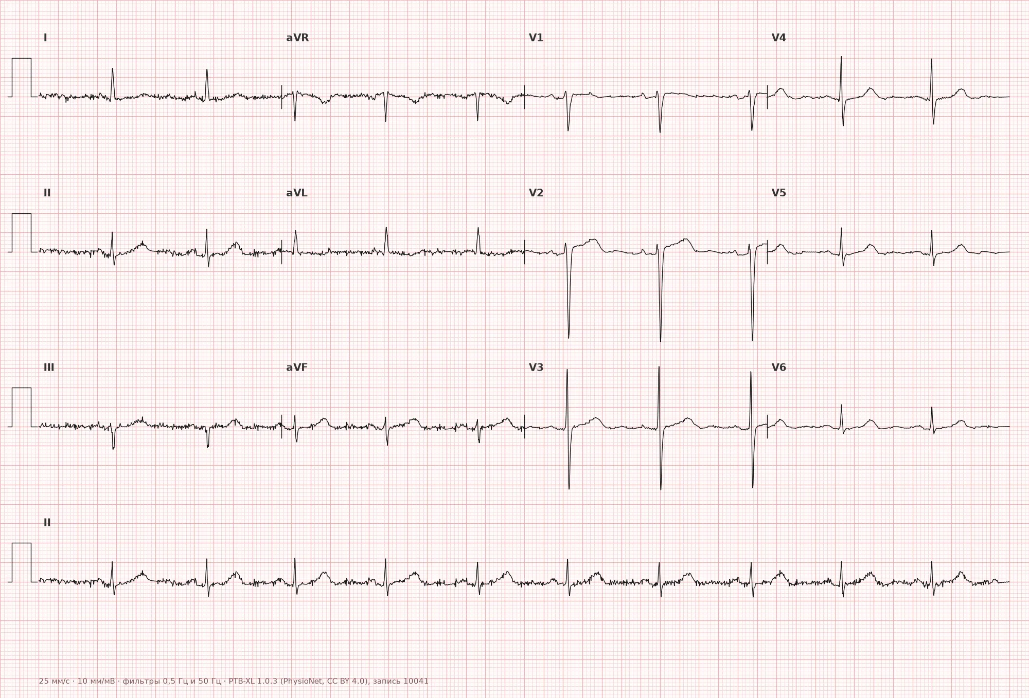
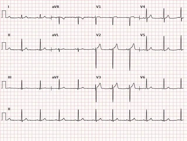
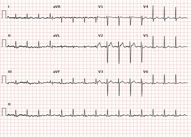
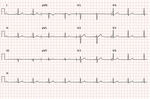
ECG from the open PTB-XL dataset (Wagner P. et al., Scientific Data, 2020; PhysioNet), CC BY 4.0 license. Rendering by WhiteCoat Pro.
Mechanism
In acute ischemia or myocardial injury, the action potential of cells in the ischemic zone changes (shortening of the plateau phase, resting potential alteration), creating a potential difference between the ischemic and healthy myocardium — the injury current. This shifts the integral ST vector away from leads facing the affected zone, producing ST elevation in those leads and often depression in the opposite leads (reciprocal). Important: ECG morphology alone does not prove the depth of injury (transmurality) or confirm necrosis — ST elevation reflects an electrical, not a histological event, and the diagnosis of infarction is established by combining clinical presentation, ECG dynamics, and biomarkers. For non-ischemic causes (pericarditis, early repolarization, hyperkalemia, Brugada syndrome), the mechanism is different — altering the epicardial action potential shape or ion channel anomalies, but the visual ECG outcome is similar.
Causes
- STEMI/classic STEMI is often caused by acute coronary occlusion, but ST elevation is non-specific; occlusive myocardial infarction (OMI) may occur without classic ST-elevation criteria
- Patterns suspicious for acute occlusion without classic ST elevation: de Winter waves, posterior myocardial infarction (ST depression in V1–V3 as a mirror image), diffuse ST depression with elevation in aVR, Aslanger pattern — require immediate expert evaluation and, upon confirmation of acute occlusion, urgent invasive strategy
- Prinzmetal angina (vasospasm)
- Pericarditis, myocarditis
- Early repolarization syndrome
- Brugada syndrome (type 1 pattern; type 2 pattern is a suggestive finding)
- Hyperkalemia
- Left ventricular aneurysm (persistent elevation following a prior infarction)
- Takotsubo cardiomyopathy
- Left bundle branch block, ventricular pacing (secondary ST changes)
- Pulmonary embolism (rarely, in right precordial leads)
- Subarachnoid hemorrhage and other cerebrovascular events
Differential diagnosis
- Early repolarization: J-point elevation with a notch or slur at the end of the QRS, usually concave ST elevation, prominent in inferior and/or lateral, sometimes anterior leads, stable dynamics, no reciprocal depression
- Pericarditis: diffuse concordant ST elevation in many leads without reciprocal changes (except aVR, sometimes V1), frequently PR-segment depression
- Brugada syndrome: the pattern is evaluated in V1–V2 (with high lead placement in the 2nd–4th intercostal spaces if necessary), V3 may be involved. Type 1 is the electrocardiographic diagnostic criterion: coved ST elevation ≥2 mm in V1 and/or V2 followed by a negative T wave; established on its own when this criterion is met. Type 2 (saddle-back, with a positive/biphasic T wave) is not a diagnostic, but a suggestive pattern requiring drug challenge testing for confirmation. The pattern may be intermittent (provoked by fever, drugs). Syncope, ventricular arrhythmias, and a family history of sudden cardiac death are not required to establish the type 1 ECG diagnosis, but are important for risk stratification and management
- Hyperkalemia: accompanied by tall narrow T waves, QT shortening, followed by QRS widening and P-wave flattening/disappearance as severity increases
- Left bundle branch block: discordant ST elevation, assessed using modified Sgarbossa (Smith) criteria
- LV aneurysm: persistent ST elevation in leads with a pathological Q wave, without dynamic changes on serial tracings and without acute pain
Clinical significance and management
An acutely developed ST elevation with ischemic symptoms requires immediate evaluation for STEMI: if criteria are met, emergency reperfusion (primary PCI within the target window, or thrombolysis per indications if unavailable). De Winter waves and posterior myocardial infarction indicate probable acute occlusion and require immediate evaluation for occlusive infarction with urgent invasive strategy. Diffuse ST depression with aVR elevation is a high-risk marker (potential multi-vessel or left main coronary artery disease), requiring urgent clinical assessment and often an early invasive strategy in instability, but it is not an automatic STEMI criterion or an indication for thrombolysis. If ST elevation does not fit STEMI and the clinical presentation is not acute, non-ischemic causes (pericarditis, early repolarization, Brugada, electrolyte abnormalities, normal variant) are sought, correlating with history, ECG dynamics, and, if necessary, biomarkers and echocardiography. The absence of chest pain does not rule out acute coronary occlusion, so any new or suspicious ST elevation requires urgent clinical assessment, serial ECGs, and troponin assay.
Common interpretation errors
- Measuring displacement not at the J point, but at the peak or middle of the ST segment — yields an incorrect value
- Using a single threshold (e.g., always 1 mm) without considering V2–V3 leads, sex, and age
- Diagnosing STEMI from a single lead without assessing contiguous leads of the same wall
- Ignoring patterns suspicious for acute occlusion in the absence of classic ST elevation
- Automatically interpreting ST elevation in aVR as proven left main occlusion without considering clinical presentation
- Failing to recognize the baseline ST morphology in young men (physiological elevation in V2–V3) as normal
1. At what point on the ECG is it correct to measure the magnitude of ST-segment elevation?
The standard for measuring ST elevation is the J point, the transition point from the QRS complex to the ST segment, relative to the baseline (TP, or PR-PQ if indistinguishable).
2. What ST-elevation threshold in leads V2–V3 in women is used as a criterion for ST elevation in suspected acute MI (AHA/ACCF/HRS, 2009)?
For women in V2–V3, the threshold is lower than for men and is ≥1.5 mm; for men according to the 2009 document, the threshold is ≥2.0 mm without age stratification (age stratification of 2.5/2.0 mm in men was introduced later in the Universal Definition of MI).
3. Which sign is most characteristic of pericarditis rather than STEMI?
In pericarditis, ST elevation is diffuse and concordant across many leads, and reciprocal changes typical of coronary occlusion are usually absent.
Frequently asked questions
How to distinguish ST elevation in myocardial infarction from early repolarization?
In myocardial infarction, ST elevation is typically convex ("tombstoning"), accompanied by reciprocal ST depression, dynamic progression over hours, and ischemic symptoms. In early repolarization, the elevation is concave, with a terminal QRS notch, stable over time, more common in young individuals without acute symptoms.
What ST-elevation threshold is considered diagnostic for acute ischemia?
In V2–V3 according to AHA/ACCF/HRS 2009 — ≥2.0 mm in men and ≥1.5 mm in women; according to later Universal Definitions of MI, a threshold of ≥2.5 mm in men under 40 years is also distinguished. In other leads — ≥1 mm in two contiguous leads. These are criteria for assessing ST elevation in suspected acute MI, not a universal normal limit, and they are always interpreted in conjunction with clinical presentation and dynamics.
Is ST elevation dangerous on its own?
The sign itself is non-specific: it can be a normal variant (early repolarization) or a sign of life-threatening coronary artery occlusion. The level of danger is determined by the clinical picture, elevation morphology, lead contiguity, and dynamics.
Should ST elevation without chest pain be treated as a myocardial infarction?
The absence of pain does not rule out myocardial infarction: acute occlusion may present with equivalent symptoms or be asymptomatic. New or suspicious ST elevation requires immediate clinical assessment, repeat ECG, troponin testing, and a decision on emergency reperfusion or invasive management in the presence of acute occlusion or instability signs.
How does ST elevation in aVR differ from typical STEMI?
ST elevation in aVR in the setting of diffuse ST depression in other leads is not an independent STEMI diagnosis, but a high-risk marker associated with severe ischemia, potential multivessel disease, or left main coronary artery disease. It requires urgent evaluation and often an early invasive strategy, but the diagnosis of left main occlusion is not made based on aVR alone.
ECG Course
Interpretation from basics to myocardial infarction — learn to read any ECG using a systematic algorithm.
- Video lessons and theory for every topic
- Simulator case studies under every lesson
- Unlimited ECG simulator practice
- Study guide: "All ECGs in 25 minutes"
ECG Strip Simulator
ECG, radiology, histology, pathomorphology, pharmacology — thousands of flashcards with detailed explanations.
- Real ECG strips, not textbook drawings
- Instant answers and explanations
- Streaks and progress tracking in your dashboard
Related terms
More in the section «Basics: waves, intervals, axes»
Sources
- AHA/ACCF/HRS Recommendations for the Standardization and Interpretation of the Electrocardiogram, 2009
- ESC Guidelines for the management of acute coronary syndromes, 2023
- Orlov, V.N. «Electrocardiography Guide»
- Surawicz B., Knilans T. Chou's Electrocardiography in Clinical Practice
Educational material for medical students and physicians. Does not replace clinical guidelines and physical examination; the ECG must always be interpreted in conjunction with the clinical picture.