ECG criteria
- T wave amplitude below baseline ≥ 1 mm (≥ 0.1 mV)
- New T wave inversion ≥ 1 mm in ≥ 2 contiguous leads with a predominant R wave is one of the potential signs of ischemia (Fourth Universal Definition of MI criteria); a separate and independent sign is new horizontal or downsloping ST-segment depression ≥ 0.5 mm in ≥ 2 contiguous leads. Symmetry of the inversion increases suspicion but is not part of the formal criterion; always correlate with clinical findings, ECG dynamics, and troponin levels.
- Deep symmetric negative T waves predominantly in V2–V4, classically often > 5 mm, although there is no strict mandatory threshold — Wellens' pattern type B; diagnosis requires an appropriate clinical context (recent chest pain, pain-free window at the time of recording) and assessment of QRS/ST dynamics, not just T wave depth.
- New dynamic inversion in ≥ 2 inferior leads (II, III, aVF) in the appropriate clinical setting may support ischemia or scar; isolated inversion in III is often non-specific.
- T wave inversion in lateral leads (I, aVL, V5–V6) — possible lateral ischemia, scar, or strain pattern in LVH.
- T wave inversion in V1–V2 (less commonly V3) without clinical symptoms can be a normal variant, especially in adolescents and young adults (juvenile pattern); in adults with inversion extending to V3 and beyond, ARVC and HCM must be ruled out.
- Isolated T wave inversion in aVR is almost always normal; in V1, it is a frequent normal finding.
- Diffuse T wave inversion (in multiple leads across different walls) — pericarditis, cardiomyopathy, pulmonary embolism, subarachnoid hemorrhage.
What it looks like on ECG
An inverted T wave points downward from the baseline. Depth is measured from the baseline to the lowest point of the wave in millimeters (1 mm = 0.1 mV at standard calibration). Morphology provides only an orientation, not a definitive diagnosis:
| Morphology | Characteristic | Possible significance |
|---|---|---|
| Symmetric (isosceles) | Smooth ascending and descending limbs | Acute ischemia, Wellens' syndrome, hypertrophic cardiomyopathy |
| Asymmetrical | Gradual onset, steeper descent | Nonspecific secondary changes, including post-ischemic states — requires correlation with QRS, ST, and prior ECGs |
| Biphasic (+/−) in V2–V3 | Initial positive part, terminal negative part | Possible Wellens' pattern type A in the appropriate clinical context |
| Biphasic (−/+) | Initial negative part, followed by positive part | Possible in hypokalemia, digitalis effect, pseudonormalization — interpret in context |
There are no rigid, unambiguous 'shape → cause' correlations: interpretation must always be combined with dynamics, clinical picture, and adjacent leads. The extent of inversion is crucial: isolated (single lead), regional (group of contiguous leads corresponding to an arterial territory), or diffuse (multiple leads across different walls).
Real ECGs: 8 examples
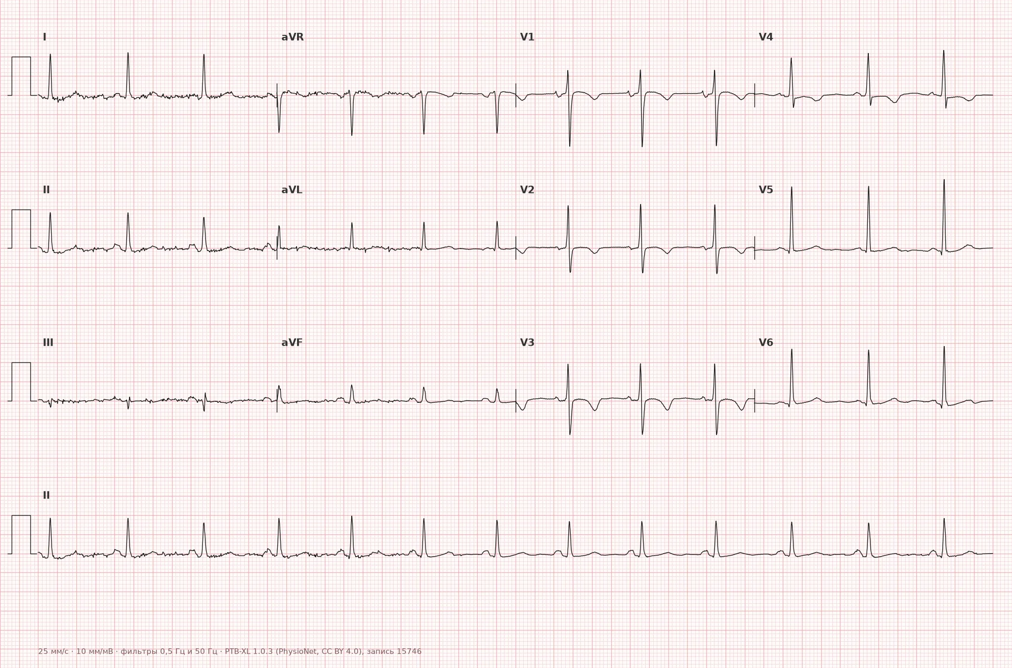
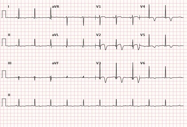
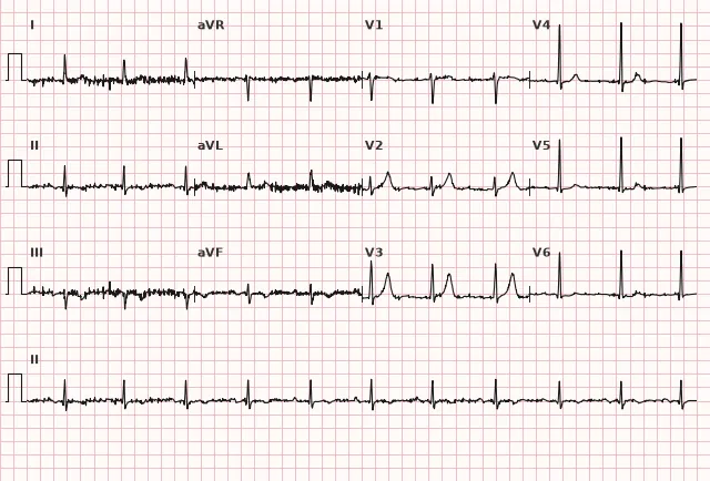
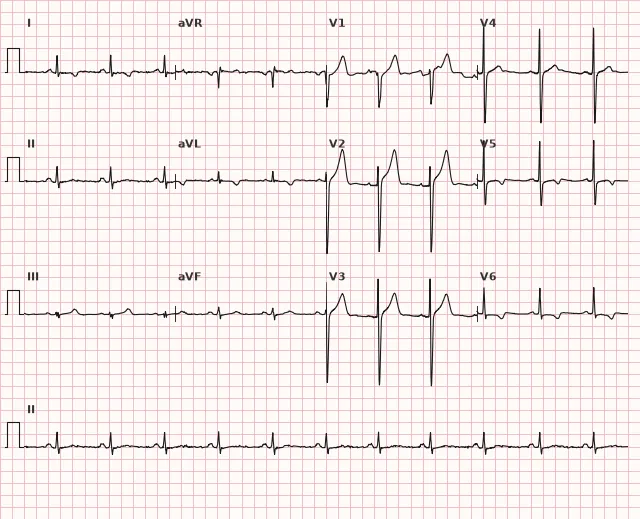
ECG from the open PTB-XL dataset (Wagner P. et al., Scientific Data, 2020; PhysioNet), CC BY 4.0 license. Rendering by WhiteCoat Pro.
Mechanism
Normally, ventricular repolarization generates a vector that produces a positive T wave in most leads. During ischemia, inflammation, hypertrophy, or scarring, the electrophysiology of myocardial cells changes, and the spatial repolarization gradient shifts — resulting in the net repolarization vector pointing away from the recording electrode, which yields a negative T wave. This is not simply a 'reversal from endocardium to epicardium': the vector direction is influenced by a combination of transmural and regional differences in action potential duration. The depth and symmetry of T wave inversion are not specific features exclusively of transmural ischemia — a similar picture occurs in Wellens' syndrome, hypertrophic cardiomyopathy, neurogenic disorders (subarachnoid hemorrhage), and other conditions.
Causes
Acute ischemia and infarction:
- High-risk Wellens' ECG pattern associated with critical left anterior descending artery stenosis and the risk of evolving anterior infarction
- Reperfusion following thrombolysis or PCI
- Posterior myocardial infarction (in conjunction with ST depression and tall R waves in V1–V3, confirmed by leads V7–V9)
Prior myocardial infarction:
- Secondary T wave changes in the scar area (nonspecific, not an independent sign)
Overload and hypertrophy:
- Left ventricular hypertrophy with systolic overload (strain pattern: T wave inversion in I, aVL, V5–V6)
- Hypertrophic cardiomyopathy (deep T waves in lateral and precordial leads)
- Cor pulmonale, pulmonary embolism (T wave inversion in III, aVF, V1–V4)
Inflammation:
- Myocarditis
- Pericarditis (diffuse inversion during the stage following ST normalization)
Electrolytes and other:
- Hypokalemia (flattening or slight inversion of T, appearance of U wave)
- Subarachnoid hemorrhage (deep, often symmetric T waves, frequently with QT prolongation)
Cardiomyopathies:
- Dilated, restrictive, Takotsubo
- Arrhythmogenic right ventricular cardiomyopathy (persistent T wave inversion in V1–V3 in adults, especially without right bundle branch block)
Normal variants:
- Juvenile pattern: T wave inversion in V1–V2, less commonly up to V3, in children and adolescents; usually resolves by late adolescence, though inversion in V1, and sometimes V2, may persist in healthy adults as a normal variant. Persistent inversion extending to V3 and beyond in an adult should be evaluated taking into account complete right bundle branch block, symptoms, and family history
- Pattern seen in some Black athletes (T wave inversion in V1–V4 combined with J-point elevation and characteristic ST elevation — not an isolated finding)
Differential diagnosis
Normal in aVR and V1: isolated T wave inversion in these leads is a frequent normal finding.
Left bundle branch block: secondary discordant ST-T changes are expected — the T wave direction is determined by the QRS polarity in each specific lead: where QRS is predominantly positive, ST is depressed and T is negative, whereas where QRS is predominantly negative, ST and T may be positive. Therefore, a negative T wave specifically in the lateral leads is not universal for LBBB. To assess ischemia in the presence of LBBB, Sgarbossa criteria and modified Sgarbossa (Smith) criteria are used.
WPW syndrome: T wave inversion in leads with a delta wave is a secondary change related to pre-excitation; it may persist for some time after the elimination of pre-excitation (electrical memory phenomenon) and does not disappear instantaneously.
Pacemaker: T wave inversion in leads opposite to the paced QRS vector is an expected secondary pattern.
Post-extrasystolic changes: transient T-wave inversion following frequent ventricular premature complexes or tachycardia paroxysms (cardiac memory, Chatterjee phenomenon) — reversible.
Hyperkalemia: typically presents with tall peaked T waves, shortened QT interval, followed by QRS widening and decreased P-wave amplitude; T-wave inversion is not a typical feature of hyperkalemia. If suspected, check potassium levels and assess serial ECGs.
Acute phase of pericarditis: at this stage, the T wave is usually positive; inversion appears later, after the ST segment returns to the baseline, and does not confirm the diagnosis on its own without meeting the appropriate criteria.
Clinical significance and management
The significance of T-wave inversion depends on the clinical context: leads involved, depth, symmetry, dynamics, and clinical presentation.
Deep symmetrical T-wave inversion, predominantly in leads V2–V4, in a patient with recent chest pain that has resolved by the time the ECG is recorded, indicates Wellens' syndrome (type B; type A is biphasic +/−). This is a high-risk ECG pattern associated with critical stenosis of the left anterior descending artery and the risk of evolving anterior myocardial infarction, though it is not direct proof of a specific stenosis or ongoing occlusion; troponin levels may be normal or slightly elevated. Urgent hospitalization and immediate consultation for coronary angiography are required; stress testing is contraindicated. The specific timeframe for coronary angiography is determined by clinical presentation, troponin and ECG dynamics, and NSTE-ACS risk stratification scores: for confirmed high-risk NSTE-ACS, an early invasive strategy within 24 hours should be considered (ESC 2023, class IIa); for very high-risk patients (ongoing or recurrent ischemia, cardiogenic shock or hemodynamic instability, life-threatening arrhythmias), an immediate invasive strategy is indicated.
New T-wave inversion in several contiguous leads in a patient with pain or dyspnea requires ruling out acute coronary syndrome (troponins, serial ECGs), pulmonary embolism (D-dimer, CT angiography), and myocarditis (troponins, cardiac MRI).
Persistent T-wave inversion in leads documenting a prior myocardial infarction is likely a secondary scar phenomenon; it requires comparison with prior ECGs rather than independently altering patient management.
Diffuse T-wave inversion is evaluated alongside the clinical picture: diagnosing acute pericarditis requires its own criteria (chest pain, pericardial friction rub, diffuse ST-elevation with PR depression in an earlier stage, effusion) rather than T-wave inversion alone. Once confirmed, treatment is determined by clinical guidelines considering contraindications. Deep inversion accompanied by signs of instability or shock requires ruling out myocarditis (troponins, cardiac MRI, hospitalization).
T-wave inversion in leads V1–V3 in asymptomatic young individuals most commonly corresponds to a juvenile pattern, but in the presence of syncope, a family history of sudden cardiac death, or persistent changes in an adult, arrhythmogenic right ventricular cardiomyopathy and hypertrophic cardiomyopathy must be ruled out (echocardiography, cardiac MRI, and Holter monitoring as indicated).
In left ventricular hypertrophy with a strain pattern (T-wave inversion in lateral leads) — monitor blood pressure, treat hypertension, and assess diastolic function.
Common interpretation errors
- Inappropriately attributing T-wave inversion to age: persistent T-wave inversion in V1–V3 in an adult (especially beyond adolescence) should not automatically be considered a normal variant — symptoms, family history, and, if ARVC/HCM is suspected, referral for echocardiography or cardiac MRI are necessary.
- Missing Wellens' syndrome: deep symmetrical negative T waves in V2–V4 in a patient whose chest pain resolved several hours or days ago are mistaken for an old scar. In reality, this is a high-risk pattern in a "pain-free window" — urgent evaluation and urgent coronary angiography are required, without a stress test.
- Failure to review prior ECGs: a new T-wave inversion is a red flag, but without comparing it to an old ECG, acute ischemia may be mistaken for a chronic finding or vice versa. Always request previous tracings.
- Overestimating T-wave inversion in aVR and V1: isolated negative T waves in these leads are a common normal variant and are not a cause for concern.
- Ignoring secondary changes: in left bundle branch block or cardiac pacing, T-wave vector is determined by QRS polarity and depends on discordance, rather than being a direct sign of ischemia. Such T waves cannot be interpreted directly — modified Sgarbossa criteria or comparison with previous ECGs are required.
"Symmetrical T wave — look for ischemia" (deep symmetrical T-wave inversion is an indication for urgent clinical evaluation, but not a standalone diagnosis)
1. A 32-year-old asymptomatic patient is found to have negative T waves in V1–V3 with a depth of 3 mm on a routine ECG. What is the correct management strategy for the physician?
T-wave inversion in V1–V3 in an adult is not automatically a juvenile normal variant — this pattern is associated, in particular, with arrhythmogenic right ventricular cardiomyopathy. Clinical presentation, history, and, when suspected, imaging are required rather than dismissing it as normal based solely on age.
2. A 58-year-old patient presented with chest pain 3 hours ago, currently pain-free. ECG shows deep (7 mm) symmetrical negative T waves in V2–V4, slightly elevated troponin, stable condition, no recurrent pain. Management?
Wellens' syndrome (type B) is associated with critical stenosis of the left anterior descending artery. Stress testing is contraindicated; urgent hospitalization and coronary angiography are required. In a stable patient without very high-risk features, an early invasive strategy within 24 hours should be considered. Thrombolysis is not indicated — there is no ST elevation ≥ 1 mm.
3. ECG shows negative T waves in I, aVL, V5–V6, ECG signs of left ventricular hypertrophy by Sokolow-Lyon voltage criteria, BP 180/100 mm Hg. What is this?
T-wave inversion in lateral leads in the presence of LVH ECG criteria and high blood pressure is a classic strain pattern. These are secondary repolarization changes due to myocardial overload, not acute ischemia.
Frequently asked questions
Does a negative T wave always indicate heart problems?
No. Isolated T-wave inversion in aVR and frequently in V1 is normal. In adolescents and some young adults, T-wave inversion in V1–V2 (infrequently up to V3) can be a normal variant (juvenile pattern). However, a new T-wave inversion, inversion in two or more contiguous leads, or deep symmetrical inversion requires ruling out acute pathology.
How to distinguish acute ischemia from an old scar based on a negative T wave?
The T-wave morphology itself (depth, symmetry) does not reliably differentiate between an acute process and a scar. The key factors are serial ECG dynamics, clinical presentation (recent chest pain, dyspnea), troponin levels, and comparison with old tracings. The presence of a pathological Q wave points more toward prior infarction but does not rule out new ischemia in the same area.
What is Wellens syndrome and why is it dangerous?
Wellens' syndrome is a high-risk ECG pattern associated with critical stenosis of the proximal left anterior descending artery: deep symmetrical negative T waves in V2–V4 (type B) or biphasic T waves +/− (type A) in a patient with recent chest pain that has resolved by the time the ECG is recorded. This pattern carries a high risk of extensive anterior myocardial infarction within the next few days and requires urgent hospitalization and coronary angiography, even if troponin is not elevated; stress testing is contraindicated.
Can T-wave inversion resolve on its own?
Yes, in reversible conditions: reperfusion following an acute ischemic episode, resolution of myocarditis or pericarditis, correction of electrolyte disturbances, resolution of cardiac memory after elimination of arrhythmia or pre-excitation (although this may take time). Secondary T-wave inversion associated with permanent structural changes (e.g., a large scar) usually remains persistent.
Is T-wave inversion in the inferior leads (II, III, aVF) dangerous?
Depends on the clinical context. A new inversion in two or more inferior leads accompanied by symptoms may indicate inferior ischemia and requires urgent evaluation (troponins, serial ECGs). Isolated shallow inversion in lead III without other changes is often nonspecific and seen in healthy individuals, especially with a vertical cardiac axis.
ECG Course
Interpretation from basics to myocardial infarction — learn to read any ECG using a systematic algorithm.
- Video lessons and theory for every topic
- Simulator case studies under every lesson
- Unlimited ECG simulator practice
- Study guide: "All ECGs in 25 minutes"
ECG Strip Simulator
ECG, radiology, histology, pathomorphology, pharmacology — thousands of flashcards with detailed explanations.
- Real ECG strips, not textbook drawings
- Instant answers and explanations
- Streaks and progress tracking in your dashboard
Related terms
More in the section «Basics: waves, intervals, axes»
Sources
- Surawicz B., Knilans T. Chou's Electrocardiography in Clinical Practice, 6th ed., 2008
- Orlov V.N. Manual of Electrocardiography, 9th ed., 2017
- ESC Guidelines for the management of acute coronary syndromes, 2023
- Goldberger A.L. Clinical Electrocardiography: A Simplified Approach, 9th ed., 2017
Educational material for medical students and physicians. Does not replace clinical guidelines and physical examination; the ECG must always be interpreted in conjunction with the clinical picture.