ECG criteria
- Evaluate QRS and signs of hypertrophy, bundle branch block, or pre-excitation; if present, ST-T changes are interpreted as secondary rather than primarily non-specific; a narrow normal QRS facilitates interpretation, but is not mandatory
- The T wave is flattened, mildly or moderately changed in shape, asymmetrically inverted — without a fixed amplitude threshold, taking into account the lead, sex, age, and previous ECGs
- The ST segment is shifted (depression or elevation) without a specific pattern; there is no strict numerical range for "non-selectivity" — for ST depression, evaluate horizontal or downsloping depression separately in two contiguous leads according to ACS criteria; for elevation, evaluate STEMI/occlusion myocardial infarction criteria taking into account the lead and sex
- Changes often do not form an anatomically convincing coronary pattern and lack the appearance of typical reciprocal changes, but this is a guiding, not mandatory sign: the absence of such a picture does not rule out ischemia or occlusion myocardial infarction
- The stability of the pattern when compared with old ECGs increases the likelihood of a chronic benign finding, but a new or dynamic pattern does not rule out non-specificity and requires ruling out ACS and other acute causes
- Clinical context (chest pain, dyspnea, instability) is not included in the ECG criteria, but determines the significance of the finding and the need for further examination — the ECG by itself does not rule out ACS, PE, or pericarditis
- Often, but not always, the changes are diffuse, across several groups of leads without anatomical focus logic
What it looks like on ECG
The term describes ST-T abnormalities that a physician cannot confidently attribute to a specific cause. Severity varies — from barely noticeable to moderate, sometimes significant; the common thread is that the pattern does not form a recognizable specific pattern. Most often these are:
| Element | Parameter | Typical leads |
|---|---|---|
| T wave | flattened (low-amplitude) or slightly/moderately negative, asymmetric | II, III, aVF, V4–V6, sometimes diffuse |
| ST segment | depression or elevation without a specific pattern, of various shapes | any, often inferolateral |
| QRS | usually normal width and shape | — |
The term means the absence of a convincing specific diagnostic pattern on a given ECG: there is no clear picture of ischemia, pericarditis, hypertrophy with overload, drug effect, or pre-excitation syndrome. Separately, it must be remembered: horizontal or downsloping ST depression in two contiguous leads is a potentially ischemic sign, not a typical safe manifestation of non-specificity, and when identified, evaluation is always carried out according to ischemia criteria regardless of the initial descriptive wording. The finding may be new, dynamic, and accompany a serious disease, so it does not rule out a pathological process and does not replace clinical evaluation with dynamic monitoring. In depolarization disorders (bundle branch block, pre-excitation, pronounced hypertrophy), ST-T changes should primarily be interpreted as secondary rather than non-specific. The amplitude of deviations is often close to the limit of measurement error (at standard calibration of 10 mm/mV, 1 mm = 0.1 mV), so the conclusion is largely descriptive and depends on the context.
Real ECGs: 8 examples
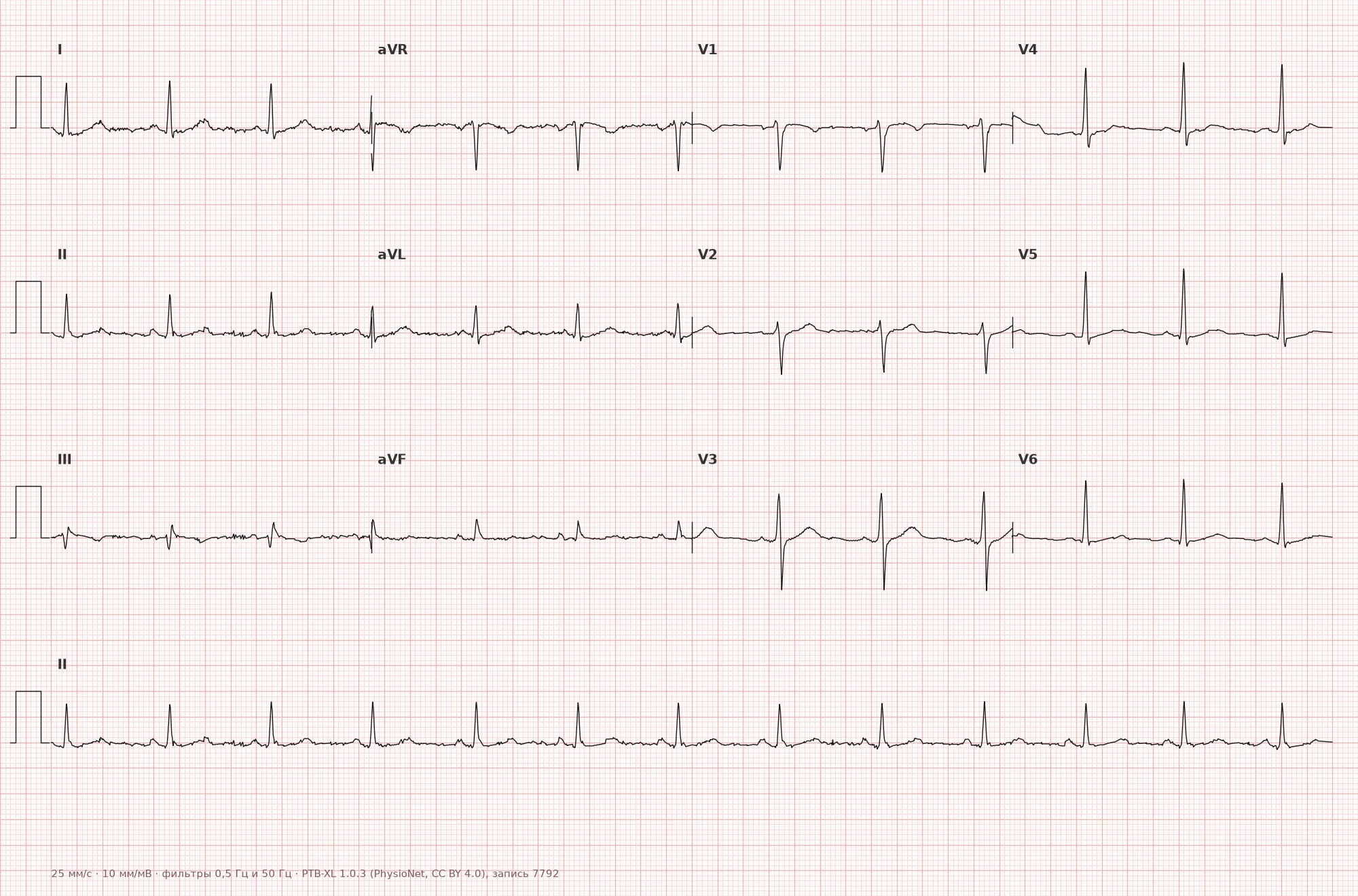
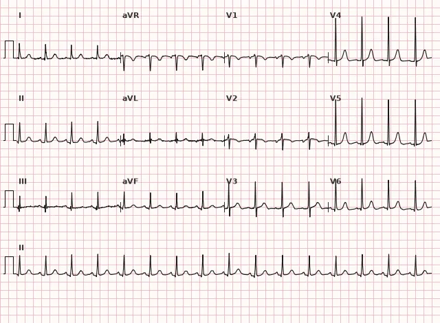
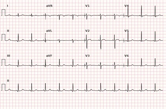
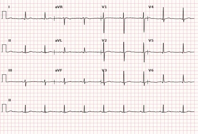
ECG from the open PTB-XL dataset (Wagner P. et al., Scientific Data, 2020; PhysioNet), CC BY 4.0 license. Rendering by WhiteCoat Pro.
Mechanism
The T wave and ST segment reflect the ventricular repolarization phase. Any factor altering the rate or homogeneity of repolarization — electrolyte shift, autonomic tone, mild hypertrophy without diagnostic voltage criteria, fibrosis, medication intake, hyperventilation, body position, electrode placement artifact — can alter T-wave shape and ST position without gross myocardial electrophysiology disruption. Depolarization disorders (bundle branch block, pre-excitation, pronounced hypertrophy) also alter repolarization, but then ST-T changes are considered secondary rather than primarily non-specific. Since there are many causes and the mechanism is non-specific, the changes do not form a recognizable pattern of a specific disease.
Causes
- Autonomic lability, hyperventilation, anxiety, pain
- Electrolyte fluctuations, including hypokalemia
- Drug effect during therapeutic administration (digoxin, diuretics, psychotropics); in suspected digoxin toxicity — separate assessment of concentration, renal function, electrolytes, and clinical picture
- Initial or minimal left ventricular hypertrophy without voltage criteria
- Obesity, chest wall deformity, diaphragmatic position
- Age-related myocardial changes, fibrosis
- Prior myocardial infarction or other structural heart disease; in the absence of specific ECG features, changes may remain indeterminate
- Infectious diseases, fever
- Technical recording artifacts (poor electrode contact, tremor)
- Idiopathic variant in healthy individuals, especially women
Differential diagnosis
- Myocardial ischemia — suspicious for new or dynamic horizontal/downsloping ST depression in two contiguous leads or progressive T-wave inversion; specific threshold and significance depend on sex, lead, baseline ECG, and clinical presentation; absence of pronounced changes does not rule out ACS
- Wellens' syndrome — high-risk clinical-ECG pattern after an episode of chest pain: deep symmetric inversion or biphasic T wave, most often in V2–V3, with preserved R-wave progression, minimal ST elevation, and usually without significant cardiac marker elevation; often associated with proximal LAD stenosis, but not an absolute sign of stenosis and requires urgent NSTE-ACS management rather than the primary PCI window adopted for STEMI
- Pericarditis — diffuse, usually concave ST elevation, typically with PR depression; in aVR (and sometimes V1) — ST depression and PR elevation; diagnosis is established by clinical criteria, ECG signs are only part of them
- LV hypertrophy with systolic overload — asymmetric T-wave inversion combined with high QRS voltage and LVH criteria
- Digoxin effect — scoop-shaped ST depression (differentiated by shape, not amplitude) during therapeutic administration
- PE — S1Q3T3 and T-wave inversion in right chest leads are possible with right ventricular overload, but have low sensitivity and specificity; diagnosis is established by clinical probability and imaging/validated algorithm
- Normal variant (juvenile T wave) — in young individuals, especially in V1–V3, without other findings
Clinical significance and management
The finding by itself does not indicate a specific disease; its significance is determined by clinical presentation, dynamics, and concomitant findings. The physician needs to: compare with previous ECGs (stability is an argument in favor of benignancy, but not absolute); evaluate clinical presentation (chest pain, dyspnea, syncope — a reason to rule out an organic cause); ensure correct electrode placement and recording quality; purposefully check electrolytes and renal function in the presence of appropriate risk factors or suspicion of a drug or metabolic mechanism, and thyroid function and magnesium according to clinical indications, rather than routinely in every asymptomatic patient. In chest pain or instability, a non-specific initial ECG should not delay treatment: immediate clinical evaluation, recording and interpretation of a 12-lead ECG without delay (typically within 10 minutes), serial high-sensitivity troponin determination using a validated algorithm, repeat ECG for persistent or recurring symptoms, and emergency tactics for high risk according to the non-ST-segment elevation ACS approach are required; comparison with old tracings is useful, but should not delay this evaluation. If the finding is incidental and asymptomatic — observation is usually sufficient; if reproducible on different tracings, it should not be overdiagnosed as pathology.
Common interpretation errors
- Write "non-specific ST-T changes" instead of looking for a specific cause (electrolytes, medications, hyperventilation)
- Miss true ischemia or occlusion myocardial infarction hidden under the wording "non-specific" when clinical presentation is present, especially with minimal or atypical ECG findings
- Uncomparable to old tracings — without comparison, it is impossible to evaluate whether a finding is new or chronic
- Misinterpreting an artifact (tremor, poor electrode contact) as an organic finding
- Using the term as a diagnosis rather than a descriptive conclusion requiring clinical interpretation
1. Which sign is most characteristic of nonspecific ST-T changes?
The defining feature of the term is the absence of a specific pattern, not necessarily a small amplitude; this distinguishes it from Wellens' syndrome, pericarditis, or the digitalis effect.
2. What should be done first when identifying nonspecific ST-T changes in a patient with chest pain?
When ACS is suspected, the priority is immediate clinical assessment and an ECG without delay, along with serial biomarkers; comparison with an old ECG is useful but does not replace or delay these actions.
3. Which statement about acute coronary occlusion is true in the context of nonspecific ST-T changes?
Occlusion myocardial infarction can manifest atypically without diagnostic ST elevation, so the absence of a typical pattern does not rule out acute coronary occlusion in the presence of a suspicious clinical picture.
Frequently asked questions
Are nonspecific ST-T changes dangerous?
By themselves, usually no — this is a descriptive conclusion, not a diagnosis. The risk is determined by the clinical context: in the presence of chest pain or instability, ACS must be ruled out using serial ECGs and troponin, whereas an asymptomatic, incidental, stable finding is most often benign.
How to distinguish nonspecific changes from ischemia?
Suspicion of ischemia is increased by new or dynamic ST-T changes in adjacent leads corresponding to a specific coronary territory, along with a matching clinical presentation. However, the absence of pronounced changes, typical localization, or dynamics does not rule out ACS — occlusion myocardial infarction sometimes presents with a minimal or atypical picture, which is why serial ECGs and high-sensitivity troponin play a decisive role.
Do nonspecific ST-T changes need to be treated?
Treatment targets the cause rather than the ECG finding itself, if identified (electrolyte imbalance, medication, anxiety). In the absence of symptoms and with a stable picture, no additional treatment is required.
Could this be a normal variant?
It can be, especially in young people during hyperventilation or anxiety, but the conclusion depends on age, symptoms, risk factors, recording quality, and comparison with previous ECGs. A new or persistent finding warrants clinical evaluation rather than automatic reassurance.
What to do if the finding appears for the first time?
Compare with previous ECGs, evaluate the clinical picture and contributing factors (electrolytes, medications, electrode placement), repeat the recording if in doubt, and determine serial troponins without delaying clinical assessment if an acute condition is suspected.
ECG Course
Interpretation from basics to myocardial infarction — learn to read any ECG using a systematic algorithm.
- Video lessons and theory for every topic
- Simulator case studies under every lesson
- Unlimited ECG simulator practice
- Study guide: "All ECGs in 25 minutes"
ECG Strip Simulator
ECG, radiology, histology, pathomorphology, pharmacology — thousands of flashcards with detailed explanations.
- Real ECG strips, not textbook drawings
- Instant answers and explanations
- Streaks and progress tracking in your dashboard
Related terms
More in the section «Basics: waves, intervals, axes»
Sources
- Orlov, V.N. «Electrocardiography Guide»
- Goldberger's Clinical Electrocardiography: A Simplified Approach
- Surawicz B., Knilans T. Chou's Electrocardiography in Clinical Practice
Educational material for medical students and physicians. Does not replace clinical guidelines and physical examination; the ECG must always be interpreted in conjunction with the clinical picture.