ECG criteria
- Sinus rhythm: P wave preceding each QRS complex, positive in II, negative in aVR
- Heart rate 60–100 bpm; in sinus arrhythmia (especially in young individuals), RR intervals vary cyclically with respiration, while P morphology and P–QRS association are preserved
- P wave: duration ≤120 ms, amplitude ≤2.5 mm in lead II
- PR interval: 120–200 ms
- QRS complex: duration typically 70–110 ms, normal <120 ms; QRS of 110–119 ms requires morphological evaluation
- Pathological Q wave excluded: criteria are ≥30 ms and ≥1 mm in depth in two contiguous leads, or a QS complex; leads V2–V3 utilize separate criteria (e.g., a QS complex can be normal within the transition zone); small q waves <30 ms and <1 mm are acceptable in I, aVL, V5–V6
- Corrected QT interval (QTc): upper limit approximately 440–450 ms in men, 450–460 ms in women (calculated using Bazett's formula; Fridericia's or Hodges' formulas are preferred in case of heart rate extremes)
- ST segment close to baseline; T wave positive in I, II, V3–V6; electrical axis of the heart between −30° and +90°
What it looks like on ECG
On a normal ECG, the P wave precedes every QRS complex and is positive in leads I, II, aVF, and most precordial leads (may be biphasic in V1), and negative in aVR. P wave duration does not exceed 120 ms, and amplitude does not exceed 2.5 mm in lead II.
The PR interval includes intra-atrial, AV nodal, and initial His-Purkinje conduction; the AV node provides the primary physiological delay. The PR interval is stable, measuring 120–200 ms. The QRS complex is narrow: typically 70–110 ms, with up to 120 ms considered acceptable. For a QRS duration of 110–119 ms, morphology should be assessed—this may be a normal variant, incomplete bundle branch block, or nonspecific intraventricular conduction delay. In the precordial leads, the R wave progresses in amplitude from V1 to V4; the transition zone is typically located in V3–V4, though variants exist. R-wave progression should be evaluated taking electrode placement, axes, and other signs into account, rather than formally.
Physiological J-point/ST elevation differs from ischemic elevation: normal ST elevation is particularly prominent in V2–V3 and depends on sex and age. In men, elevation up to 2–2.5 mm is permissible in V2–V3, while thresholds are lower in women. When acute myocardial infarction is suspected, the J-point is evaluated in two contiguous leads using specific criteria for each localization, accounting for sex, age, and clinical presentation; fixed values of 0.5–1 mm are not applicable to all leads. The T wave is positive in leads I, II, and the left precordial leads; it may be negative in III, aVR, and V1 as a normal variant. The corrected QT interval does not exceed normal limits for age and sex. The electrical axis of the heart normally ranges from −30° to +90°, although vertical and horizontal positions are also found in healthy individuals.
| Parameter | Normal |
|---|---|
| HR | 60–100/min |
| PQ | 120–200 ms |
| QRS | 70–110 ms, <120 ms |
| QTc | up to 440–460 ms |
| QRS Axis | −30°…+90° |
Real ECGs: 8 examples
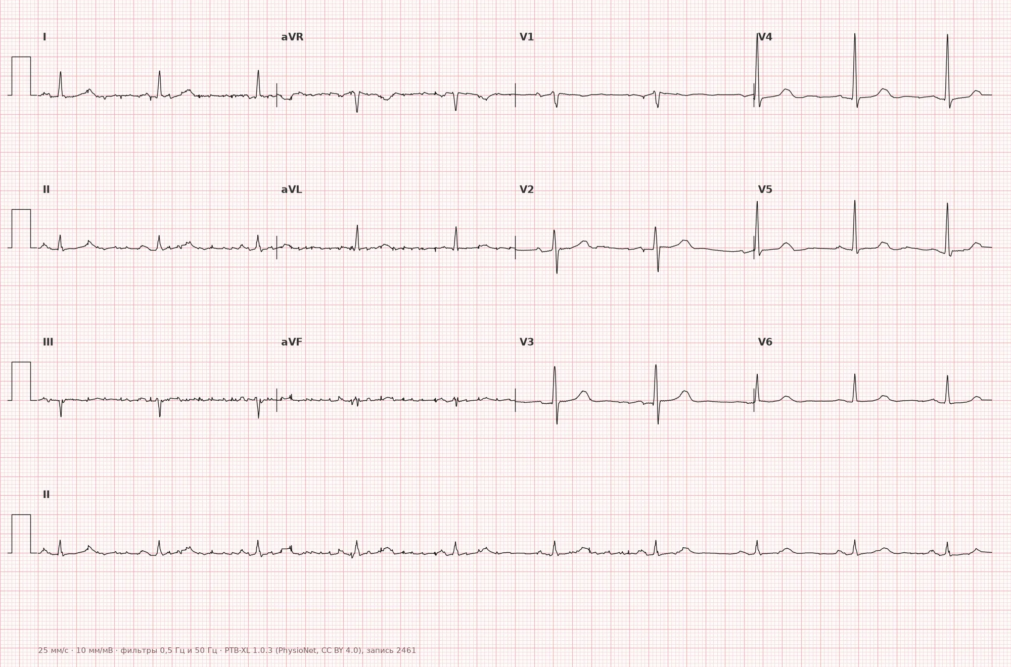
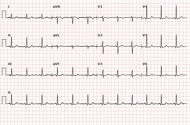
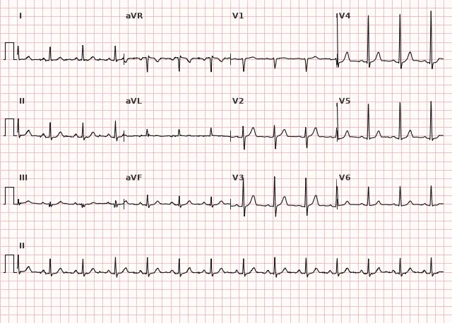
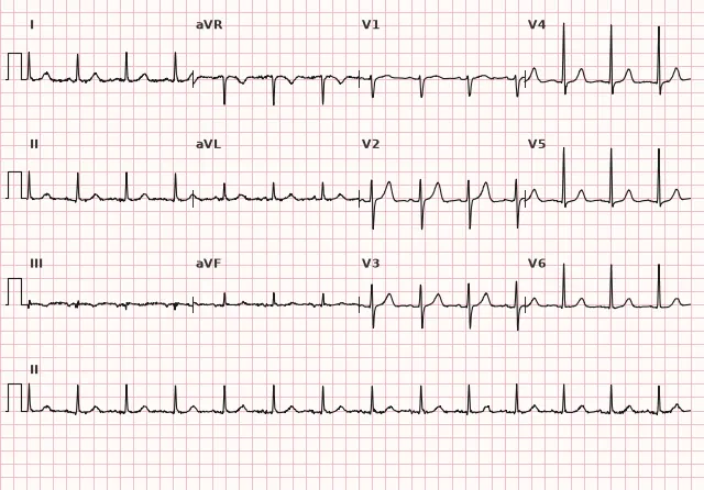
ECG from the open PTB-XL dataset (Wagner P. et al., Scientific Data, 2020; PhysioNet), CC BY 4.0 license. Rendering by WhiteCoat Pro.
Mechanism
A normal ECG reflects the physiological sequence of cardiac activation: the impulse originates in the sinus node, propagates through the atria (P wave), is then delayed in the AV node allowing the atria to contract fully, and rapidly spreads through the His-Purkinje system to the ventricles (QRS complex). The ST segment corresponds to the plateau phase of the ventricular action potential (Phase 2), while the primary visible ventricular repolarization is represented by the T wave. When the anatomy of the conduction system and myocardium is intact, all these stages fall within standard time and amplitude parameters.
Normal Variants
- Sinus (respiratory) arrhythmia, especially in young individuals and children — RR intervals vary with respiration (increasing during inspiration, decreasing during expiration), while P and QRS morphology remain unchanged
- Vertical or horizontal electrical axis without signs of hypertrophy
- Early ventricular repolarization — J-point elevation/notching at the end of the QRS complex, typically ≥1 mm in at least two contiguous leads, most often inferior and/or lateral, frequently accompanied by concave ST elevation and tall T waves; in the absence of reciprocal ST depression and in the presence of ischemic symptoms, serial ECGs, clinical context, and troponin levels are required to rule out ACS
- Juvenile T-wave pattern — negative symmetrical T waves in V1–V3 in children and young women, shallow, without dynamic changes between ECGs or clinical symptoms
- Normal rSr' variant in V1–V2 with a narrow QRS — differentiation from incomplete RBBB requires morphological criteria (presence of terminal S in I/V6), electrical axis position, and clinical information; a QRS <110 ms can correspond to either a normal variant or incomplete RBBB; morphology, not just QRS width, is the decisive feature
- Low voltage associated with obesity and emphysema: <5 mm in all limb leads and/or <10 mm in all precordial leads; supplemented by echocardiography and clinical context to rule out effusion, infiltrative diseases, or amyloidosis
Differential diagnosis
- Sinus arrhythmia vs. sinoatrial block — in arrhythmia, RR fluctuations are smooth and respiration-dependent, the P wave does not change in morphology or position, and it follows a predictable pattern aligned with the respiratory cycle
- Early repolarization vs. ischemic ST elevation — in early repolarization, the elevation is concave, the T wave is tall, there is no reciprocal ST depression in opposite leads, and symptoms are absent; ACS presents with dynamic ECG changes, clinical signs, and elevated troponin
- Normal rSr' variant vs. incomplete RBBB — incomplete block is identified by characteristic QRS morphology in V1–V2 (rSr') and a terminal S wave in I/V6 with a QRS <120 ms; the QRS duration typically ranges from 110–119 ms, but rSr' with a QRS of 100–110 ms may also represent incomplete block; morphology and electrical axis are evaluated, not just width
- Juvenile T wave vs. ischemia — negative T waves are symmetrical, shallow, most commonly in V1–V3, without dynamic evolution between serial ECGs, and without clinical symptoms
- Low voltage due to obesity/emphysema vs. pathological low voltage (effusion, amyloidosis, infiltration) — differentiated based on clinical presentation, echocardiography, and laboratory findings
Clinical significance and management
A normal resting ECG does not rule out structural heart disease — early-stage valvular defects, cardiomyopathies, stable coronary artery disease between attacks, or paroxysmal arrhythmias. It is a snapshot in time, not a definitive exclusion of disease. In the presence of typical clinical symptoms (chest pain, syncope, palpitations), a normal ECG warrants further investigation: serial ECGs (at 0–3–6 hours or later if symptoms persist when ACS is suspected), Holter monitoring (for palpitations, syncope), stress testing (only after ruling out acute coronary syndrome and under appropriate indications), and echocardiography as indicated. When acute coronary syndrome is suspected, prompt clinical evaluation, repeat ECGs, and high-sensitivity troponin measurement according to an algorithmic approach (typically 0–3 hours) are indicated; in the absence of STEMI and with an initially normal ECG, early rule-out algorithms for NSTE-ACS are used rather than immediate exercise testing, which is contraindicated in acute syndromes.
Common interpretation errors
- Failing to check calibration (speed 25 mm/s, amplitude 1 mV = 10 mm) prior to evaluating amplitudes and intervals, leading to erroneous conclusions regarding high voltage, low voltage, or complex durations
- Measuring intervals and amplitudes "by eye" without using a caliper or dividers, resulting in overestimation or underestimation of durations; for example, a QRS complex may appear to be 120 ms when it is actually 110 ms, or vice versa
- Mistaking normal variants (early repolarization, juvenile T waves, moderate sinus arrhythmia, normal rSr' in V1–V2) for pathology, and vice versa; diagnosing incomplete RBBB in the presence of an rSr' pattern and a QRS <110 ms without assessing morphology and S waves in lateral leads
- Ignoring artifacts (baseline drift, 60 Hz interference, motion tremor) and misinterpreting them as ischemic ST-T changes or arrhythmias
- Making conclusions based on a single lead without comparing the pattern across all 12; for example, evaluating ST in V2–V3 without considering adjacent leads, sex, age, and clinical context; mistaking physiological elevation for STEMI
1. Which QRS complex duration corresponds to the normal width?
The normal QRS width is usually 70–110 ms. Complete bundle branch block is defined at QRS ≥120 ms. The 110–119 ms range requires evaluation of QRS morphology and may correspond to incomplete block or nonspecific conduction delay.
2. Which ECG finding in a young, healthy individual is not considered pathological?
Early ventricular repolarization is a classic normal variant characterized by J-point elevation, concavity-up ST elevation, and tall T waves, particularly in inferior and lateral leads, without reciprocal changes and in the absence of symptoms.
3. Which feature indicates a normal sinus rhythm on an ECG?
Sinus rhythm is characterized by a P wave preceding each QRS, its positivity in lead II and negativity in aVR, and stable morphology. Minor RR fluctuations (physiological sinus arrhythmia) are permissible and respiration-related.
Frequently asked questions
Can a normal ECG occur in heart disease?
Yes. A normal resting ECG does not rule out coronary artery disease outside of an episode, structural heart disease, early-stage cardiomyopathy, and certain arrhythmias if they do not manifest at the time of recording. If pathology is suspected, supplement with serial ECGs, Holter monitoring, stress testing, and echocardiography.
What heart rate is considered normal on an ECG?
60–100 bpm for sinus rhythm in adults. The range is wider in children. In athletes and conditioned individuals, a resting heart rate below 60 (down to 40) without clinical symptoms is a normal variant.
What is sinus arrhythmia and is it dangerous?
Sinus arrhythmia is a regular variation of RR intervals with respiration (increases during inspiration, decreases during expiration). The morphology of P and QRS remains unchanged. In young individuals and children, this is a normal variant, requires no treatment, and does not indicate pathology.
How to distinguish a normal variant from pathology on an ECG?
A normal variant is a finding without clinical significance seen in healthy individuals (early repolarization, juvenile T wave, mild sinus arrhythmia). Pathology falls outside established quantitative criteria; the absence of symptoms does not rule out disease, and quantitative thresholds do not apply to all findings; evaluate taking into account age, sex, clinical picture, and dynamics.
Is a repeat ECG necessary if it is normal but symptoms are present?
Yes, if symptoms persist (chest pain, syncope, palpitations). A single normal ECG does not rule out the diagnosis — dynamic monitoring, serial ECGs in suspected ACS, Holter monitoring, or stress testing as clinically indicated are warranted.
ECG Course
Interpretation from basics to myocardial infarction — learn to read any ECG using a systematic algorithm.
- Video lessons and theory for every topic
- Simulator case studies under every lesson
- Unlimited ECG simulator practice
- Study guide: "All ECGs in 25 minutes"
ECG Strip Simulator
ECG, radiology, histology, pathomorphology, pharmacology — thousands of flashcards with detailed explanations.
- Real ECG strips, not textbook drawings
- Instant answers and explanations
- Streaks and progress tracking in your dashboard
Related terms
More in the section «Basics: waves, intervals, axes»
Sources
- Orlov V.N. Guidelines for Electrocardiography, 2018
- Goldberger A.L. Clinical Electrocardiography: A Simplified Approach, 2018
- AHA/ACCF/HRS Recommendations for the Standardization and Interpretation of the Electrocardiogram, 2009
- Surawicz B., Knilans T. Chou's Electrocardiography in Clinical Practice, 2008
Educational material for medical students and physicians. Does not replace clinical guidelines and physical examination; the ECG must always be interpreted in conjunction with the clinical picture.