ECG criteria
- P wave of sinus morphology — positive in II, negative in aVR — preceding each QRS complex
- The shape of the P wave remains constant within a single lead (unlike wandering atrial pacemaker)
- The PQ (PR) interval is usually constant; its duration is assessed separately, and the range of 0.12–0.20 s is not a mandatory criterion for diagnosing sinus arrhythmia
- PP interval dispersion between the longest and shortest cycle — an approximate threshold in various sources ranges from 0.12 s (120 ms) to 0.16 s (160 ms); this alone is not a sufficient criterion: the diagnosis is made based on a combination of features — consecutive sinus P waves, smooth cyclic variability of PP, and the exclusion of extrasystoles, pauses, and blocks
- The change in PP intervals is gradual and cyclic rather than sudden (unlike a sinus pause)
- A relationship with respiration is often visible: PP is shorter during inspiration and longer during expiration (best seen on a synchronous respiratory recording)
- The QRS complex is narrow and of unchanging morphology (unless there is concomitant intraventricular conduction block)
What it looks like on ECG
Sinus rhythm: each QRS complex is preceded by a P wave with the morphology expected for sinus rhythm, usually positive in I, II, and aVF and negative in aVR, with a constant shape in each lead. Unlike normal sinus rhythm, the PP intervals are not equal but fluctuate in a wave-like manner: the dispersion between the longest and shortest PP is cited in various sources as 0.12 s (120 ms) to 0.16 s (160 ms); the exact threshold is not universal, and the diagnosis is established not by a single numerical value, but by a combination of signs — sequential sinus P-wave morphology, smooth cyclic variability of PP, and the exclusion of extrasystoles, pauses, and blocks.
In the respiratory form (the most frequent), PP intervals predictably shorten during inspiration and lengthen during expiration — this is clearly visible on a long rhythm strip in lead II, especially in young individuals and children. The PQ interval usually remains constant, but its duration is assessed independently of the arrhythmia diagnosis itself; the QRS is narrow and morphologically stable.
| Parameter | Significance |
|---|---|
| P wave | sinus, stable morphology in each lead |
| PQ interval | usually constant, duration evaluated separately |
| PP dispersion | approximately 0.12 s to 0.16 s between min and max cycle, according to various sources |
| Nature of changes | smooth, cyclic |
| QRS | narrow, unchanged |
Respiratory sinus arrhythmia typically decreases with age due to a decline in vagal variability. Non-respiratory (non-respiratory) irregularity in an elderly patient requires evaluation for medications, ischemia, heart disease, and signs of sick sinus syndrome; by itself, it does not establish this diagnosis.
Real ECGs: 8 examples
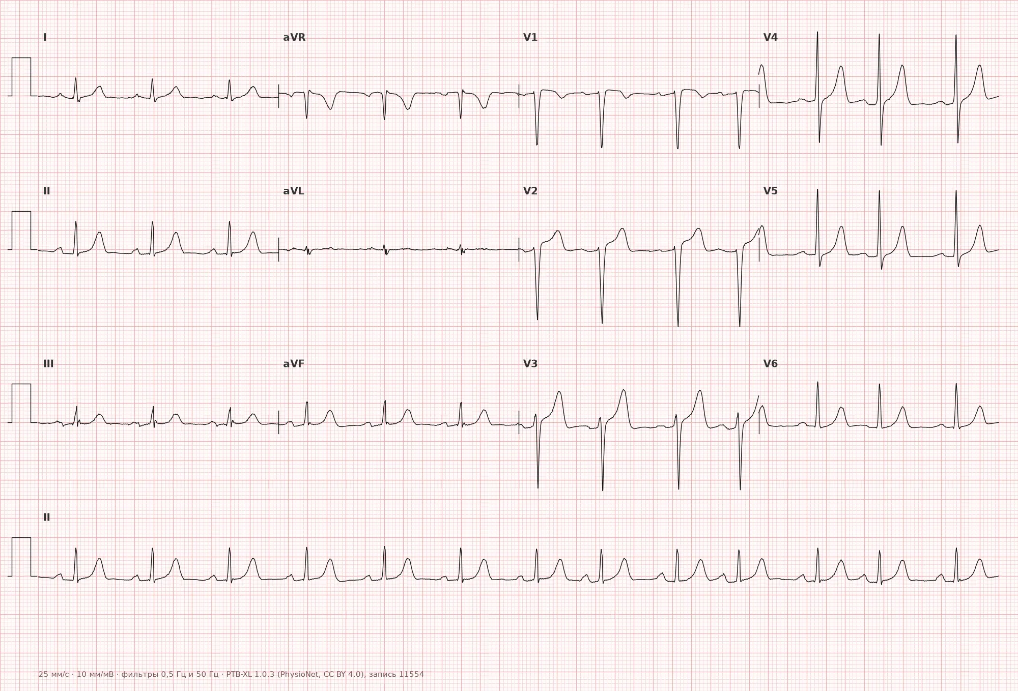
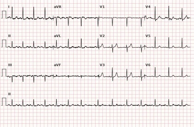
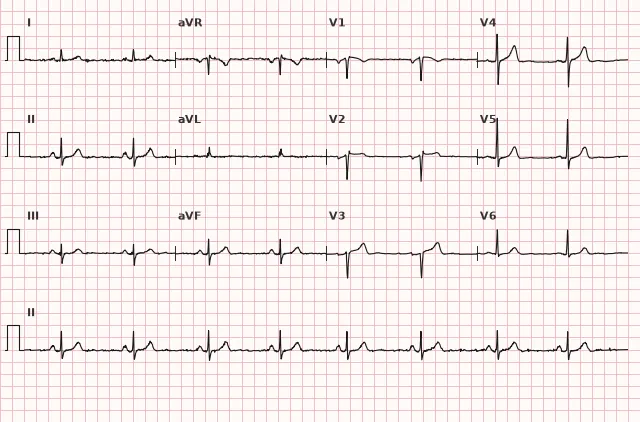
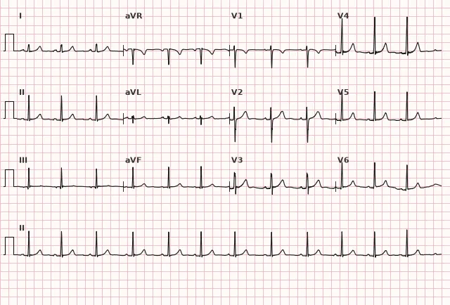
ECG from the open PTB-XL dataset (Wagner P. et al., Scientific Data, 2020; PhysioNet), CC BY 4.0 license. Rendering by WhiteCoat Pro.
Mechanism
Sinoatrial node automatism is regulated by the autonomic nervous system. In respiratory sinus arrhythmia, vagal tone decreases during inspiration (reflexively via pulmonary stretch receptors and changes in venous return), which accelerates sinus node discharges and shortens PP; during expiration, vagal tone increases, and PP lengthens. This is a normal physiological response, more pronounced in young individuals with high vagal tone. The non-respiratory form is associated with other fluctuations in autonomic balance or early changes within the sinus node itself, without a clear relationship to respiratory phases.
Causes
- Normal variant in children, adolescents, and young adults with high parasympathetic tone
- High physical fitness (athletes)
- Increased vagal tone of any origin
- Use of certain medications that enhance vagal tone
- In the elderly and in organic sinus node disease — non-respiratory form as a non-specific finding requiring evaluation for sick sinus syndrome
- Sinus rhythm variability can change in autonomic, metabolic, and systemic conditions, but a causal relationship must be assessed clinically in each case
Differential diagnosis
- Sinus arrhythmia vs sinus pause: in arrhythmia, PP changes are smooth and repetitive; in sinus pause, the expected sinus impulse is suddenly absent and a pause occurs, with episodes that may be single or repeated
- Sinus arrhythmia vs wandering atrial pacemaker: in arrhythmia, the P-wave shape remains constant across all cycles; in wandering pacemaker, P morphology changes, typically describing at least three different P forms with PR and PP variability, and the origin may shift between the sinus node, other atrial regions, and, in some classifications, the AV junction
- Sinus arrhythmia vs Mobitz type II second-degree sinoatrial block: in type II block, the pause is usually a multiple of the baseline PP (2 or 3 times); in type I, Wenckebach periodicity is observed with progressive PP shortening before an dropped beat; in sinus arrhythmia, PP changes are gradual and non-multiples, and the diagnosis is evaluated in the context of respiratory variability
- Sinus arrhythmia vs atrial extrasystole with irregularity: extrasystoles present with a premature altered or hidden P wave, a premature PP interval, and a subsequent pause that is often less than fully compensatory; the QRS morphology and the duration of the following interval are also evaluated. In sinus arrhythmia, the P wave remains sinus in all cycles
Clinical significance and management
In healthy asymptomatic children, adolescents, and trained young adults with typical respiratory sinus arrhythmia, workup and treatment are generally unnecessary, and simply describing the finding in the report is sufficient. In the presence of symptoms (fatigue, presyncope), atypical irregularity, marked bradycardia, pauses, or concomitant heart disease, further clinical evaluation is indicated. In elderly patients, especially with the non-respiratory form, the ECG should be additionally evaluated for signs of sick sinus syndrome (sinus bradycardia, sinus pauses, sinoatrial block) and, if clinical symptoms are present, referred for Holter monitoring. There is no specific treatment for sinus arrhythmia itself — management is determined by the underlying condition, if present.
Common interpretation errors
- Mistaking pronounced sinus arrhythmia for atrial fibrillation due to rhythm irregularity — in arrhythmia, P waves are preserved and maintain a constant shape
- Failing to measure the actual PP dispersion and limiting oneself to a subjective «irregular rhythm»
- Confusing it with a wandering atrial pacemaker by not comparing the P-wave shape across different cycles
- Interpreting a non-respiratory form in the elderly as an unconditional early sign of sinus node dysfunction without assessing the clinical context
- Describing it as «arrhythmia» without specifying its sinus origin, which causes unnecessary anxiety for the patient
1. Which criterion confirms the sinus origin of the rhythm arrhythmia?
Constant sinus P morphology and a smooth cyclic pattern of PP fluctuations are characteristic of sinus arrhythmia, but interpretation also requires the exclusion of extrasystoles, pauses, and blocks — a single numerical PP threshold is insufficient.
2. How does respiratory sinus arrhythmia differ from a wandering atrial pacemaker?
Wandering atrial pacemaker is accompanied by changes in P morphology (usually at least three forms) and PR/PP variability due to a shift in the rhythm origin, whereas in sinus arrhythmia, the P wave remains sinus.
3. In which group of patients does non-respiratory sinus arrhythmia require additional attention?
In elderly patients, the non-respiratory form alone does not prove sick sinus syndrome, but it requires clinical evaluation for symptoms and other ECG signs of sinus node dysfunction.
Frequently asked questions
Is sinus arrhythmia dangerous?
The typical respiratory form in healthy children and young adults is benign and considered a variant of norm. The non-respiratory or symptomatic form, especially in the elderly, requires evaluation of the cause and may be associated with sinus node dysfunction.
How to distinguish sinus arrhythmia from atrial fibrillation?
In sinus arrhythmia, a distinct P wave of constant sinus morphology precedes each QRS complex. In atrial fibrillation, organized P waves are absent; fine or coarse fibrillatory activity may be present between complexes, but it is not always clearly visible, and the rhythm remains irregularly irregular.
Does sinus arrhythmia require treatment?
No specific treatment is required; this is not an independent disease. Management depends on whether there is concomitant sinus node pathology, symptoms, or another underlying condition.
Who is most commonly affected by sinus arrhythmia?
More common in children, adolescents, and physically conditioned young adults due to high vagal tone. With age, the respiratory form usually decreases; non-respiratory irregularity in the elderly warrants attention to possible sinus node dysfunction.
How to confirm that the arrhythmia is indeed sinus in origin?
It is necessary to verify that the P wave has the expected sinus morphology (typically positive in I, II, aVF and negative in aVR), maintains the same shape across all cycles, and precedes every QRS complex, while the PP interval changes are smooth and cyclic rather than related to extrasystoles, pauses, or block.
ECG Course
Interpretation from basics to myocardial infarction — learn to read any ECG using a systematic algorithm.
- Video lessons and theory for every topic
- Simulator case studies under every lesson
- Unlimited ECG simulator practice
- Study guide: "All ECGs in 25 minutes"
ECG Strip Simulator
ECG, radiology, histology, pathomorphology, pharmacology — thousands of flashcards with detailed explanations.
- Real ECG strips, not textbook drawings
- Instant answers and explanations
- Streaks and progress tracking in your dashboard
Related terms
More in the «Sinus Node» section
Sources
- Goldberger A. Clinical Electrocardiography
- Hampton J. The ECG Made Easy
- Orlov V.N. Guide to Electrocardiography
- AHA/ACCF/HRS Recommendations for the Standardization and Interpretation of the Electrocardiogram, 2009
Educational material for medical students and physicians. Does not replace clinical guidelines and physical examination; the ECG must always be interpreted in conjunction with the clinical picture.