ECG criteria
- HR >100 bpm (in adults), predominantly at rest; no fixed upper limit — during exertion, fever, and other states, the rate can be significantly higher
- A P wave is present before every QRS
- P wave is positive in II, negative in aVR — normal P axis (as in sinus rhythm)
- P wave morphology is identical from cycle to cycle
- PQ (PR) interval is usually constant, 120–200 ms; it may slightly shorten as the rhythm accelerates, but marked shortening requires ruling out pre-excitation and other causes
- Rhythm is regular; RR intervals may slightly shorten and lengthen smoothly (respiratory modulation)
- QRS is usually narrow; may be wide in intraventricular conduction disturbance or pre-excitation
What it looks like on ECG
The P wave retains sinus morphology: positive in I, II, aVF, negative in aVR, often biphasic in V1 with a small initial positive component. The only difference from a normal sinus rhythm is the rate — P waves occur more frequently, which can cause them to superimpose on the preceding T wave, forming a pseudo-notched T-P complex.
The PQ (PR) interval in sinus tachycardia is usually constant at 120–200 ms; with rate acceleration due to sympathetic activation, it may shorten slightly, but a PR interval of <120 ms should not be automatically attributed to the heart rate alone — it requires evaluating the causes of a short PR, including pre-excitation.
Nonspecific ST-segment and T-wave changes, including ST depression and T-wave flattening, can occur during tachycardia. Their significance should be assessed in the clinical context, based on dynamics and after rate correction — ischemia cannot be ruled out a priori solely on the assumption of "rate-related" changes.
| Parameter | Normal | Sinus tachycardia |
|---|---|---|
| HR | 60–100 bpm | >100 bpm |
| P wave | sinus | sinus, same morphology |
| PQ | 120–200 ms | usually normal, may slightly shorten |
| QRS | narrow | usually narrow; may be wide in bundle branch block or pre-excitation |
At high heart rates, the P wave may merge with the preceding T wave, making identification difficult and requiring comparison with a resting ECG or a longer recording.
Real ECGs: 8 examples
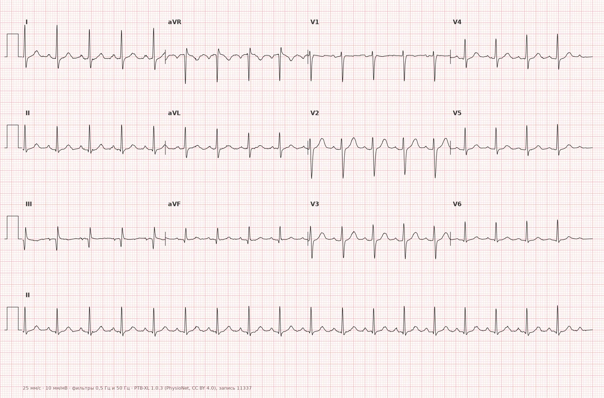
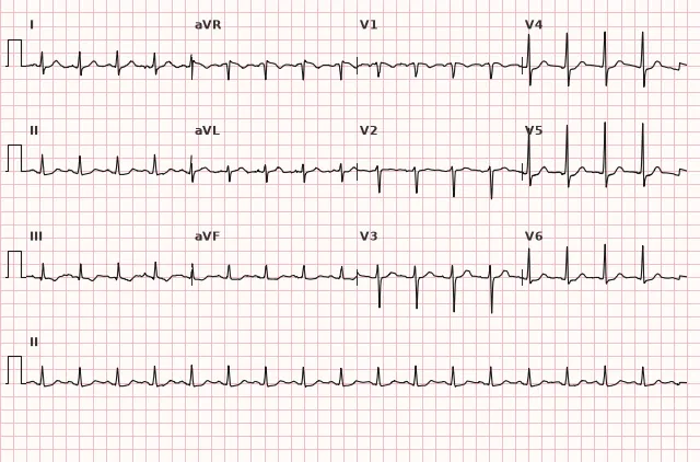
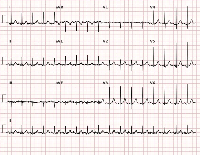
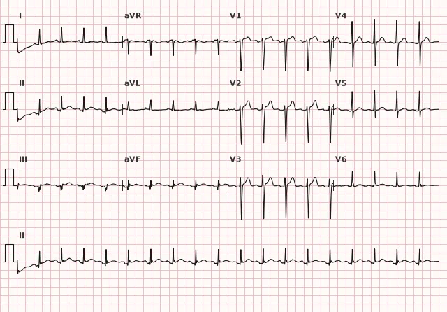
ECG from the open PTB-XL dataset (Wagner P. et al., Scientific Data, 2020; PhysioNet), CC BY 4.0 license. Rendering by WhiteCoat Pro.
Mechanism
The sinus node is the pacemaker with its own automatism; its discharge rate is determined by the balance of sympathetic and parasympathetic influences. Increased sympathetic nervous system tone or decreased vagal tone accelerates the depolarization of sinus node pacemaker cells, shortening the time required to reach threshold potential. As a result, impulses are generated more frequently. The activation pathway through the atria, AV node, bundle of His, and ventricles generally remains the same, so P and QRS morphology largely remain sinus-like, although PR and ST-T characteristics may change under the influence of the same sympathetic activation and AV conduction.
Causes
- Physiological: physical exertion, emotional excitement, pain, anxiety
- Fever and infections
- Hypovolemia, blood loss, dehydration
- Anemia
- Thyrotoxicosis (hypothyroidism, conversely, is more often accompanied by bradycardia)
- Heart failure, compensatory tachycardia in decreased cardiac output
- Pulmonary embolism
- Shock of any etiology
- Use of stimulants: caffeine, nicotine, sympathomimetics, cocaine
- Medications: beta-agonists, theophylline, atropine, and other vagolytics
- Beta-blocker withdrawal syndrome
- Pregnancy
- Pain, postoperative period
- Inappropriate sinus tachycardia (a separate nosology, diagnosis of exclusion)
Differential diagnosis
- Atrial flutter 2:1 — regular F-wave atrial activity is detected, often with a sawtooth appearance, usually around 250–350 per minute, rather than discrete P waves; with 2:1 conduction, some waves may be hidden within the QRS or T wave; vagal maneuvers or adenosine can slow AV conduction and reveal flutter waves
- Atrial tachycardia — P-wave morphology and axis often differ from sinus, but the degree of difference depends on the focus location (a focus in the upper right atrium can yield a P wave close to sinus); sudden onset/offset, an isoelectric interval between P waves, and a constant atrial activation pattern are also important
- Sinus node reentrant tachycardia — sudden onset and offset (paroxysmal), unlike the gradual acceleration and deceleration of sinus tachycardia
- Inappropriate sinus tachycardia — persistently high resting heart rate without an apparent cause, diagnosed after ruling out secondary causes
- Supraventricular tachycardia (AVNRT/AVRT) — rhythm is usually regular with sudden onset and offset; retrograde P waves may be absent, buried in the QRS complex, or appear immediately after it, and their polarity depends on the direction of retrograde activation
Clinical significance and management
Sinus tachycardia is an ECG finding rather than a standalone diagnosis: it reflects the sinus node's response to an external or internal stimulus. First, treat the underlying cause (fever, pain, hypovolemia, thyrotoxicosis, anemia, pulmonary embolism, etc.). Persistent sinus tachycardia without an obvious cause requires further workup: complete blood count, thyroid function tests, volume status assessment, source of infection screening, and an appropriate diagnostic algorithm if pulmonary embolism is suspected. If secondary causes are ruled out and symptomatic inappropriate sinus tachycardia persists, specialized therapy according to guidelines is possible, usually in consultation with a cardiologist.
Common interpretation errors
- Mistaking a P wave fused with a T wave for its absence and diagnosing a different tachycardia
- Failing to notice 2:1 atrial flutter and confusing the F wave with a P wave
- Initiating antiarrhythmic therapy instead of searching for the cause of tachycardia
- Failing to compare P-wave morphology with the patient's baseline resting ECG when its sinus origin is in doubt
- Interpreting ST depression during tachycardia as inherently non-ischemic without clinical context and serial changes
1. Which ECG feature differentiates sinus tachycardia from 2:1 atrial flutter?
In atrial flutter, regular atrial activity (F waves) is detected, often sawtooth-like, usually at about 250–350 per minute, rather than discrete sinus P waves.
2. What is the correct physician tactic when discovering sinus tachycardia of unclear etiology?
Sinus tachycardia is a reaction of the sinus node to an underlying cause; treatment should target the cause primarily rather than the tachycardia itself.
3. What P wave characteristic is typical for sinus tachycardia?
This is the normal sinus P wave axis, which remains unchanged during sinus tachycardia; only the rate changes.
Frequently asked questions
At what heart rate is sinus tachycardia diagnosed?
In adults, defined as a heart rate greater than 100 beats per minute on a sinus rhythm background, predominantly at rest; there is no fixed upper limit. In children, the upper limit of normal heart rate is higher and age-dependent, so the criteria for sinus tachycardia in children are also age-adjusted.
Is sinus tachycardia dangerous?
Sinus tachycardia is often a physiological or compensatory response, but its clinical significance depends on the cause, severity, and duration. The underlying cause may pose a danger, such as blood loss, pulmonary embolism, shock, or thyrotoxicosis. Persistent pronounced tachycardia of any origin can be detrimental and eventually lead to tachycardia-induced cardiomyopathy.
How to distinguish sinus tachycardia from supraventricular tachycardia?
In sinus tachycardia, the P wave maintains its normal sinus morphology and axis, onset and offset are gradual, and the rate changes smoothly. In supraventricular tachycardia, onset and offset are sudden, the P wave may be absent, buried in the QRS, retrograde, or have a different morphology.
Should sinus tachycardia be treated with rate-lowering medications?
Not always. First, the underlying cause is addressed (pain, fever, hypovolemia, anemia, etc.). Rate-lowering medications are used only when indicated, particularly in symptomatic inappropriate sinus tachycardia, after excluding secondary causes.
What is inappropriate sinus tachycardia?
This is a persistent elevation of resting heart rate or an inappropriate heart rate increase with minimal exertion without an apparent physiological or pathological cause. The diagnosis is made by exclusion after a thorough evaluation.
ECG Course
Interpretation from basics to myocardial infarction — learn to read any ECG using a systematic algorithm.
- Video lessons and theory for every topic
- Simulator case studies under every lesson
- Unlimited ECG simulator practice
- Study guide: "All ECGs in 25 minutes"
ECG Strip Simulator
ECG, radiology, histology, pathomorphology, pharmacology — thousands of flashcards with detailed explanations.
- Real ECG strips, not textbook drawings
- Instant answers and explanations
- Streaks and progress tracking in your dashboard
Related terms
More in the «Sinus Node» section
Sources
- Orlov, V.N. «Electrocardiography Guide»
- Hampton J.R. «The ECG Made Easy»
- Surawicz B., Knilans T. «Chou's Electrocardiography in Clinical Practice»
- AHA/ACCF/HRS Recommendations for the Standardization and Interpretation of the Electrocardiogram, 2009
Educational material for medical students and physicians. Does not replace clinical guidelines and physical examination; the ECG must always be interpreted in conjunction with the clinical picture.