ECG criteria
- The P wave is typically positive in I, II, and aVF, negative in aVR; usually biphasic in V1, with a predominantly positive variant also possible — normal P-axis; evaluate the overall pattern rather than a single lead
- Each P wave is followed by a QRS complex (1:1 AV conduction)
- The PQ (PR) interval is generally constant from cycle to cycle; typically 120–200 ms, but may be prolonged in concomitant first-degree AV block — this does not rule out sinus bradycardia
- Heart rate less than 60 beats per minute, calculated from RR intervals (typically based on the mean rate over the recording)
- Sinus rhythm may be regular or exhibit physiological respiratory RR variability (differentiate marked cyclic variability from stably prolonged intervals — see "Differential Diagnosis")
- P wave morphology is identical across all cycles within the same lead
What it looks like on ECG
On the ECG in sinus bradycardia, the normal P wave → QRS complex → T wave sequence is preserved, but RR intervals are prolonged, and the heart rate drops below 60 beats per minute.
The P wave has normal morphology for sinus rhythm: positive in I, II, and aVF, negative in aVR, and usually biphasic in V1 (a predominantly positive variant is also possible). P wave amplitude and shape do not change from complex to complex. The PQ interval is generally constant from cycle to cycle and typically falls within 120–200 ms, but may be prolonged, for example, in concomitant first-degree AV block — conduction remains 1:1 with no dropped QRS complexes, however, the mere presence of a PR prolongation exceeding 200 ms is classified as first-degree AV block and is described separately alongside sinus bradycardia.
The QRS complex is narrow unless accompanied by a bundle branch block. In isolated sinus bradycardia, QRS and ST-T segment morphology is normal; abnormalities affect only the rate and cycle length, though this does not rule out concomitant changes — bundle branch block, ischemic ST-T changes, signs of hypertrophy, or electrolyte imbalances. These should be assessed independently rather than attributed to bradycardia itself.
The rhythm in sinus bradycardia may be regular or show physiological respiratory RR variability (acceleration during inspiration, deceleration during expiration) — this does not contradict the diagnosis. When there is marked cyclic PP/RR variability, particularly respiration-related, with unchanged P wave morphology, concomitant sinus arrhythmia is diagnosed; other causes of irregularity (extrasystoles, including blocked atrial extrasystoles) should be ruled out separately.
| Parameter | Normal findings in sinus bradycardia |
|---|---|
| HR | less than 60 bpm |
| P wave | sinus morphology, 1:1 with QRS |
| PQ | typically 120–200 ms, may be prolonged in first-degree AV block |
| QRS | narrow, normal shape (unless concomitant bundle branch block is present) |
| Rhythm | regular or with respiratory variability |
There is no well-established, universally accepted grading of sinus bradycardia severity by heart rate (such as "moderate" or "marked") in guidelines — clinical significance is determined by symptoms, hemodynamics, and the underlying cause rather than the heart rate value alone. At any heart rate, including marked bradycardia, it is important to carefully check for dropped QRS complexes (a sign of sinoatrial block or sick sinus syndrome) and escape beats from lower pacemakers — pauses and dropped beats require differentiation from AV block regardless of the baseline heart rate.
Real ECGs: 8 examples
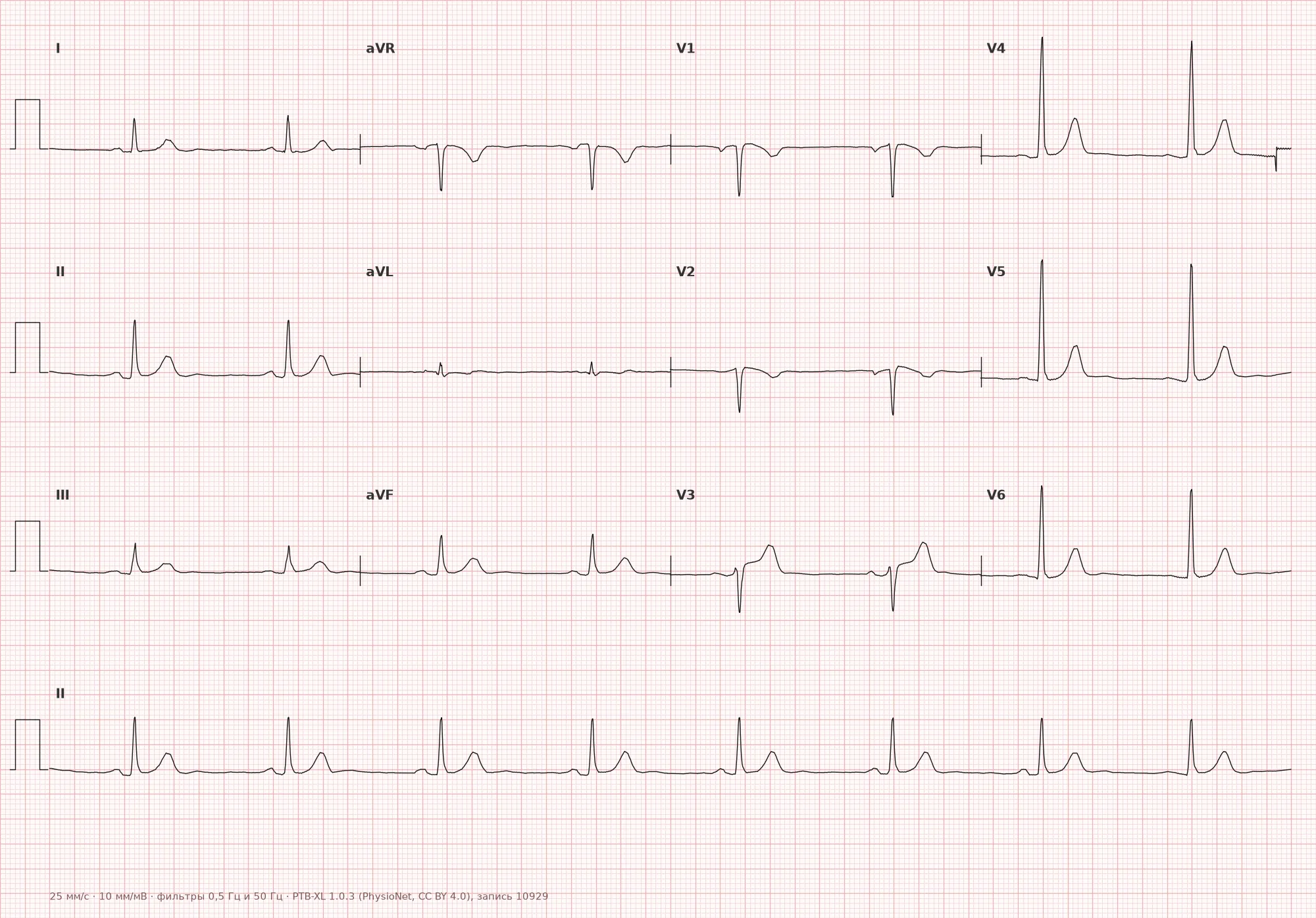
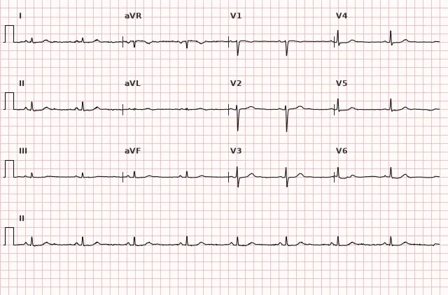
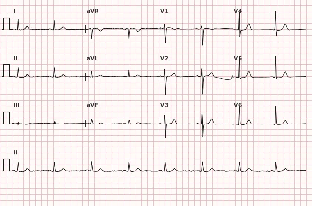
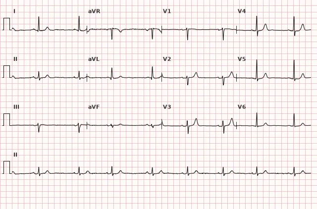
ECG from the open PTB-XL dataset (Wagner P. et al., Scientific Data, 2020; PhysioNet), CC BY 4.0 license. Rendering by WhiteCoat Pro.
Mechanism
The sinus node sets the rhythm through spontaneous depolarization of pacemaker cells (automaticity). The rate of this depolarization depends on the balance of sympathetic and parasympathetic influences, as well as the intrinsic properties of the node cells.
In sinus bradycardia, the rate of spontaneous diastolic depolarization of sinus cells slows down — most commonly due to increased parasympathetic (vagal) tone, and less frequently due to organic disease of the node itself or impaired blood supply. The impulse still originates in the sinus node and propagates along the normal pathway, so P wave morphology and the sequence of atrial and ventricular activation remain unchanged — only the impulse generation rate is affected.
Causes
- Increased vagal tone: sleep, trained athletes, vagal maneuvers, vomiting, pain
- Physiological variant of normal in healthy individuals, especially young people and athletes
- Medication use: beta-blockers, non-dihydropyridine calcium channel blockers (verapamil, diltiazem), digoxin, antiarrhythmics (amiodarone, etc.), clonidine
- Hypothyroidism
- Hypothermia
- Increased intracranial pressure (Cushing's reflex)
- Hyperkalemia and other electrolyte imbalances
- Myocardial ischemia or infarction, particularly of the inferior wall (vagal reflex and/or sinus node ischemia)
- Sick sinus syndrome, age-related degenerative changes of the node
- Obstructive sleep apnea, cholestasis, jaundice (reflex mechanisms)
Differential diagnosis
- Sick sinus syndrome — bradycardia combined with pauses, sinoatrial block, tachy-brady syndrome; to confirm the association between symptoms and rhythm in stable patients, ambulatory ECG monitoring of appropriate duration is used — ranging from 24–48-hour Holter monitoring to longer-term patch monitoring, external or implantable loop recorders, depending on the frequency of episodes
- Sinus arrhythmia — HR may be low, but RR intervals vary noticeably from cycle to cycle (often respiration-related) rather than being stably prolonged
- 2:1 AV block — at first glance a slow rhythm, but upon careful analysis, some P waves fail to conduct to the ventricles (P to QRS ratio is not 1:1)
- Junctional (AV) rhythm — P wave is absent, inverted, or hidden within the QRS, P-axis is non-sinus
- Second-degree sinoatrial block — dropped P-QRS cycles with a pause that is a multiple of the baseline PP interval
Clinical significance and management
Sinus bradycardia in itself is benign and often requires no intervention, especially in young, trained, and asymptomatic individuals. Clinical significance is determined by the presence of symptoms (fatigue, presyncope, syncope, reduced exercise tolerance), hemodynamic impact, and the underlying cause of the bradycardia.
In hemodynamically significant or acutely presenting symptomatic bradycardia (hypotension, signs of hypoperfusion, ischemia, acute heart failure, syncope with persistent marked bradycardia), management should not be delayed until ambulatory monitoring: immediate rhythm and blood pressure monitoring, 12-lead ECG acquisition, identification and correction of reversible causes (medications, electrolyte imbalances, hypothyroidism, inferior wall ischemia), atropine as indicated, and if ineffective, temporary cardiac pacing (transcutaneous or transvenous) according to emergency bradycardia algorithms.
In stable patients without acute symptoms, the approach differs: hemodynamic significance is assessed, reversible causes are ruled out, and heart rate is correlated with the clinical picture — a well-rested athlete with an HR of 45 bpm and no symptoms requires no treatment. For chronic or episodic symptoms, correlation with rhythm is established via ambulatory ECG monitoring (Holter monitoring for frequent symptoms, longer monitoring methods for rare episodes), the need to discontinue or adjust negative chronotropic drugs is evaluated, and sick sinus syndrome is ruled out.
Permanent cardiac pacing is considered in symptomatic sinus node dysfunction if symptoms correlate with bradycardia or pauses and reversible causes have been excluded; there is no universal heart rate threshold or pause duration that automatically serves as an indication. Asymptomatic sinus bradycardia, including in athletes and during sleep, is not an indication for pacing by itself.
Common interpretation errors
- Diagnosing "bradycardia" without checking P wave morphology — junctional rhythm or high-grade AV block may be missed
- Failure to evaluate the pattern of RR intervals — marked sinus arrhythmia with a low mean heart rate may be mistaken for stable bradycardia, and PR prolongation exceeding 200 ms may not be recognized as concomitant first-degree AV block
- Failure to count the P to QRS ratio — concealed 2:1 or 3:1 AV block can be masked as simple bradycardia
- Forgot to check for pauses and dropped beats — missing sinoatrial block or sick sinus syndrome
- Heart rate estimated "by eye" from a single RR interval rather than averaged over the tracing
- In hemodynamically significant bradycardia, time is wasted on outpatient monitoring instead of immediate monitoring and emergency measures
1. Which feature is mandatory for the diagnosis of sinus bradycardia?
The diagnosis requires a preserved sinus P wave (positive in II, negative in aVR) preceding each QRS complex and a heart rate under 60 bpm.
2. What differentiates sinus bradycardia from sinus arrhythmia?
In bradycardia, RR intervals are consistently prolonged; in sinus arrhythmia, they fluctuate noticeably from cycle to cycle, often synchronously with respiration.
3. Which cause of sinus bradycardia is associated with myocardial infarction of a specific localization?
Inferior myocardial infarction may be accompanied by bradycardia due to a vagal reflex and/or ischemia of the sinus node, which is supplied in most cases by the right coronary artery.
Frequently asked questions
What heart rate is considered sinus bradycardia in adults?
Heart rate under 60 bpm with a sinus rhythm (a normal P wave is preserved before each QRS complex). Threshold values of the age norm differ in children and across various age groups.
Is sinus bradycardia dangerous?
Not in itself, especially in trained and asymptomatic individuals during sleep. Danger arises when there are symptoms of hypoperfusion (syncope, weakness) or when bradycardia is a manifestation of sick sinus syndrome or acute ischemia.
How to distinguish sinus bradycardia from sinus arrhythmia?
In sinus bradycardia, RR intervals are consistently prolonged and vary little between cycles. In sinus arrhythmia, RR intervals fluctuate noticeably (often synchronously with respiration), and the average heart rate may be normal or reduced.
Does sinus bradycardia require treatment?
Asymptomatic bradycardia, especially in athletes and during sleep, requires no treatment. Acute hemodynamically significant bradycardia requires immediate monitoring, search for a reversible cause, atropine, and temporary cardiac pacing if ineffective. For chronic symptomatic bradycardia without a reversible cause, permanent cardiac pacing is considered.
Can sinus bradycardia be a sign of myocardial infarction?
Yes, especially in inferior myocardial infarction — bradycardia in this situation may be associated with a vagal reflex and/or ischemia of the sinus node, whose blood supply in most cases comes from the right coronary artery. Such bradycardia is evaluated alongside characteristic ST-T changes in leads II, III, and aVF.
ECG Course
Interpretation from basics to myocardial infarction — learn to read any ECG using a systematic algorithm.
- Video lessons and theory for every topic
- Simulator case studies under every lesson
- Unlimited ECG simulator practice
- Study guide: "All ECGs in 25 minutes"
ECG Strip Simulator
ECG, radiology, histology, pathomorphology, pharmacology — thousands of flashcards with detailed explanations.
- Real ECG strips, not textbook drawings
- Instant answers and explanations
- Streaks and progress tracking in your dashboard
Related terms
More in the «Sinus Node» section
Sources
- Goldberger's Clinical Electrocardiography, 9th ed.
- Hampton. The ECG Made Easy
- Orlov V.N. Guide to Electrocardiography
- Surawicz B., Knilans T. Chou's Electrocardiography in Clinical Practice
Educational material for medical students and physicians. Does not replace clinical guidelines and physical examination; the ECG must always be interpreted in conjunction with the clinical picture.