ECG criteria
- Determine the earliest QRS onset and the latest QRS offset across all leads; in practice, measure in the lead with the maximum duration, usually additionally checking lead II and the precordial leads
- QRS ≥0.12 s (120 ms) — wide complex according to the 2009 AHA/ACCF/HRS criteria
- QRS 0.11–0.119 s — intermediate/borderline widening; does not diagnose incomplete bundle branch block on its own, characteristic morphology is required
- QRS approximately 0.07–0.11 s is usually normal in adults (exact limits depend on the measurement method and context)
- Evaluate morphology: is it typical for RBBB (rsR' in V1, wide S in I, V6) or LBBB (wide notched R in I, aVL, V6, absence of Q in V5–V6)
- Check regularity and relationship to rhythm — wide QRS during sinus rhythm (block/IVB) or tachycardia (VT, SVT with aberration, antidromic tachycardia in WPW)
- Correlate with clinical status and history: drugs (class I antiarrhythmics, TCAs), electrolytes (potassium), pacemaker
- If QRS is wide during tachycardia — apply differential diagnosis algorithms for VT/SVT with aberration (Brugada algorithm, Vereckei algorithm)
What it looks like on ECG
The QRS width is measured from the onset of the first wave of the complex (Q or R) to the end of the last wave (S or R). For standardized evaluation, determine the earliest QRS onset and the latest QRS offset across all leads; in practice, measure in the lead where the complex appears widest — often V2–V3 or II. At a paper speed of 25 mm/s, 1 mm corresponds to 0.04 s, so the threshold of 0.12 s is 3 mm.
| QRS width | Interpretation |
|---|---|
| approximately 0.07–0.11 s | usually normal (depends on method and context) |
| 0.11–0.119 s | intermediate/borderline widening; diagnosis of incomplete block requires characteristic morphology, not just duration |
| ≥0.12 s | wide QRS (complete bundle branch block, IVB, VT, pre-excitation) |
The morphology of the complex suggests the mechanism. In right bundle branch block — a terminal wide R' wave in V1 (rsR' or RSR'), a wide notched S wave in I and V6. In left bundle branch block — a wide, often notched R wave in I, aVL, V5–V6, absence of a normal Q wave in V5–V6, discordant ST-T segments. In ventricular tachycardia, QRS morphology may resemble typical RBBB or LBBB, so a "typical" shape does not rule out VT; VT is indicated by a morphology atypical for bundle branch blocks, axis deviation, absence of normal R wave progression, QRS concordance in the precordial leads, as well as AV dissociation, capture and fusion beats. Any wide complex during tachycardia should be considered VT until proven otherwise. In antidromic tachycardia associated with WPW, the QRS is wide because the ventricles are activated primarily anterogradely via an accessory pathway; a typical delta wave as a distinct initial slurring of the complex during the tachycardia itself is usually not identifiable — to confirm, look for signs of manifest pre-excitation on the baseline ECG outside of an episode, but its absence does not rule out the diagnosis (intermittent pre-excitation is possible). In hyperkalemia and the action of sodium channel blockers, the QRS widens diffusely, but the shape of the widening is variable and may mimic intraventricular conduction disturbances; the absence of a classic bundle branch block pattern does not rule out these conditions.
Real ECGs: 8 examples
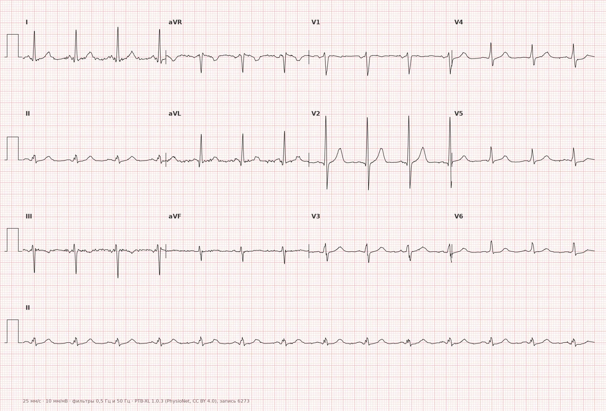
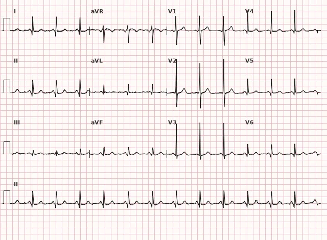
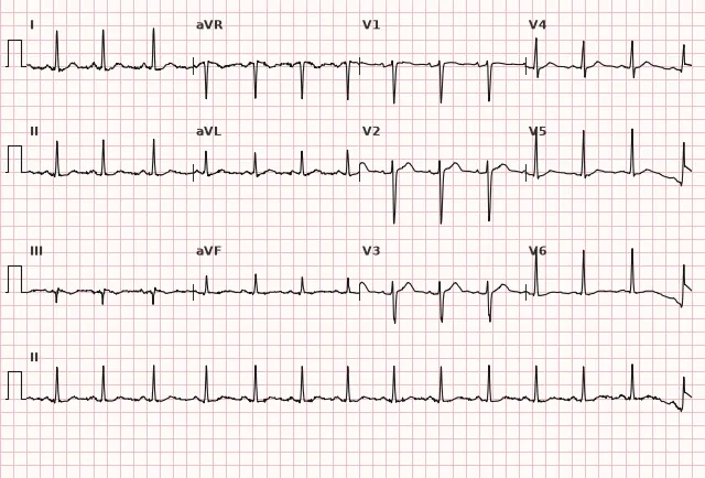
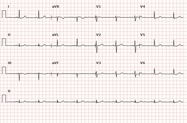
ECG from the open PTB-XL dataset (Wagner P. et al., Scientific Data, 2020; PhysioNet), CC BY 4.0 license. Rendering by WhiteCoat Pro.
Mechanism
Normal QRS width is ensured by rapid synchronous conduction through the His-Purkinje system to both ventricles. Widening occurs when conduction is slowed along one of the branches or fascicles (block), when there is an abnormal propagation pathway outside the conduction system (ectopic ventricular focus in PVCs/VT, pre-excitation via an accessory pathway in WPW), or due to diffuse slowing of conduction through the ventricular myocardium (hyperkalemia, sodium channel blockers, prominent structural myocardial pathology). In any scenario, the time required for complete ventricular depolarization increases, lengthening the complex on the ECG.
Causes
- Right bundle branch block (complete)
- Left bundle branch block (complete)
- Nonspecific intraventricular conduction block
- Premature ventricular contractions and ventricular tachycardia
- WPW syndrome (pre-excitation, delta wave)
- Ventricular pacing (pacemaker, PM)
- Hyperkalemia (diffuse QRS widening, variable morphology)
- Sodium channel blocker toxicity, TCAs, carbamazepine, cocaine — slowing of phase 0
- Severe ventricular hypertrophy with intraventricular block
- Dilated or other structural cardiomyopathy with non-specific intraventricular delay/bundle branch block
Differential diagnosis
- Wide QRS due to electrode placement artifact or tremor — check calibration and repeat recording
- Wide QRS during tachycardia: VT vs. SVT with aberrant conduction — look for AV dissociation, fusion/capture beats, precordial concordance, Brugada or Vereckei algorithm
- Wide QRS in WPW with antidromic tachycardia — look for signs of manifest pre-excitation on baseline ECG outside of episodes
- Diffuse widening in hyperkalemia — pay attention to tall peaked T waves, slowing or disappearance of the P wave, PR prolongation; changes are variable and may lack a typical pattern
- Wide QRS in the presence of a pacemaker — presence of pacing spikes preceding the complex
Clinical significance and management
The danger of a wide QRS is entirely determined by the cause. Isolated right bundle branch block in an asymptomatic patient without other abnormalities is frequently found in healthy individuals; clinical evaluation and observation are usually sufficient. Newly diagnosed left bundle branch block, especially with symptoms, AV block, syncope, or other signs of structural heart disease, is much more frequently associated with myocardial pathology and requires echocardiography and further workup as indicated. A wide QRS during tachycardia with hemodynamic instability is considered VT until proven otherwise — corresponding emergency management protocols for life-threatening tachyarrhythmias are indicated.
If ECG signs of hyperkalemia are present, immediate monitoring, intravenous calcium, and potassium-lowering measures should be initiated without waiting for laboratory confirmation; if sodium channel blocker/TCA poisoning is suspected and the QRS is widened, sodium bicarbonate should be promptly administered per toxicology treatment protocols while ensuring resuscitation monitoring and toxicological consultation. Treatment of these potentially life-threatening conditions should not be delayed due to atypical QRS morphology, as the ECG picture is variable.
In newly diagnosed left bundle branch block combined with ischemic symptoms, modified Sgarbossa (Smith) criteria are applied to assess possible STEMI, as standard ST criteria are unreliable in the presence of a wide QRS.
Common interpretation errors
- Measuring QRS in only a single lead without finding the lead with maximum width and checking complex onset/offset across all leads
- Assuming a wide QRS during tachycardia is SVT with aberration without applying VT differential diagnosis algorithms, including when the morphology resembles a typical bundle branch block
- Failing to consider the possibility of intermittent pre-excitation and confusing WPW with bundle branch block
- Ruling out hyperkalemia or sodium channel blocker toxicity simply because the QRS morphology does not look "typical"
- Missing pacemaker spikes, leading to misinterpretation of a wide QRS as IVB rather than ventricular pacing
1. Which QRS width threshold corresponds to the "wide complex" criterion according to the 2009 AHA/ACCF/HRS guidelines?
The wide QRS threshold is ≥0.12 s (120 ms), which equals 3 mm at a recording speed of 25 mm/s.
2. Which sign most reliably indicates ventricular tachycardia rather than SVT with aberration in the presence of a wide QRS?
AV dissociation, fusion and capture beats are specific signs of independent ventricular activity characteristic of ventricular tachycardia (VT). A typical bundle branch block morphology does not rule out VT.
3. Which cause produces diffuse QRS widening that can mimic intraventricular conduction delays without necessarily presenting a typical bundle branch block pattern?
Hyperkalemia diffusely slows ventricular myocardial depolarization; the ECG picture is variable and may mimic a block without having to match its typical morphology.
Frequently asked questions
What QRS duration is considered wide?
A QRS duration ≥0.12 s (120 ms, 3 mm at a paper speed of 25 mm/s) is considered wide. The 0.11–0.119 s range is borderline; by itself, it does not prove incomplete bundle branch block, which requires the appropriate complex morphology.
How to distinguish VT from SVT with aberrancy in wide QRS tachycardia?
Use signs of AV dissociation, capture and fusion beats, QRS concordance in the chest leads, and specialized algorithms (Brugada, Vereckei). A morphology resembling typical RBBB or LBBB does not rule out VT. When in doubt and in the presence of hemodynamic instability, wide QRS tachycardia should be managed as VT.
Is a wide QRS complex dangerous by itself?
The QRS widening itself is not inherently dangerous—the underlying cause determines the risk. Isolated, asymptomatic right bundle branch block is common in healthy individuals, whereas VT, severe hyperkalemia, or sodium channel blocker toxicity requires emergency intervention.
Can ST-segment elevation be evaluated in the presence of a wide QRS due to bundle branch block?
In left bundle branch block, standard ST-segment criteria are unreliable due to discordant ST-T changes; modified Sgarbossa (Smith) criteria are used to assess potential coronary artery occlusion.
What causes diffuse QRS widening without typical bundle branch block morphology?
Most commonly hyperkalemia or toxicity from sodium channel-blocking drugs (tricyclic antidepressants, carbamazepine, certain antiarrhythmics). Changes are variable: ranging from mild uniform widening to a picture mimicking bundle branch block, and progressing to a sine wave pattern in severe hyperkalemia.
ECG Course
Interpretation from basics to myocardial infarction — learn to read any ECG using a systematic algorithm.
- Video lessons and theory for every topic
- Simulator case studies under every lesson
- Unlimited ECG simulator practice
- Study guide: "All ECGs in 25 minutes"
ECG Strip Simulator
ECG, radiology, histology, pathomorphology, pharmacology — thousands of flashcards with detailed explanations.
- Real ECG strips, not textbook drawings
- Instant answers and explanations
- Streaks and progress tracking in your dashboard
Related terms
More in the section «Basics: waves, intervals, axes»
Sources
- AHA/ACCF/HRS. Recommendations for the Standardization and Interpretation of the Electrocardiogram, 2009
- Hampton J. The ECG Made Easy
- Surawicz B., Knilans T. Chou's Electrocardiography in Clinical Practice
- Orlov V.N. Guide to Electrocardiography
Educational material for medical students and physicians. Does not replace clinical guidelines and physical examination; the ECG must always be interpreted in conjunction with the clinical picture.