EKG-Kriterien
- The rhythm is usually regular, with RR intervals close to each other; there is no strict universal threshold for RR variability — regularity is assessed using consecutive intervals and clinically.
- The heart rate is most frequently 150–220 bpm — this is a typical guideline rather than a diagnostic criterion: values both below and above this range are possible in adults.
- The QRS complex is usually narrow (less than 120 ms) unless there is a bundle branch block, aberrant conduction, or pre-excitation.
- P wave: either not visible (merged with QRS in typical AVNRT), located immediately after QRS (pseudo-r' in V1, pseudo-S in II, III, aVF in AVNRT), or before QRS with a short RP in orthodromic AVRT.
- Sudden onset and sudden termination of the episode (on a long recording or by history) is a sign typical of re-entrant mechanisms, but not mandatory for all supraventricular tachycardias.
- Absence of a preceding gradual "warm-up" phase characteristic of sinus tachycardia.
- Response to vagal maneuvers or adenosine: termination of tachycardia (in re-entry involving the AV node) or transient slowing with unmasking of atrial activity (in atrial tachycardia); the response is evaluated together with the ECG pattern, not in isolation.
Wie es im EKG aussieht
On the ECG during an episode — tachycardia with a narrow QRS and a rate typically of 150–220 bpm. The rhythm is usually strictly regular, with RR intervals close from complex to complex; there is no strict variability threshold, and minor RR fluctuations do not exclude PSVT on their own. The regularity of narrow-complex tachycardia helps in differential diagnosis, but is not specific to PSVT: a similar picture is produced by sinus tachycardia, atrial flutter with fixed AV conduction, and certain atrial tachycardias. Therefore, the onset/offset of the episode, visible atrial activity, and the response to vagal maneuvers or adenosine are additionally evaluated.
The P wave is a key yet often elusive element. In typical AV nodal re-entrant tachycardia (AVNRT), the atrium and ventricle are activated almost simultaneously, so the P wave merges with the QRS or is visible as a small notched component at its end: in lead V1 this looks like a pseudo-r' (a small false wave after R), and in the inferior leads II, III, aVF like a pseudo-S (a shallow wave after R). The RP interval in this case is short, less than 70 ms.
In orthodromic AV re-entrant tachycardia (AVRT, involving an accessory pathway), the retrograde P wave is usually visible after the QRS, but may merge with its terminal portion; RP is frequently greater than 70 ms and often shorter than PR, but these ratios are approximate and overlap noticeably with other mechanisms — a short RP is also possible in AVRT, and in atypical AVNRT the RP may be long. Determining mechanisms reliably by RP/PR is often only possible via intracardiac EPS.
In atrial tachycardia, the P wave precedes the QRS, its morphology differs from sinus rhythm, and the PR interval may be prolonged depending on the heart rate.
The transition zone and P wave axis help suggest the focus localization, but on a standard 12-lead ECG the P wave is frequently hidden by tachycardia, and confidently differentiating the mechanism without intracardiac EPS is not always possible. The ST segment and T wave during tachycardia often exhibit secondary rate-dependent changes (ST depression, T inversion), but ischemia cannot be excluded based on the ECG appearance alone: it can coexist with PSVT, especially with symptoms, risk factors, persistent changes after rhythm restoration, or elevated troponin — this requires clinical evaluation.
Echte EKGs: 8 Beispiele
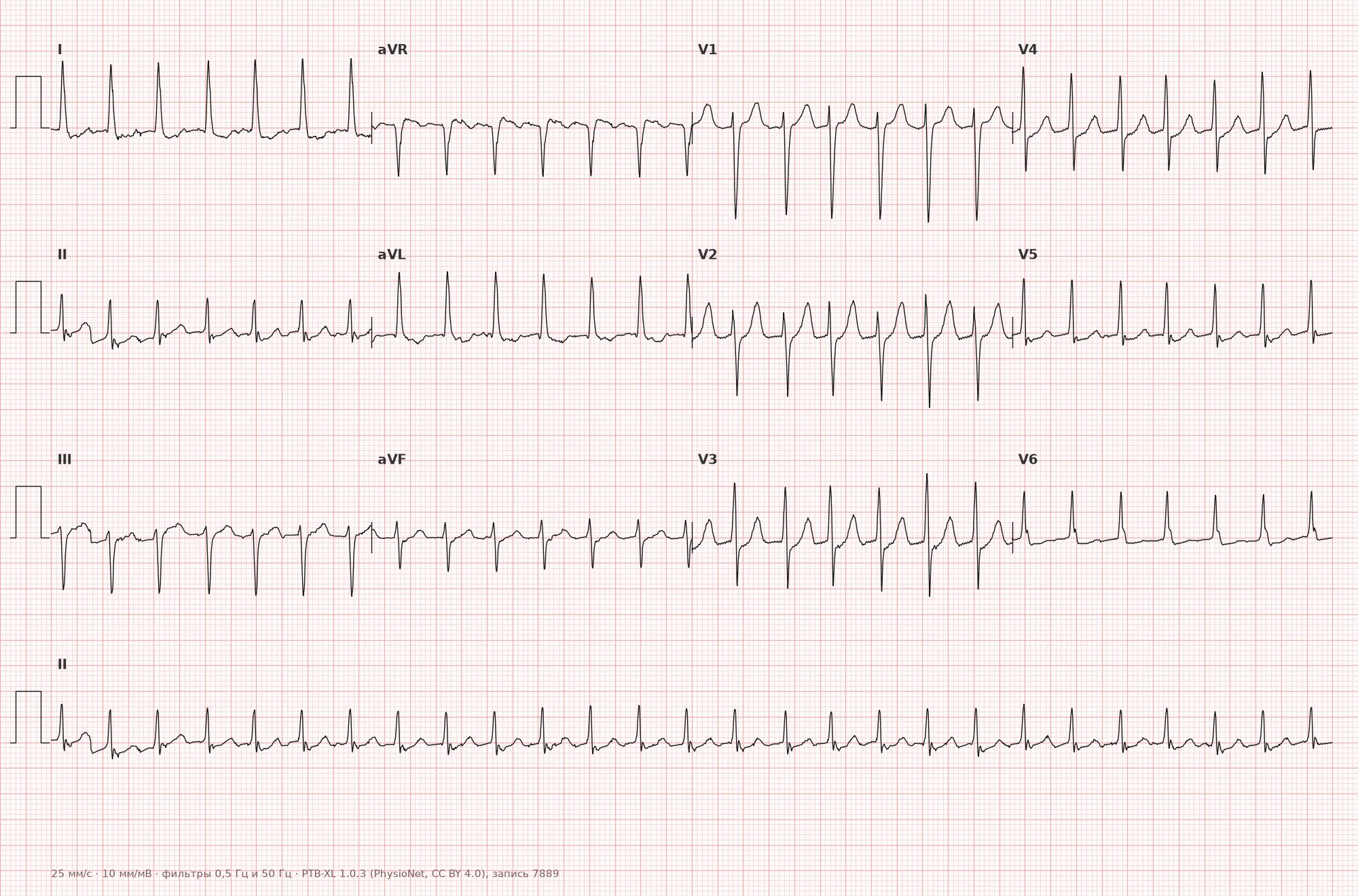
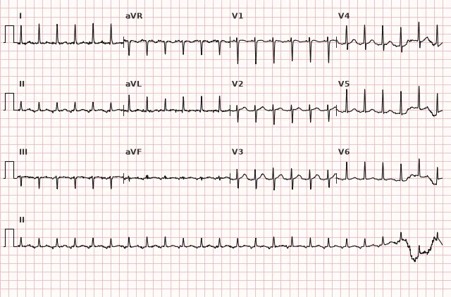
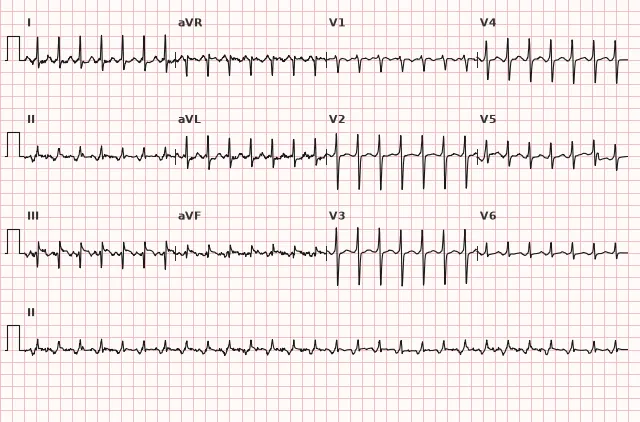
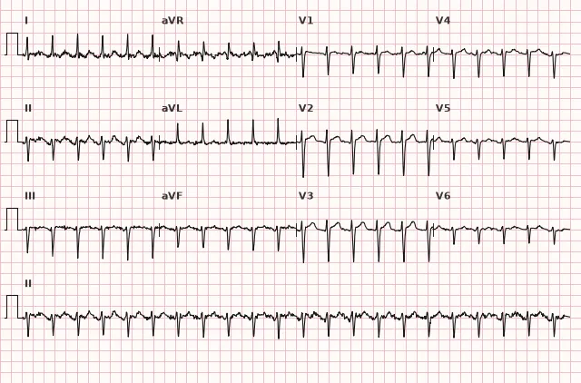
EKG aus dem öffentlich zugänglichen PTB-XL-Datensatz (Wagner P. et al., Scientific Data, 2020; PhysioNet), Lizenz CC BY 4.0. Visualisierung durch WhiteCoat Pro.
Mechanismus
Most paroxysmal SVTs are based on a re-entry mechanism (re-entry of the excitation wave). Most commonly this is AV nodal re-entrant tachycardia: in the AV node, two functional conduction pathways exist with different velocities and refractoriness (fast and slow), forming a circuit for the excitation loop. Less frequently, AV re-entrant tachycardia occurs involving an anatomical accessory pathway between the atrium and ventricle (orthodromic variant — conduction down through the AV node, up through the accessory pathway). The third mechanism is atrial tachycardia caused by focal ectopic activity or micro-re-entry in the atrium, not directly involving the AV node. Ventricular activation normally proceeds via the normal His-Purkinje system, so the QRS remains narrow, but it can be wide with aberrant conduction, preexisting bundle branch block, electrolyte disturbances, or pre-excitation (antidromic AVRT is typically wide-complex). Therefore, any wide-QRS tachycardia is initially considered ventricular until proven otherwise.
Ursachen
- Functionally separate fast and slow AV nodal pathways (dual physiology) — the basis of AVNRT, often without structural heart disease.
- Accessory atrioventricular pathways (manifest in WPW syndrome or concealed) — the basis of AVRT.
- Structurally normal heart — in the majority of patients, often young and middle-aged.
- Triggering factors: emotional stress, physical exertion, caffeine, alcohol, sleep deprivation.
- Organic heart disease (CAD, valvular heart disease, cardiomyopathies) — less frequent as a background.
- Pregnancy (increased frequency of paroxysms due to hormonal and hemodynamic changes).
- Electrolyte disturbances, thyrotoxicosis — as triggering rather than causative factors.
Differenzialdiagnose
- Sinus tachycardia — P wave of sinus morphology before each QRS, gradual onset and termination, HR variability with respiration and exertion; heart rate alone (even above 150–160 bpm) does not allow diagnosing sinus tachycardia or excluding PSVT — one must evaluate P morphology, regularity, context, and the dynamics of episode onset/termination.
- Atrial flutter with 2:1 conduction — F waves at a rate of about 300 bpm; sawtooth activity in II, III, aVF is typical primarily of typical counterclockwise flutter, in atypical flutter the morphology of F waves may differ, and with 2:1 conduction the waves are often poorly discernible; with slowed AV conduction (vagal maneuver, adenosine), flutter waves are unmasked.
- Atrial tachycardia with irregular conduction (multifocal atrial tachycardia) — irregular rhythm, different P wave morphologies.
- Ventricular tachycardia with retrograde atrial activation — usually wide QRS; a narrow QRS makes VT rare, but does not exclude it entirely (fascicular and other narrow-complex ventricular tachycardias have been described) — the diagnosis is determined by a combination of signs and clinical context. In SVT with concomitant bundle branch block or antidromic AVRT, the QRS may be wide; in this case, it is safer to initially interpret the rhythm as VT until proven otherwise and apply wide-complex tachycardia differentiation criteria (Brugada algorithm, Vereckei).
- Inappropriate sinus tachycardia — persistently elevated HR without a paroxysmal character, the P wave retains sinus morphology.
Klinische Bedeutung und Taktik
Most PSVT paroxysms are not directly life-threatening, but cause pronounced symptoms: palpitations, anxiety, dyspnea, presyncope, and in patients with organic heart disease or a very high heart rate — arterial hypotension and myocardial ischemia.
The first step is to record a 12-lead ECG during the episode if possible; this is important for subsequent verification of the mechanism. In stable hemodynamics, the preferred first maneuver is the modified Valsalva maneuver. Carotid sinus massage is performed only by trained medical personnel, sequentially on one side, after excluding contraindications (bruit over the carotid artery without ruling out significant stenosis, prior stroke or TIA) and with continuous rhythm monitoring. Vagal maneuvers can terminate tachycardia involving the AV node in the re-entry circuit, or transiently slow AV conduction and unmask P waves in atrial tachycardia or atrial flutter.
Adenosin wird bei hämodynamisch stabiler, regelmäßiger Tachykardie mit schmalen QRS-Komplexen und nach erfolglosen Vagusmanövern angewendet: Es wird als schneller intravenöser Bolus unter kontinuierlicher EKG-Registrierung, Blutdrucküberwachung und in Bereitschaft für Defibrillation und Reanimationsmaßnahmen verabreicht. Es wird nicht bei unregelmäßiger Breitkomplextachykardie und bei Verdacht auf Vorhofflimmern mit Präexzitation verabreicht – die AV-Knoten-Blockade kann die Leitung über eine akzessorische Bahn verstärken und Kammerflimmern provozieren. Mit Vorsicht oder nach speziellen Protokollen ist es anzuwenden bei schwerem Asthma bronchiale und ausgeprägter Bronchoobstruktion, bei Sinusknotendysfunktion oder AV-Blockierung höheren Grades ohne implantierten Schrittmacher, nach Herztransplantation (erhöhte Empfindlichkeit) sowie bei gleichzeitiger Einnahme von Dipyridamol (Potenzierung der Wirkung). Unter Einhaltung der Bedingungen blockiert Adenosin die Überleitung über den AV-Knoten und terminiert gewöhnlich AVNRT und orthodromer AVRT; bei Vorhoftachykardie verlangsamt es in der Regel nur die AV-Überleitung und demaskiert die Vorhofaktivität, ohne die Arrhythmie zu beseitigen, jedoch können einzelne fokale Vorhoftachykardien (insbesondere adenosinsensitive) dadurch beendet werden – daher beweist die Beendigung der Tachykardie an sich nicht automatisch eine AVNRT oder AVRT, der Befund ist zusammen mit dem EKG zu interpretieren.
Bei instabiler Hämodynamik (Hypotonie, Lungenödem, Ischämie, Bewusstseinsstörung) erfolgt eine kardiochirurgische bzw. Notfall-Kardioversion (synkrone elektrische Kardioversion).
Zur Rezidivprophylaxe und radikalen Behandlung sollte eine Katheterablation in Betracht gezogen werden, die bei der Mehrzahl der Patienten mit AVNRT und AVRT zur vollständigen Beseitigung der Arrhythmie führt.
Häufige Fehler bei der Auswertung
- Übersehen der versteckten P-Welle im QRS-Komplex und Fehlinterpretation des Rhythmus als „bloße Tachykardie“ ohne Klärung des Mechanismus
- Diagnostik einer Sinustachykardie oder Ausschluss einer SVT ausschließlich anhand der Herzfrequenz ohne Berücksichtigung der P-Wellen-Morphologie, der Regelmäßigkeit und der Dynamik des Anfallsbeginns/-endes
- Nichtberücksichtigung der Möglichkeit eines breiten QRS-Komplexes bei antidromer AVRT oder begleitendem Schenkelblock – fälschlicherweise als ventrikuläre Tachykardie interpretiert oder umgekehrt; im Zweifelsfall ist es sicherer, eine breite Tachykardie als ventrikulär einzustufen
- Interpretation einer sekundären ST-Strecken-Depression während der Tachykardie als absolutes Ischämiezeichen ohne klinischen Kontext und Beurteilung nach Beendigung des Anfalls
- Verabreichung von Adenosin ohne Bereitschaft zur Dokumentation der Antwort (EKG-Aufzeichnung zum Zeitpunkt der Verabreichung) und ohne Berücksichtigung von Kontraindikationen (unregelmäßige Breitkomplextachykardie / Verdacht auf Vorhofflimmern mit Präexzitation, schweres Asthma, Sinusknotendysfunktion)
1. Welches Merkmal ist für eine typische paroxysmale supraventrikuläre Tachykardie im EKG am charakteristischsten?
Eine typische regelmäßige SVT mit schmalen QRS-Komplexen ist durch einen plötzlichen Beginn und ein plötzliches Ende des Anfalls gekennzeichnet, im Gegensatz zur Sinustachykardie mit gradueller Herzfrequenzdynamik; ein breiter QRS-Komplex schließt einen supraventrikulären Mechanismus bei Aberration oder Präexzitation nicht aus.
2. Warum ist die P-Welle bei einer typischen AV-Knoten-Reentry-Tachykardie im EKG oft nicht sichtbar?
Bei typischer AVNRT führt der Reentry-Kreis innerhalb des AV-Knotens zu einer nahezu synchronen Erregung von Vorhöfen und Ventrikeln, weshalb die P-Welle vom QRS-Komplex maskiert wird.
3. Welche Wirkung von Adenosin ist für die Differenzierung des Mechanismus einer Schmalqrs-Tachykardie am aussagekräftigsten?
Die Blockade der AV-Überleitung durch Adenosin terminiert gewöhnlich Tachykardien, die den AV-Knoten in den Reentry-Kreis einbeziehen, während sie bei Vorhoftachykardie oder Vorhofflattern meist nur vorübergehend die ventrikuläre Antwort verlangsamt, was hilft, die wahre Vorhofaktivität zu erkennen.
Häufig gestellte Fragen
Wie unterscheidet man eine SVT von einer Sinustachykardie?
Eine SVT beginnt und endet gewöhnlich abrupt, und die P-Welle ist oft verborgen oder weist eine abnorme Morphologie auf. Die Sinustachykardie steigt und sinkt allmählich, die P-Welle behält vor jedem QRS-Komplex eine normale Sinusmorphologie bei, und die Herzfrequenz verändert sich mit der Atmung und Belastung. Die Frequenz an sich ist kein zuverlässiges Kriterium: Eine Sinustachykardie kann 150 Schläge pro Minute überschreiten, und einige SVTs verlaufen langsamer.
Ist eine paroxysmale supraventrikuläre Tachykardie gefährlich?
Bei den meisten Patienten mit strukturell gesundem Herzen bedroht eine SVT das Leben nicht direkt, kann jedoch erhebliche Symptome und einen Blutdruckabfall verursachen. Bei organischen Herzerkrankungen oder sehr hoher Frequenz sind Ischämie und Dekompensation möglich, weshalb eine dringende Terminiertung erforderlich ist.
Warum ist bei einer SVT keine P-Welle zu sehen?
Bei einer typischen AV-Knoten-Reentry-Tachykardie erregen sich Vorhöfe und Ventrikel durch einen Reentry-Mechanismus fast gleichzeitig, weshalb sich die P-Welle dem QRS-Komplex überlagert und unsichtbar wird oder nur als kleine Inklination (Zacke) an dessen Ende zu erkennen ist.
Kann man AVNRT und AVRT im Standard-EKG unterscheiden?
Ungefähr ja – anhand der Lage und des Abstands der P-Welle zum QRS-Komplex (RP-Intervall), jedoch weisen diese Verhältnisse erhebliche Überschneidungen zwischen den Mechanismen auf, sodass eine zuverlässige Unterscheidung oft nur durch eine intrakardiale elektrophysiologische Untersuchung möglich ist.
Was bewirkt Adenosin bei einer SVT?
Adenosin blockiert vorübergehend die Überleitung durch den AV-Knoten. Wenn die Tachykardie den AV-Knoten als Teil des Reentry-Kreises nutzt (AVNRT, orthodromer AVRT), wird sie in der Regel terminiert. Wenn der AV-Knoten nicht Bestandteil des Kreises ist (Vorhoftachykardie, Vorhofflattern), verlangsamt Adenosin in der Regel nur die ventrikuläre Antwort und demaskiert die Vorhofaktivität, obwohl einzelne fokale Vorhoftachykardien ebenfalls terminiert werden können.
EKG-Kurs
Auswertung von den Grundlagen bis zum Infarkt — um jedes EKG systematisch zu beurteilen.
- Videolektionen und Theorie zu jedem Thema
- Trainingsfälle nach jeder Lektion
- EKG-Trainer ohne Tageslimit
- Leitfaden „Alle EKG-Befunde in 25 Minuten“
EKG-Trainer
EKG, Radiologie, Histologie, Pathologie, Pharmakologie — Tausende Lernkarten mit Erklärungen.
- Echte EKG-Streifen statt Lehrbuchgrafiken
- Sofortige Antwort und Auswertung
- Serien und Lernfortschritt im Profil
Verwandte Begriffe
Mehr im Bereich „Supraventrikuläre Arrhythmien“
Quellen
- Goldberger's Clinical Electrocardiography, aktuelle Ausgabe
- Hampton. The ECG in Practice
- Surawicz B., Knilans T. Chou's Electrocardiography in Clinical Practice
- Orlow W. N. Leitfaden der Elektrokardiographie
Lehrmaterial für Medizinstudierende und Ärzte. Ersetzt weder klinische Leitlinien noch die ärztliche Untersuchung; ein EKG wird stets im klinischen Kontext interpretiert.