ECG criteria
- Regular, organized atrial activity — F waves, often forming a sawtooth pattern in the inferior leads; absence of an isoelectric baseline is characteristic but not mandatory (isoelectric segments are possible in atypical AFL and in certain leads)
- F-wave rate is usually 250–350 per minute (typical AFL is around 300/min)
- In leads II, III, aVF — negative sawtooth F waves in counterclockwise typical (isoventricular-dependent) AFL
- In lead V1 — F waves are often positive, frequently resembling a P wave
- Atrial activity is usually regular; F-F variability is assessed over several cycles without using a fixed numerical threshold
- AV conduction is fixed (2:1, 3:1, 4:1) or variable, resulting in a regular or irregular ventricular rhythm
- During 2:1 conduction, some F waves may be masked by the QRS complex or T wave — suspect this when the heart rate is around 150/min
- The QRS complex is narrow unless there is bundle branch block or aberrant conduction
What it looks like on ECG
The basis of diagnosis is flutter waves (F waves) occurring regularly one after another, usually without returning to the isoelectric baseline. In typical (isthmus-dependent, counterclockwise) AFL in leads II, III, and aVF, the F waves have a characteristic sawtooth appearance: a slow downslope and a sharper upstroke. In lead V1, the F wave is often positive and may resemble a P wave, while in lead V6 it is negative.
The atrial wave rate is typically 250–350 per minute, classically around 300/min, but the range is variable, especially in atypical AFL and in patients on antiarrhythmic drugs, and therefore it does not serve as an absolute diagnostic criterion by itself. Due to the refractoriness of the AV node, functional conduction block of 2:1 and higher usually occurs (yielding a ventricular rate of around 150/min with 2:1 block); however, 1:1 conduction is possible — especially with high sympathetic tone, in young patients, and with class IC drugs — and requires urgent evaluation. A 2:1 block is one of the most common reasons for failing to recognize AFL: half of the F waves are 'hidden' in the QRS complex or T wave, and the rhythm looks like regular narrow-complex tachycardia.
| Parameter | Typical AFL |
|---|---|
| F-wave rate | 250–350/min |
| Morphology in II, III, aVF | sawtooth, negative |
| Morphology in V1 | positive |
| QRS | narrow, unless aberrancy is present |
| Ventricular rhythm | regular (fixed block) or irregular |
When hidden F waves are suspected, vagal maneuvers or administration of adenosine under monitoring can help — transient enhancement of AV block can make the sawtooth pattern in the limb leads more distinct. This is a diagnostic maneuver, not a treatment for AFL: the effect is short-lived, not guaranteed, and does not replace synchronized cardioversion or rate control.
Real ECGs: 8 examples
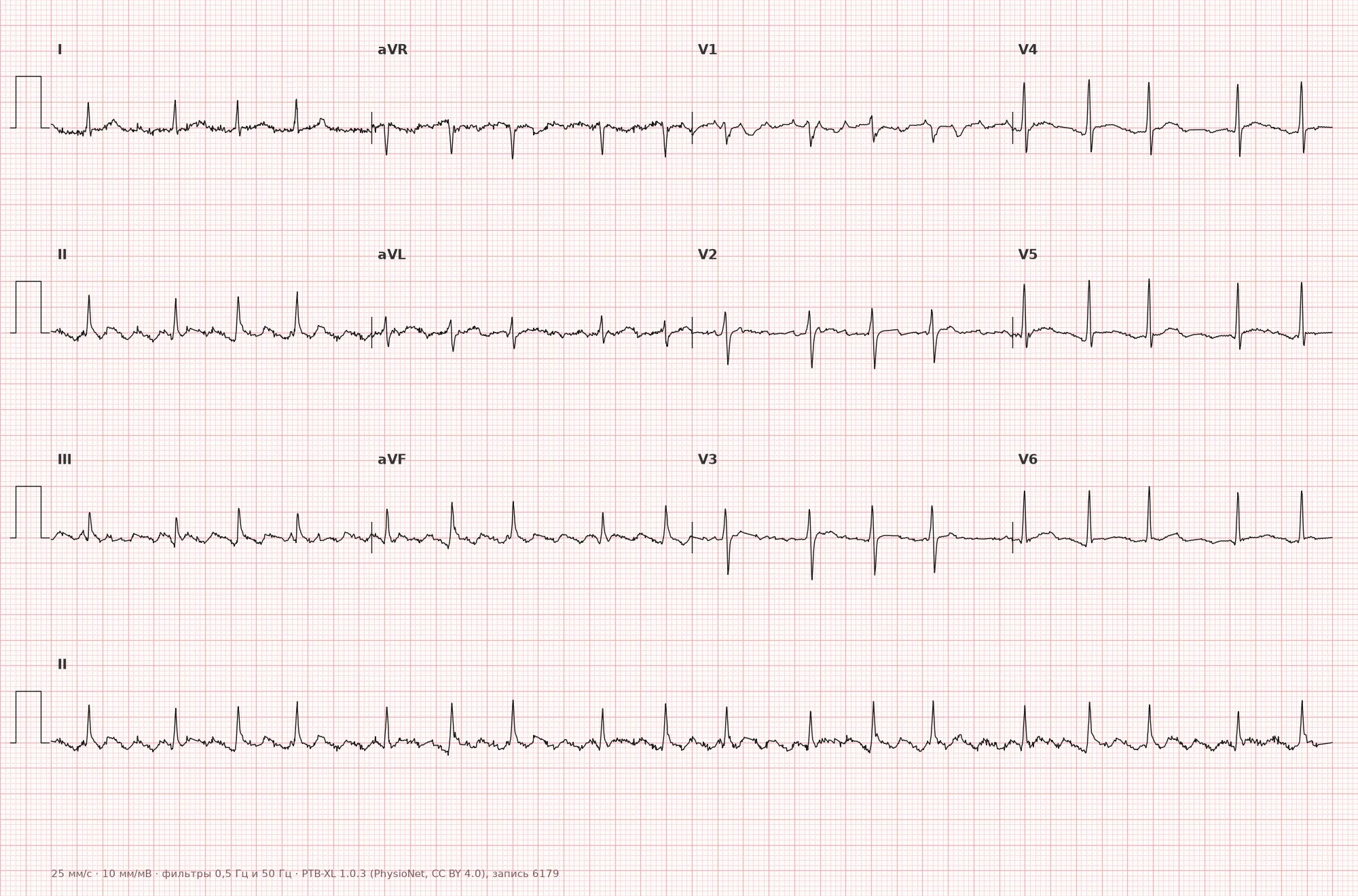
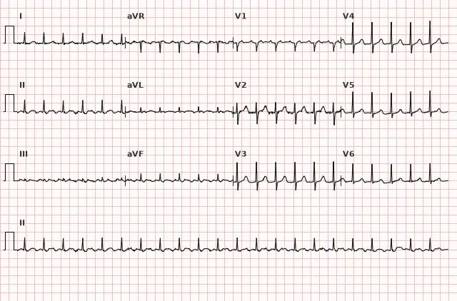
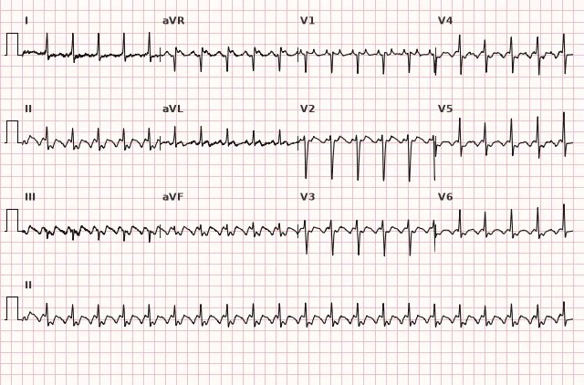
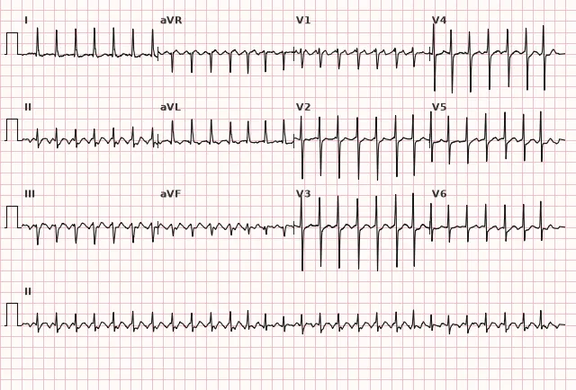
ECG from the open PTB-XL dataset (Wagner P. et al., Scientific Data, 2020; PhysioNet), CC BY 4.0 license. Rendering by WhiteCoat Pro.
Mechanism
Typical AFL is driven by macro-re-entry in the right atrium, the circuit of which passes through the cavotricuspid isthmus (the narrow region between the inferior vena cava and the tricuspid annulus). The excitation wave circulates continuously, which is why the ECG typically shows no isoelectric line between F waves — the atrium is activated with virtually no rest. The direction of circulation (counterclockwise or clockwise) determines the polarity of the F waves in the inferior leads. Due to AV node refractoriness, functional conduction block of 2:1, 3:1, 4:1, or variable is usually present, but 1:1 conduction is possible and requires urgent evaluation.
Causes
- Structural heart diseases: CAD, valvular heart disease, cardiomyopathies
- Arterial hypertension with atrial enlargement
- COPD and chronic cor pulmonale
- History of cardiac surgery, especially involving the atria
- Hyperthyroidism
- Myocarditis, pericarditis
- As a transitional form during treatment of atrial fibrillation with class IC antiarrhythmics (pharmacological conversion of AF to organized AFL)
- AFL without identifiable structural heart disease
Differential diagnosis
- Atrial fibrillation — irregular, chaotic activity without consistent wave morphology, unlike the regular sawtooth F waves in AFL
- Atrial tachycardia — usually discrete P waves with a possible isoelectric interval, but the rate and presence of an isoelectric line overlap with AFL; a single numerical rate range does not reliably distinguish these arrhythmias, differentiation is based on mechanism and atrial activity morphology/axis
- Sinus tachycardia — preserves sinus P-wave morphology and physiological rate variability; very regular tachycardia with a rate around 250 per minute and higher requires ruling out SVT or AFL, especially if P waves are unconvincing
- Narrow-complex tachycardia with a rate of about 150/min without clear P waves — always rule out 2:1 AFL as a frequent masquerading cause
- Atypical atrial flutter — the re-entry circuit is not related to the cavotricuspid isthmus, and the morphology of F waves in the inferior leads may differ from the classic sawtooth
Clinical significance and management
AFL carries a significant risk of thromboembolism; the risk may be lower than in AF, but due to limited data, anticoagulation is generally considered based on similar risk factors and in accordance with current guidelines. The specific risk score is chosen according to local guidelines (e.g., CHA2DS2-VA in ESC 2024 or CHA2DS2-VASc in other documents, including a number of Russian recommendations). With a high ventricular rate and prolonged duration, tachycardiomyopathy may develop.
Textbook-level clinical management: assess hemodynamic instability. In case of instability (hypotension, pulmonary edema, ischemia, altered mental status) — emergency synchronized electrical cardioversion. In stable patients — rate control (AV nodal blockers), and when indicated, rhythm restoration (synchronized cardioversion, antiarrhythmics) or catheter ablation of the isthmus as a definitive treatment for typical AFL. Peri-cardioversion anticoagulation is managed using the same principles as for AF: if the episode duration exceeds 24 hours or is unknown, at least 3 weeks of effective anticoagulation prior to cardioversion is usually required, or a transesophageal echocardiography-guided strategy to rule out thrombus, and anticoagulation is generally continued for at least 4 weeks after cardioversion; further duration depends on the stroke risk according to the chosen score. Specific thresholds and preferred risk scores depend on the version of guidelines used. Administration of class IC drugs without prior slowing of AV conduction is dangerous — it can provoke acceleration of the ventricular rate due to transition to 1:1 conduction.
Common interpretation errors
- Missing 2:1 AFL in a narrow tachycardia around 150/min without clear P waves — F waves are hidden in the QRS or T wave
- Mistaking sawtooth F waves for artifacts or baseline tremor
- Diagnosing atrial fibrillation instead of AFL due to inadequate analysis of wave regularity
- Ignoring variable AV conduction, leading to the rhythm being mistakenly considered irregular like AF
- Underestimating the risk of ventricular rate acceleration when prescribing antiarrhythmic drugs without controlling AV conduction
Sawtooth without rest — flutter; atrial rate around 300, ventricular rate is often exactly half — 150
1. What atrial F-wave rate is most typical for atrial flutter?
The typical F-wave rate in atrial flutter is approximately 250–350 per minute, classically around 300/min, although the range is variable.
2. Which feature differentiates atrial flutter from atrial fibrillation on an ECG?
In atrial flutter, atrial activity is organized and regular, unlike the chaotic, irregular activity in atrial fibrillation.
3. Why is 2:1 atrial flutter easily missed?
In fixed 2:1 block, one of the two F waves is superimposed on the QRS or T wave and is visually obscured, masking the sawtooth pattern.
Frequently asked questions
How to distinguish atrial flutter from atrial fibrillation?
In atrial flutter, F waves are regular, identical in morphology, and form an organized, often sawtooth pattern with a rate typically around 300 bpm. In atrial fibrillation, activity is chaotic, irregular in amplitude and morphology, with no consistent repeating pattern.
Why is the heart rate in atrial flutter frequently exactly 150 bpm?
This is a consequence of fixed 2:1 AV conduction at an atrial rate of approximately 300 beats per minute. Half of the impulses are blocked in the AV node, so the ventricles contract at half the rate of the atria. At a different F-wave rate or block ratio, this relationship may differ.
Is atrial flutter dangerous?
Yes, it increases the risk of thromboembolism—significant, though perhaps slightly lower than in atrial fibrillation—and requires risk assessment to guide anticoagulation according to the score recommended by the guidelines in use. Prolonged tachysystole can lead to tachycardia-induced cardiomyopathy.
How to detect hidden F waves during 2:1 conduction?
Carefully inspect leads II, III, and aVF for an additional notch within the QRS complex or T wave. Vagal maneuvers or adenosine under monitoring can transiently slow AV conduction and make F waves more distinct, but this is a diagnostic maneuver rather than a treatment.
What is the difference between typical and atypical atrial flutter?
Typical atrial flutter involves a re-entry circuit around the cavotricuspid isthmus and most commonly produces a sawtooth pattern in the inferior leads. Atypical atrial flutter arises from other circuits, frequently following cardiac surgery or ablation, and the morphology of F waves may differ from the classic appearance.
ECG Course
Interpretation from basics to myocardial infarction — learn to read any ECG using a systematic algorithm.
- Video lessons and theory for every topic
- Simulator case studies under every lesson
- Unlimited ECG simulator practice
- Study guide: "All ECGs in 25 minutes"
ECG Strip Simulator
ECG, radiology, histology, pathomorphology, pharmacology — thousands of flashcards with detailed explanations.
- Real ECG strips, not textbook drawings
- Instant answers and explanations
- Streaks and progress tracking in your dashboard
Related terms
More in the "Supraventricular Arrhythmias" section
Sources
- Hampton J. The ECG Made Easy
- Surawicz B., Knilans T. Chou's Electrocardiography in Clinical Practice
- Orlov V.N. Guide to Electrocardiography
- ESC Guidelines for the management of atrial fibrillation and supraventricular tachyarrhythmias
Educational material for medical students and physicians. Does not replace clinical guidelines and physical examination; the ECG must always be interpreted in conjunction with the clinical picture.