ECG criteria
- Stable discrete P waves are absent in all leads; instead, there is disorganized fibrillatory activity, the visibility of which depends on the lead and the quality of the recording
- Instead of the isoelectric line, there are continuous undulating oscillations (f waves) of irregular shape and varying amplitude
- Historically, AF with f-waves of approximately ≥1 mm (most commonly assessed in V1) is termed coarse; the threshold and lead selection vary across sources, making this a descriptive rather than a formal diagnostic characteristic
- Atrial activation is very rapid and irregular, roughly in the range of 350–600/min; exact counting on the surface ECG is unreliable and is not used for diagnosis
- Ventricular rhythm is typically irregularly irregular (RR without a repeating pattern); RR irregularity is not entirely specific for AF and can also occur with atrial flutter with variable AV conduction—all features must be evaluated together
- QRS complexes are usually narrow unless there is underlying bundle branch block, nonspecific intraventricular conduction delay, aberrant conduction, ventricular extrasystoles, or ventricular tachycardia
- The morphology of f waves can vary from cycle to cycle and across different leads
- To confirm AF on monitoring data, an arrhythmia episode lasting at least 30 seconds combined with the listed features is taken into account
What it looks like on ECG
Stable discrete P waves are not identified — instead, the isoelectric line is replaced by continuous f waves of irregular shape, varying amplitude, and duration. At the same time, the complete absence of any P-like deflection in all 12 leads is not mandatory: in some leads small f waves may be barely discernible, while in others individual coarse waves may externally resemble P waves. In the coarse form, the oscillations are sufficiently large and clearly distinguishable by eye, unlike fine AF, where the isoelectric oscillations are small and resemble a mere uneven line.
Coarse f waves are most noticeable in lead V1 and the inferior leads (II, III, aVF), which is due to the direction and projection of the summary atrial vector onto these leads. Nevertheless, all 12 leads must be evaluated because wave visibility is uneven, and classifying the form of AF based on a single lead alone is incorrect. Atrial activation is very rapid, roughly 350–600/min, but counting the exact number of waves by eye is usually impossible and unnecessary for diagnosis—the mere fact of disorganized fibrillatory activity is what matters.
Ventricular rhythm in AF is typically irregularly irregular, with no repeating RR pattern. This is a frequent but not absolute sign: with concomitant high-degree AV block, cardiac pacing, certain drug effects, or the presence of an accessory pathway, the rhythm may be relatively regular. In typical atrial flutter with a fixed conduction ratio, RR intervals are often regular, but with variable conduction they can also be irregular—therefore, RR irregularity itself is not a reliable differentiating feature, and the diagnosis is based on the totality of data. QRS complexes are usually normal in shape and width (narrow) unless there is underlying bundle branch block or nonspecific intraventricular conduction delay, aberrant conduction (Ashman phenomenon), ventricular extrasystolic beats, or ventricular tachycardia.
| Sign | Coarse AF | Fine AF |
|---|---|---|
| f-wave amplitude | roughly ≥ 1 mm (descriptive, most often in V1) | < 1 mm |
| Visual distinctness | Pronounced undulation | Almost flat isoelectric line |
| Association with substrate | Inconsistent, historically associated with less fibrosis/enlarged atrium | Inconsistent, historically associated with longstanding AF and fibrosis |
Real ECGs: 8 examples
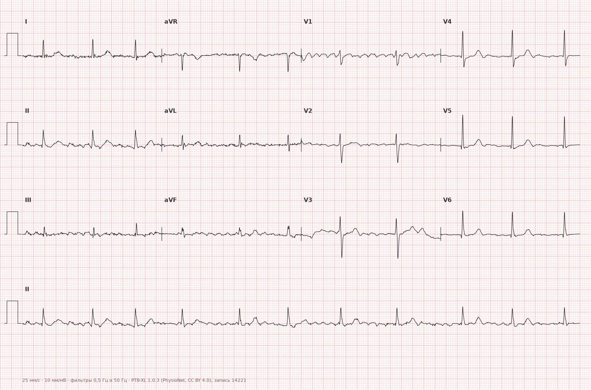
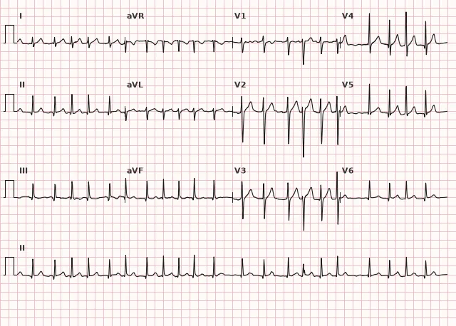
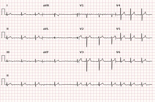
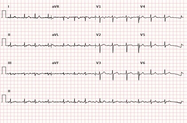
ECG from the open PTB-XL dataset (Wagner P. et al., Scientific Data, 2020; PhysioNet), CC BY 4.0 license. Rendering by WhiteCoat Pro.
Mechanism
Atrial fibrillation is based on multiple chaotic micro-reentry wavelets and foci of ectopic activity, predominantly in the area of the pulmonary veins and adjacent left atrial tissue. The lack of a single coordinated wavefront of depolarization prevents the formation of a normal P wave.
The amplitude of f waves on the surface ECG is presumably related to the mass and degree of synchronization of the atrial tissue involved in fibrillation: larger waves are sometimes associated with relatively preserved atrial myocardium, while smaller ones are associated with marked fibrosis and a long history of arrhythmia. However, this is not a reliable diagnostic rule: the amplitude of f waves on the ECG also depends on the lead, recording calibration, heart position, atrial size, conductivity of surrounding tissues, and technical factors. One cannot reliably judge the degree of fibrosis, duration of AF, or atrial status based on the coarse or fine form—this is not a validated marker.
Causes
The division into coarse and fine forms has only historical and inconsistent associations with the AF substrate, but does not allow determining the etiology. Among the conditions in which AF occurs (including its coarse form):
- Mitral valve disease (stenosis, regurgitation)
- Hypertension with left atrial enlargement
- Hyperthyroidism
- Chronic heart failure
- Dilated cardiomyopathy
- Constrictive pericarditis
- Idiopathic (lone) atrial fibrillation in relatively young patients without structural heart disease
- Postoperative period of cardiac surgery
Differential diagnosis
Atrial flutter — typical flutter is characterized by organized regular atrial activity with repeating F waves and an atrial cycle typically around 240–340/min, often sawtooth in inferior leads; with a fixed AV conduction ratio, the RR rhythm is regular. In atypical flutter, wave morphology may differ from classic sawtooth, and variable AV conduction makes the RR intervals irregular. In AF, atrial activity is disorganized and has no stable repeating cycle. Wave morphology and RR regularity are evaluated only in combination and are not absolute signs individually.
Fine atrial fibrillation — the same mechanism, f-wave amplitude less than ~1 mm; the distinction is descriptive (amplitude-based) and carries no independent clinical significance by itself.
Sinus arrhythmia with artifact (tremor artifact) — against the background of tremor or mains interference, the isoelectric line may appear wavy, but upon careful analysis, true P waves and a regular or slightly varying, but not chaotic, RR rhythm can be found.
Multifocal atrial tachycardia — discernible P waves of at least three different morphologies with an isoelectric interval between them, irregular rhythm, and an atrial rate typically greater than 100/min (at a rate of ≤100/min, it is termed multifocal atrial rhythm), in contrast to the absence of discrete P waves in AF.
Clinical significance and management
The division into coarse and fine forms is a purely descriptive characteristic of the ECG pattern, which does not affect the classification of AF (first diagnosed, paroxysmal, persistent, long-standing persistent, permanent) and does not change management tactics. f-wave amplitude is not used as an independent prognostic or therapeutic criterion.
It is important for the physician to: confirm the diagnosis of AF (absence of stable P waves, RR irregularity, or other variants listed above), assess ventricular rate and hemodynamic stability, search for a structural cause (echocardiography — atrial dimensions, valve apparatus, LV function), assess thromboembolic risk using the applicable scale (CHA2DS2-VASc or its modern variants according to current national and ESC guidelines) and indications for anticoagulant therapy, and determine the rhythm or rate control strategy. In hemodynamic instability (hypotension, pulmonary edema, ischemia, syncope), immediate synchronized electrical cardioversion is indicated; anticoagulation should be initiated as early as possible. In stable patients with an AF duration of more than 24–48 hours or of unknown duration, the cardioversion strategy is determined taking into account anticoagulation and/or exclusion of a left atrial thrombus (transesophageal echocardiography) according to current guidelines. The coarse form per se does not change these decisions.
Common interpretation errors
- Mistaking large f waves for P waves and falsely diagnosing sinus rhythm or atrial tachycardia
- Confusing coarse atrial fibrillation (AF) with atrial flutter due to visually similar undulation — the key sign: organized regular atrial activity with repeating F waves and a stable cycle length in flutter versus disorganized activity without a stable cycle length in AF; RR regularity by itself is not an absolute differentiating feature
- Attributing independent prognostic significance to f-wave amplitude or drawing conclusions from it regarding the duration of AF and the degree of atrial fibrosis, which it does not have
- Failing to check all leads — large f waves may be visible in only one or two leads (most often V1, II, III, aVF) and appear fine-wave in the rest; the AF form should not be classified based on a single lead
- Missing aberrant conduction (Ashman phenomenon — aberration following a long RR cycle immediately followed by a short cycle) against the background of a rapid irregular rhythm, mistaking wide complexes for ventricular extrasystoles; with wide complexes, ventricular extrasystole and ventricular tachycardia must always be ruled out based on the totality of signs
1. Which amplitude benchmark descriptively distinguishes coarse atrial fibrillation from fine atrial fibrillation?
The coarse form is historically described by a fibrillation wave amplitude of approximately 1 mm or more, most often evaluated in V1; the threshold and lead are not standardized, and this is not a formal diagnostic criterion.
2. Which sign most reliably distinguishes coarse AF from typical atrial flutter?
RR irregularity by itself does not exclude flutter (possible with variable AV conduction), so the key sign is the organization and presence of a stable repeating cycle of atrial activity in flutter versus chaotic activity without a stable cycle in AF.
3. Does the amplitude of f waves (coarse or fine form) affect the choice of anticoagulant therapy in atrial fibrillation?
f-wave amplitude is a purely descriptive ECG characteristic not included in the thromboembolic risk assessment criteria.
Frequently asked questions
How does coarse AF differ from fine AF?
Only by the amplitude of f waves on the ECG: in the coarse form, it is approximately 1 mm (0.1 mV) or more, most often evaluated in V1, while in the fine form, it is less. This is a descriptive criterion not unified across all sources, rather than a formal AF classification. The mechanism, classification, and management of AF do not change based on this.
Is coarse atrial fibrillation dangerous?
The danger is determined by atrial fibrillation itself — the risk of thromboembolic complications and potential tachysystole, rather than the f-wave amplitude. The coarse form does not inherently increase or decrease this risk.
How to distinguish coarse AF from atrial flutter?
In typical atrial flutter, atrial activity is organized: F waves are repeating, with a stable cycle typically at a rate of about 240–340/min, often with a constant conduction ratio to the ventricles. In AF, atrial activity is disorganized, there is no stable repeating cycle, and the ventricular rhythm is usually irregularly irregular. It is important to remember that with variable AV conduction, flutter can also produce an irregular RR, so the focus should be on the organization of atrial activity rather than RR alone.
Is it necessary to indicate «coarse» in the ECG conclusion?
This is an optional descriptive clarification that may help during visual tracing analysis, but it is not a mandatory element of the atrial fibrillation diagnosis and does not affect further management.
In which leads are large f waves best visible?
Most often in V1 and inferior leads (II, III, aVF) — this is due to the direction of the atrial electrical vector relative to these leads. All 12 leads should be analyzed, as wave visibility is uneven and the AF form cannot be determined from a single lead.
ECG Course
Interpretation from basics to myocardial infarction — learn to read any ECG using a systematic algorithm.
- Video lessons and theory for every topic
- Simulator case studies under every lesson
- Unlimited ECG simulator practice
- Study guide: "All ECGs in 25 minutes"
ECG Strip Simulator
ECG, radiology, histology, pathomorphology, pharmacology — thousands of flashcards with detailed explanations.
- Real ECG strips, not textbook drawings
- Instant answers and explanations
- Streaks and progress tracking in your dashboard
Related terms
More in the "Supraventricular Arrhythmias" section
Sources
- Hampton J.R. The ECG Made Easy
- Orlov V.N. Guide to Electrocardiography
- Surawicz B., Knilans T. Chou's Electrocardiography in Clinical Practice
- ESC Guidelines for the diagnosis and management of atrial fibrillation
Educational material for medical students and physicians. Does not replace clinical guidelines and physical examination; the ECG must always be interpreted in conjunction with the clinical picture.