معايير تخطيط القلب
- New ST elevation at the J point ≥1 mm (0.1 mV) in two contiguous leads among II, III, aVF — regardless of sex and age; assess in the absence of conditions mimicking STEMI and in the clinical context
- For comparison: in V2–V3 the ST elevation threshold is different and depends on sex and age — at least 1.5 mm in women, at least 2 mm in men aged 40 and older, and at least 2.5 mm in men younger than 40; in the remaining leads (including II, III, aVF) — at least 1 mm in two contiguous leads
- Reciprocal ST depression in aVL often supports the diagnosis of inferior STEMI, but its absence does not exclude infarction — it may be absent, especially with circumflex artery occlusion; depression in I is less common and less pronounced
- The ST III/II ratio and depression in I/aVL help suggest the likely artery but are not a strict diagnostic rule — the culprit artery is ultimately determined by coronary angiography
- ST in III greater than in II, with marked depression in I and aVL — more often corresponds to right coronary artery occlusion; ST in II ≥ ST in III with elevation in I and aVL — more often seen with circumflex branch occlusion
- Pathological Q waves or QS in two contiguous inferior leads support a diagnosis of prior or evolving infarction, but are not mandatory and may appear early in acute ischemia; as a guide — Q ≥0.03 s and ≥1 mm or QS, allowing for normal variants and dynamics on serial ECGs
- Record right precordial leads V3R–V4R when right ventricular involvement is suspected — ST elevation ≥1 mm supports the diagnosis of RV infarction
- With ST depression in V1–V3, tall R waves, or clinical suspicion of posterior infarction, record V7–V9; ST elevation ≥0.5 mm in two contiguous posterior leads supports posterior STEMI, taking into account sex, age and clinical picture
- Assess AV conduction and rhythm — sinus bradycardia is common, as are AV blocks of varying degree
كيف يظهر في التخطيط
The main sign is ST-segment elevation, convex or upsloping, in leads II, III and aVF. It is often accompanied by reciprocal ST depression in aVL, which supports the diagnosis of ischemic rather than pericarditic ST elevation, although the absence of such depression does not exclude infarction. Depression in I is less common and less pronounced.
The ratio of ST elevation in III and II helps suggest the likely artery, but coronary angiography ultimately determines it:
| النمط | Likely artery |
|---|---|
| ST in III > ST in II, marked depression in I and aVL | more often the right coronary artery |
| ST in II ≥ ST in III, elevation also in I, aVL | more often the circumflex branch of the left coronary artery |
With occlusion of the proximal right coronary artery, the right ventricle is often affected simultaneously — leads V3R–V4R are needed. With ST depression and tall R waves in V1–V3 (mirror image of posterior ST elevation) or clinical suspicion of posterior infarction, V7–V9 are recorded.
Over time, a sequence is possible: hyperacute T waves → ST elevation → formation of pathological Q → ST decline → T inversion. The timing and order of this evolution are individual and depend on the duration of occlusion, collateral flow and reperfusion — it is impossible to date the infarction by ECG shape alone without clinical data, serial ECGs and troponin.
مخططات حقيقية: 8 أمثلة
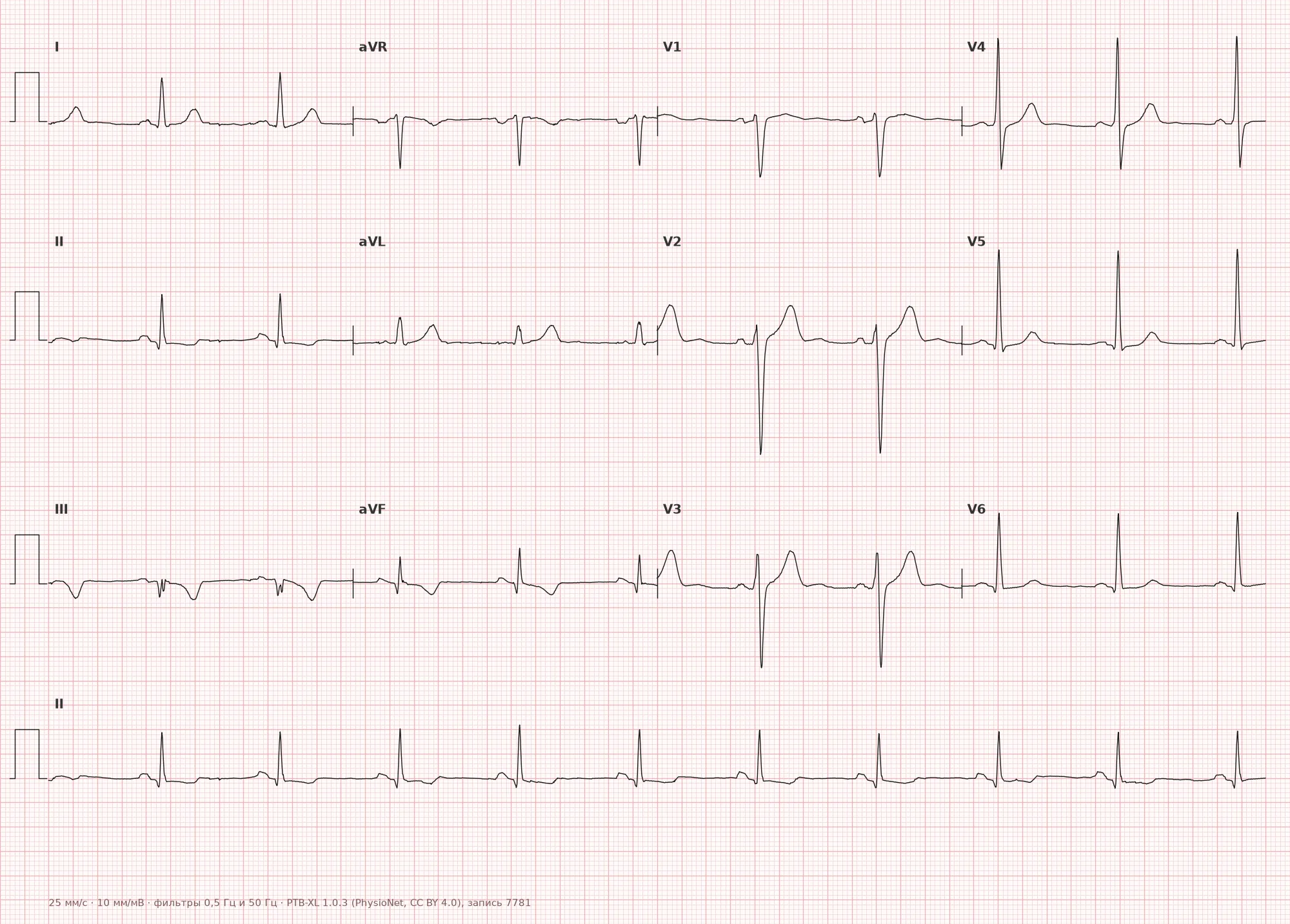
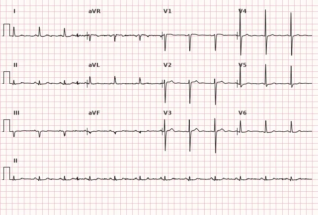
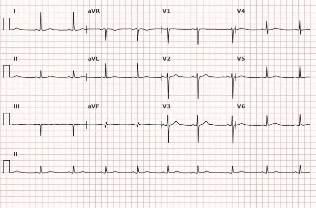
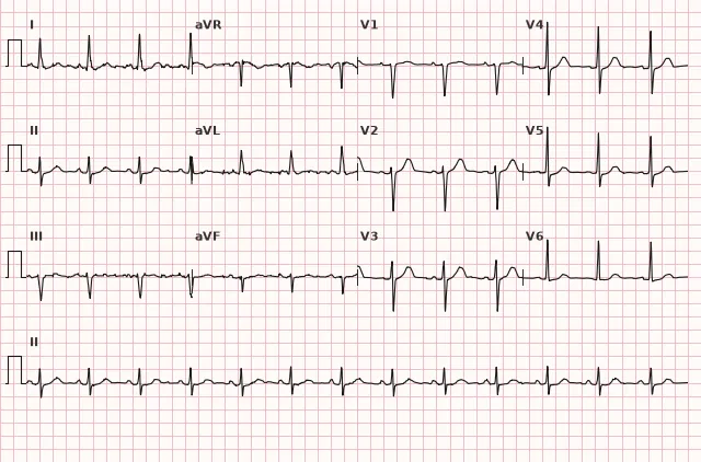
مخطط قلب من قاعدة البيانات المفتوحة PTB-XL (Wagner P. et al., Scientific Data, 2020; PhysioNet)، ترخيص CC BY 4.0. الرسم - منصة WhiteCoat Pro.
الآلية
The inferior wall of the left ventricle is in most cases supplied by the right coronary artery (right dominance), less often by the circumflex branch of the left coronary artery (left dominance). Thrombotic occlusion causes acute injury and then myocardial necrosis. The injury vector is directed downward, so the EMF shifts toward leads II, III, aVF (positive deflection) and often opposite to aVL (negative deflection, reciprocal depression). The right coronary artery often supplies both the sinus node and the AV node — hence the frequent bradyarrhythmias and AV blocks in inferior MI.
الأسباب
- Thrombotic occlusion of the right coronary artery (more common)
- Thrombotic occlusion of the circumflex branch of the left coronary artery
- Coronary artery spasm (Prinzmetal variant)
- Coronary artery dissection
- Embolism into the coronary circulation
- Multivessel disease in chronic CAD with acute decompensation of blood flow
التشخيص التفريقي
- Pericarditis: diffuse ST elevation, concordant, usually without local reciprocal changes (except aVR and sometimes V1), often PR depression
- Early repolarization: ST elevation with a characteristic notched QRS-ST transition, stable pattern without dynamics, no reciprocal changes and no Q wave
- Brugada syndrome and other patterns involve the right precordial leads, not the inferior wall
- Inferolateral infarction — changes in I, aVL, V5–V6 are added to the inferior leads
- Hyperkalemia can mimic or alter the ischemic pattern, including pseudoelevation of ST, tall peaked T waves and QRS widening; this combination requires urgent electrolyte assessment and clinical correlation, and with genuine suspicion of occlusion — treatment as STEMI without delaying reperfusion
الأهمية السريرية والتكتيكات
Inferior STEMI is an indication for immediate activation of the reperfusion network. Primary PCI is preferred if wire crossing is expected within 120 minutes from STEMI diagnosis. If this is not possible within 120 minutes, symptoms usually began within 12 hours and there are no contraindications — immediate fibrinolysis is considered with mandatory transfer to a PCI center: rescue PCI for failed reperfusion and routine coronary angiography/PCI usually within 2–24 hours after successful lysis.
It is advisable to record right precordial leads (V3R–V4R) when RV involvement is suspected and posterior leads (V7–V9) when concomitant posterior infarction is suspected — this changes infusion strategy and prognosis. When RV infarction is suspected, hemodynamics must be assessed: avoid nitrates and other vasodilators in hypotension or when cardiac output depends on preload, and do not prescribe them without assessing the condition. In hypotension without pulmonary edema, cautious infusion is possible, and in persistent shock intensive hemodynamic support is needed.
Continuous rhythm monitoring is needed — sinus bradycardia, second- and third-degree AV block are likely; these are often reversible after reperfusion but may require temporary pacing.
الأخطاء الشائعة أثناء قراءة تخطيط القلب
- Assess only II, III, aVF and fail to consider indications for V3R–V4R and V7–V9
- Consider the absence of ST depression in aVL a reason to exclude inferior MI
- Confuse isolated ST elevation in III with inferior MI without confirmation in a contiguous lead (criterion — at least two contiguous leads)
- Fail to consider that bradycardia and AV block in inferior MI are often reflex (vagal) or related to AV node ischemia, and do not always reflect organic damage to the conduction system
- Miss concomitant posterior infarction because in the standard 12 leads it is visible only as a mirror image
1. Which sign more often supports the diagnosis of inferior myocardial infarction as opposed to pericarditis?
Reciprocal depression in aVL often accompanies ischemic ST elevation of the inferior wall and is usually absent in pericarditis, although its absence does not exclude infarction.
2. The ratio of ST elevation in which leads more often suggests right coronary artery occlusion in inferior MI?
Predominance of ST elevation in III over II with marked depression in I and aVL more often corresponds to occlusion of the right coronary artery, but coronary angiography definitively identifies the artery.
3. Which additional leads should be recorded in inferior MI to detect right ventricular infarction?
Right precordial leads V3R–V4R allow detection of ST elevation with right ventricular involvement, which changes treatment strategy.
الأسئلة الشائعة
How to distinguish inferior MI from pericarditis on ECG?
In inferior MI, ST elevation is limited to the inferior leads and is often accompanied by reciprocal depression in aVL. In pericarditis, ST elevation is usually diffuse and concordant, with no local reciprocal depression (except in aVR and sometimes V1), and PR depression is often seen.
Why do bradycardia and AV block occur in inferior MI?
The right coronary artery in most cases supplies the sinus node and AV node, so its occlusion can cause ischemia of the conduction system. Reflex vagal activation from the inferior ventricular wall (Bezold–Jarisch reflex) also plays a role.
Why record right precordial leads in inferior MI?
To detect concomitant right ventricular infarction — ST elevation in V3R–V4R. This changes management: when RV MI is suspected, hemodynamics are assessed and nitrates and other preload-reducing drugs are used with caution due to the risk of severe hypotension.
How to determine the culprit artery on ECG in inferior MI?
If ST elevation in III is higher than in II, with marked depression in I and aVL — occlusion of the right coronary artery is more often suspected. If ST elevation in II is equal to or higher than III with elevation in I, aVL — occlusion of the circumflex branch is more often suspected. These are only probabilistic signs; coronary angiography definitively identifies the artery.
Is isolated inferior MI dangerous compared with anterior?
Overall, inferior MI without right ventricular involvement and without concomitant posterior or lateral damage has a more favorable prognosis than anterior. But the addition of RV infarction or multivessel disease substantially increases the risk of complications and mortality.
دورة تخطيط القلب الكهربائي
التفسير من الأساسيات وحتى احتساء الاحتشاءات — لقراءة أي مخطط وفق خوارزمية دقيقة.
- دروس فيديو ونظرية لكل موضوع
- حالات تدريبية تحت كل درس
- جهاز محاكاة تخطيط القلب بلا حدود يومية
- دليل «جميع تخطيطات القلب في 25 دقيقة»
جهاز محاكاة التخطيطات
تخطيط القلب، الأشعة، علم الأنسجة، علم الأمراض، الصيدلة — آلاف البطاقات مع الشرح.
- مخططات حقيقية وليست مجرد صور من الكتب
- الإجابة والشرح الفوري
- السلاسل والتقدم في الحساب الشخصي
المصطلحات ذات الصلة
المزيد في قسم «نقص تروية واحباط عضلة القلب»
المصادر
- Goldberger's Clinical Electrocardiography, 9th ed.
- Hampton, The ECG Made Easy
- ESC Guidelines for the management of STEMI, 2023
- أورلوف ف. ن.، دليل تخطيط القلب الكهربائي
مادة تعليمية لطلاب كليات الطب والأطباء. لا تحل محل التوصيات السريرية والفحص الطبي؛ يُفسر تخطيط القلب دائماً جنباً إلى جنبกับการحالة السريرية.