معايير تخطيط القلب
- ST elevation at the J point in two contiguous leads: in V2–V3 — ≥2.5 mm in men younger than 40 years, ≥2 mm in men 40 years and older, ≥1.5 mm in women; in other leads — ≥1 mm (AHA/ACCF/HRS 2009 criteria)
- Any Q wave in V2–V3 with duration ≥0.02 s or a QS complex in V2–V3; or Q ≥0.03 s and depth ≥1 mm or QS in I, II, aVL, aVF, V4–V6 (Universal Definition criteria); these signs indicate a pathological Q wave, but by themselves do not prove acuity of the infarction
- Poor R-wave progression from V1 to V3 — a nonspecific finding; the diagnosis of prior MI requires correlation with previous ECGs, clinical picture, imaging, and other signs of scarring
- Reciprocal ST depression in inferior leads (II, III, aVF) is not obligatory and does not always occur
- Evolution over time: acute phase — ST elevation and possible hyperacute T waves; subacute — Q-wave formation and T-wave inversion; scar phase — persistent Q with normalization of ST-T
- Correlation with clinical picture and troponin to distinguish STEMI from other causes of ST elevation in V1–V3; interpretation of pathological Q together with clinical picture and evolution
كيف يظهر في التخطيط
The area of interest is leads V1–V3, sometimes involving V4. In the acute phase, hyperacute broad T waves are possible followed by or nearly simultaneous with ST-segment elevation; the sequence is variable, and hyperacute T waves may be absent or short-lived — ST elevation often becomes the first noticeable sign. Pathological Q/QS waves may appear in the acute phase or later, reflect changes in excitation spread, and cannot by themselves determine transmurality of necrosis.
An important feature: QS in V1 is sometimes normal, whereas QS in V2 and especially in V1–V2 requires checking electrode position, comparison with previous ECGs, and assessment of other signs. A pathological Q wave in these leads is considered to be a change compared with the baseline ECG, appearance of notching, increase in width or depth, and most importantly — combination with ST-T elevation/evolution and clinical picture.
| الاتجاه | What we look at | Typical finding |
|---|---|---|
| V1–V2 | ST, Q | ST elevation, QS/Q |
| V3 (±V4) | ST, Q, R | ST elevation, decreased R, sometimes Q |
| II, III, aVF | reciprocity | possible small ST depression, not obligatory |
| V7–V9 | rule out posterior MI | normally without ST elevation |
In isolated anteroseptal involvement, a Q wave in I, aVL, V5–V6 is usually absent — this distinguishes it from extensive anterior or anterolateral infarction.
مخططات حقيقية: 8 أمثلة
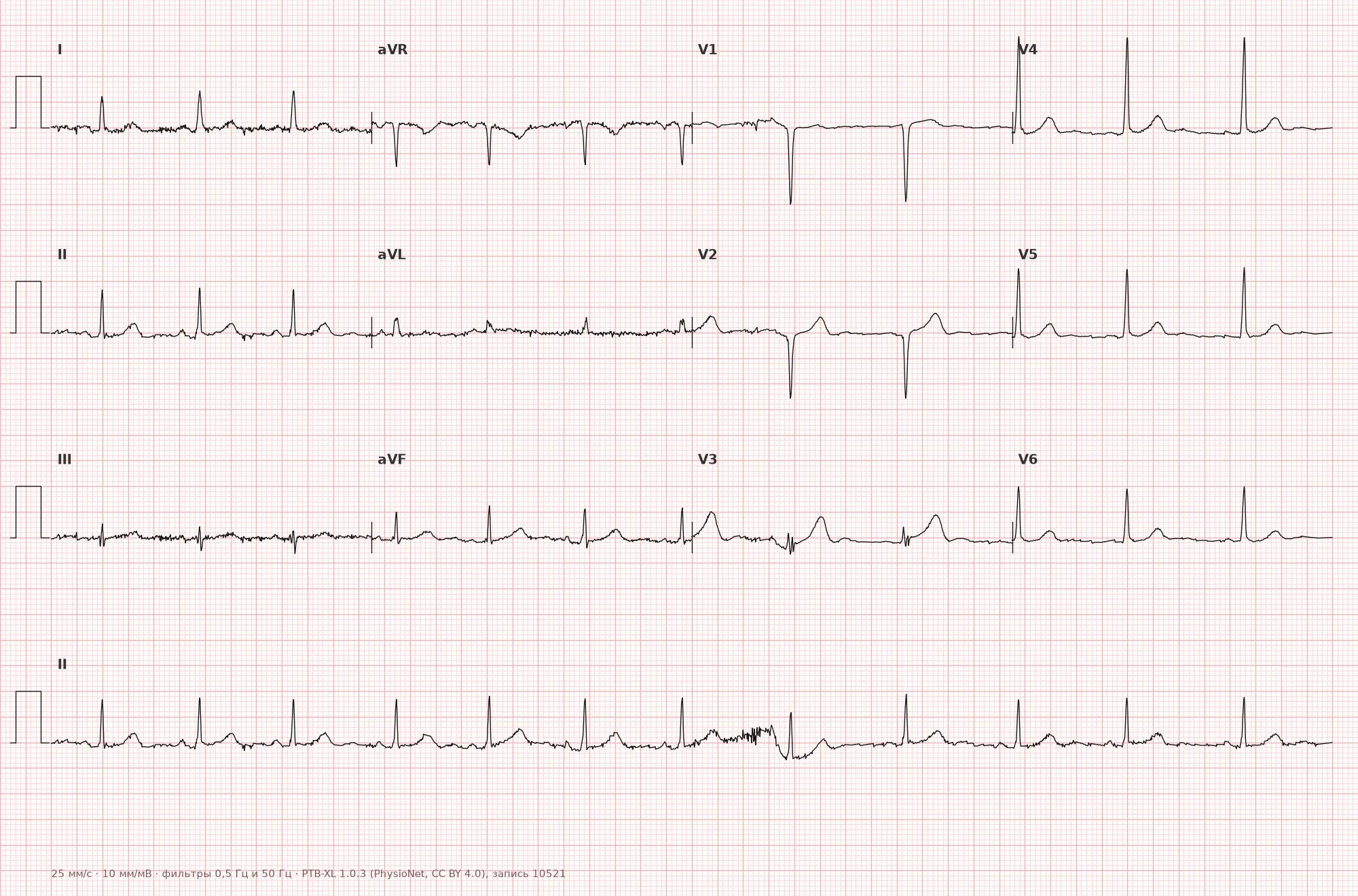
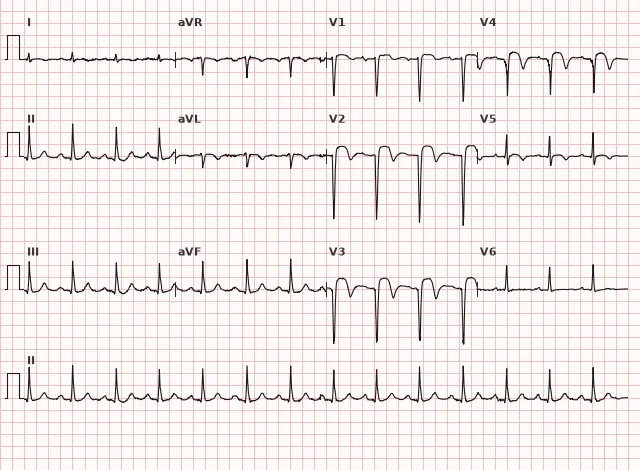
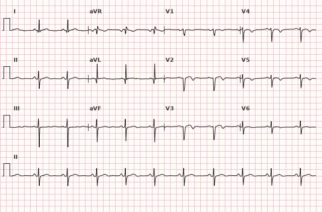
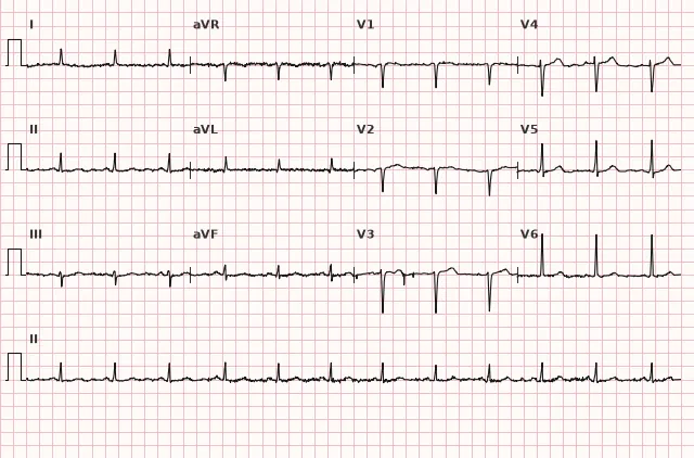
مخطط قلب من قاعدة البيانات المفتوحة PTB-XL (Wagner P. et al., Scientific Data, 2020; PhysioNet)، ترخيص CC BY 4.0. الرسم - منصة WhiteCoat Pro.
الآلية
The interventricular septum and the adjacent anterior wall of the LV are supplied by septal perforating branches and proximal anterior septal branches of the LAD. Occlusion of this LAD segment causes ischemia and necrosis of the septum, which on ECG is reflected by ST elevation and Q-wave formation specifically in the leads electrically facing the septum — V1–V3. Since the septum contains the proximal portions of the right and left bundle branches, extensive septal necrosis may cause intraventricular conduction disturbances (right bundle branch block, rarely bifascicular blocks).
الأسباب
- Atherothrombotic occlusion or critical stenosis of the proximal/mid LAD involving septal branches
- Atherosclerotic plaque rupture with thrombosis (the most common mechanism of STEMI)
- Coronary spasm in the LAD territory (variant angina)
- تسلخ الشريان التاجي (بما في ذلك التسلخ العفوي، SCAD)
- Embolism into the LAD (rare)
- Iatrogenic occlusion of a septal branch during coronary interventions
التشخيص التفريقي
- Normal variant QS in V1: QS in V1 is sometimes normal, without ST-T evolution, without clinical picture, stable on previous ECGs; QS in V2 and wider requires checking electrode position and ruling out pathology
- Left bundle branch block: wide QRS, typical morphology in V1 and V5–V6, masks and mimics the Q wave; infarction is assessed by Sgarbossa criteria (modified — Smith)
- Brugada syndrome (type 1 pattern): coved elevation of the J point/ST usually ≥2 mm in at least one of leads V1–V2 recorded in the standard (4th intercostal space) or high (2nd–3rd intercostal space) position, followed by a negative T wave; requires clinical-electrocardiographic assessment — an isolated Brugada-like pattern without the appropriate context does not equal a clinical syndrome
- Hypertrophic cardiomyopathy with septal hypertrophy: deep narrow Q waves combined with tall R waves, without ST-T evolution
- Myocarditis: diffuse ST-T changes without strict relation to a single artery territory, often together with changes in inferior and lateral leads
- Early repolarization: ST elevation with J-point notch, concave, without reciprocal depression and without Q-wave formation
الأهمية السريرية والتكتيكات
New ST elevation in V1–V3(V4) in two contiguous leads with clinical evidence of ischemia is STEMI requiring emergency reperfusion (primary PCI within the recommended window, and if unavailable — fibrinolysis according to current criteria and time frames). One should actively look for extension to the lateral wall (I, aVL, V5–V6) and the at-risk territory of the right ventricle/conduction system, and consider the appearance of new bundle branch blocks as a marker of extensive involvement.
ST elevation in aVR with diffuse ST depression indicates high ischemic risk and possible left main/multivessel disease, but does not allow an anatomical diagnosis; an immediate invasive strategy is indicated under the appropriate very high-risk criteria. In the absence of ST elevation but with suspected ACS (e.g., a pattern resembling Wellens syndrome in precordial leads), the approach is different — an early invasive strategy for NSTE-ACS, not emergency PCI "as STEMI".
Prior anteroseptal infarction (persistent Q without acute evolution) requires assessment of LV ejection fraction and LV thrombus, risk of anterior wall/apex aneurysm, and prescription of antithrombotic and other secondary prevention as indicated. Anticoagulation is prescribed for documented LV thrombus, atrial fibrillation, or another established indication; antiarrhythmic drugs — for a specific arrhythmia and taking risk into account; in reduced EF, indications for ICD implantation are assessed for primary prevention of sudden death after optimal medical therapy and in accordance with recommended waiting periods.
الأخطاء الشائعة أثناء قراءة تخطيط القلب
- Mistaking a physiological QS in V1 for a pathological Q without assessing evolution and clinical picture; forgetting that QS in V2 and wider requires checking
- Interpreting a pathological Q as a sign of acute STEMI by itself — without new ST elevation or occlusion equivalent and clinical picture, it only indicates possible necrosis/scarring
- Not comparing the current ECG with previous ones — missing the new appearance of Q or ST elevation
- Ignoring left bundle branch block as a possible masking factor of infarction
- Not assessing lead contiguity — the diagnosis of STEMI requires ST elevation in two or more contiguous leads, not in one random lead
- Forgetting gender and age differences in ST elevation thresholds in V2–V3 when interpreting borderline values
1. In which leads is ST elevation expected in anteroseptal infarction?
The septum and adjacent anterior wall project onto leads V1–V3, sometimes up to V4.
2. Which artery is most often responsible for anteroseptal infarction?
تنشأ الفروع الانثقابية الحاجزية غالبًا من الشريان الأمامي النازل الأيسر (LAD) فتغذّي الحاجز بين البطينين، لكن تخطيط القلب لا يسمح بتحديد الجزء والفرع المصابين بدقة موثوقة.
3. ما الذي يميّز موجة QS الفسيولوجية في V1–V2 عن موجة Q المرضية في الاحتشاء؟
قد تكون موجة QS الثابتة في V1 أحد أشكال الطبيعي؛ وتُقيَّم موجات Q/QS المرضية وفق المعايير المورفولوجية والمقارنة بتخطيط سابق، ويُستخدم التغيّر الحاد في ST-T والصورة السريرية لتحديد حدة الاحتشاء، لكنها ليست شرطًا لوجود موجة Q المرضية بحد ذاتها.
الأسئلة الشائعة
كيف نميّز الاحتشاء الأمامي الحاجزي عن شكل طبيعي لموجة QS في V1–V2؟
موجة QS في V1 قد تكون طبيعية أحيانًا، بينما QS في V2 وخاصة في V1–V2 تستدعي التحقق من موضع الأقطاب ومقارنة بتخطيطات سابقة وتقييم علامات أخرى. في الاحتشاء الحاد تترافق تغيّرات Q بارتفاع جديد في ST وديناميكيته وصورة سريرية موافقة، ويُؤكَّد ذلك بالمقارنة مع تخطيطات سابقة.
أي شريان يُصاب عادةً في الاحتشاء الأمامي الحاجزي؟
السبب الأكثر شيوعًا هو انسداد الشريان الأمامي النازل الأيسر (LAD)، لكن تخطيط القلب لا يسمح بتحديد الجزء والفرع المصابين بدقة موثوقة. ومصطلح «احتشاء أمامي حاجزي» هو توطين تخطيطي تقليدي؛ وفي الأدلة الحديثة يُستخدم غالبًا «احتشاء أمامي/احتشاء أمامي مع ارتفاع ST»، لأن حدود المناطق والتطابق التشريحي متغيّرة.
هل يُعدّ الاحتشاء الأمامي الحاجزي خطيرًا بسبب إحصارات التوصيل؟
نعم، يحتوي الحاجز على الأجزاء الدانية من حزمتي فرعي حزمة هيس، لذا قد يؤدي الضرر الحاجزي الواسع إلى ظهور إحصار الفرع الأيمن وإحصار ثنائي الفصيلة وحتى إحصار AV عالي الدرجة، مما يسيء إلى الإنذار.
هل يلزم رأب وعائي أولي إسعافي عند ارتفاع ST أمامي حاجزي؟
نعم، إذا كان الارتفاع الجديد في ST في V1–V3 يستوفي معايير احتشاء عضلة القلب مع ارتفاع ST وكانت الصورة السريرية موافقة، فتُستطب إعادة التروية الإسعافية — رأب وعائي أولي عبر الجلد (PCI) في المدة الموصى بها أو حلّ فيبريني عند تعذّر PCI.
لماذا تختلف عتبات ارتفاع ST في V2–V3 حسب الجنس والعمر؟
يعود ذلك إلى فروق فسيولوجية في سعة ST في هذه المسارات بين الرجال والنساء مختلفي الأعمار؛ وتقترح AHA/ACCF/HRS 2009 عتبات متمايزة (مثلًا أعلى لدى الرجال دون 40 عامًا) لتقليل التشخيصات الإيجابية الكاذبة لاحتشاء عضلة القلب مع ارتفاع ST.
دورة تخطيط القلب الكهربائي
التفسير من الأساسيات وحتى احتساء الاحتشاءات — لقراءة أي مخطط وفق خوارزمية دقيقة.
- دروس فيديو ونظرية لكل موضوع
- حالات تدريبية تحت كل درس
- جهاز محاكاة تخطيط القلب بلا حدود يومية
- دليل «جميع تخطيطات القلب في 25 دقيقة»
جهاز محاكاة التخطيطات
تخطيط القلب، الأشعة، علم الأنسجة، علم الأمراض، الصيدلة — آلاف البطاقات مع الشرح.
- مخططات حقيقية وليست مجرد صور من الكتب
- الإجابة والشرح الفوري
- السلاسل والتقدم في الحساب الشخصي
المصطلحات ذات الصلة
المزيد في قسم «نقص تروية واحباط عضلة القلب»
المصادر
- Goldberger's Clinical Electrocardiography، الطبعة الحالية
- AHA/ACCF/HRS Recommendations for the Standardization and Interpretation of the Electrocardiogram, 2009
- Surawicz B., Knilans T. Chou's Electrocardiography in Clinical Practice
- أورلوف ف.ن. دليل تخطيط القلب الكهربائي
مادة تعليمية لطلاب كليات الطب والأطباء. لا تحل محل التوصيات السريرية والفحص الطبي؛ يُفسر تخطيط القلب دائماً جنباً إلى جنبกับการحالة السريرية.