معايير تخطيط القلب
- New ST elevation at the J point in two anatomically contiguous leads: ≥ 1 mm — except V2–V3, where separate sex- and age-specific thresholds apply
- In men ≥40 years: ST elevation in V2–V3 ≥ 2 mm; in men <40 years: ≥ 2.5 mm
- In women: ST elevation in V2–V3 ≥ 1.5 mm regardless of age
- In the remaining leads (V1, V4–V6, I, aVL): ST elevation ≥ 1 mm in two contiguous leads
- The shape of the ST segment (concave, straight, convex upward) and hyperacute tall T waves are additional signs and do not replace the criterion of ST-elevation magnitude
- Reciprocal ST depression in the inferior leads (II, III, aVF) in extensive anterior MI; not required for the diagnosis
- Over time — pathological Q wave: in I, II, aVL, aVF, V4–V6 — Q ≥ 0.03 s and ≥ 1 mm deep (or QS) in two contiguous leads; in V2–V3 — any Q wave ≥ 0.02 s or a QS complex is already pathological (one of these two leads is sufficient); the appearance of Q is not required for the diagnosis of acute STEMI
- Assess the extent of involvement by the number of involved leads: V1–V2 (septal) vs V1–V6 + I, aVL (extensive anterior) — as a guide to the ischemic zone, without a precise conclusion about the level of LAD occlusion
كيف يظهر في التخطيط
Anterior STEMI reflects ischemia of the anterior wall and interventricular septum, usually supplied by the LAD. The key finding is new ST-segment elevation at the J point in two contiguous precordial leads (± I, aVL); its distribution relates to the extent of involvement:
| المنطقة | المسارات | Approximate territory |
|---|---|---|
| Septal | V1–V2 | septal perforators |
| الأمامي | V3–V4 | LAD territory |
| Anterolateral | V5–V6, I, aVL | LAD, often with diagonal branches |
| Extensive anterior | V1–V6, I, aVL | large LAD territory |
This is a guide to the area of ischemia, not a precise anatomical localization of the occlusion level or a specific branch — this is unreliable on a standard ECG; the exact level of the lesion is determined by clinical data and coronary angiography. In the hyperacute phase, tall hyperacute T waves in V2–V4 may appear even before clear ST elevation. New right bundle branch block, especially combined with ischemic symptoms, is associated with extensive anterior ischemia and an unfavorable prognosis and requires immediate assessment for urgent reperfusion strategy; it is not a precise marker of the LAD occlusion level and does not replace ST analysis as the main diagnostic criterion. Reciprocal ST depression in the inferior leads is not always present, but when present in anterior MI it indicates a larger area of damage. ST elevation in aVR against a background of diffuse ST depression in other leads is a sign of extensive high-risk subendocardial ischemia (seen in multivessel disease, left main coronary artery occlusion, but also in tachycardia, anemia, hypotension and other conditions of imbalance between oxygen supply and demand); it requires urgent clinical assessment but by itself does not prove left main coronary artery involvement. Over hours to days a pathological Q wave forms and the T wave becomes negative (reperfusion-related or as part of the natural evolution of necrosis).
مخططات حقيقية: 8 أمثلة
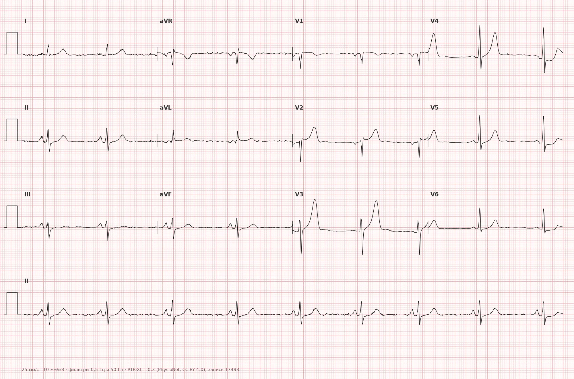
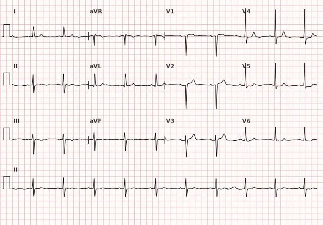
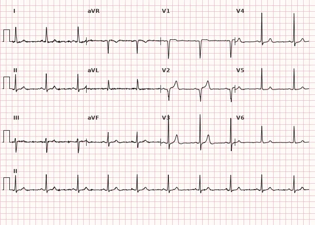
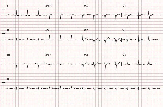
مخطط قلب من قاعدة البيانات المفتوحة PTB-XL (Wagner P. et al., Scientific Data, 2020; PhysioNet)، ترخيص CC BY 4.0. الرسم - منصة WhiteCoat Pro.
الآلية
Acute thrombotic occlusion of the LAD (or another culprit artery) stops blood flow to the anterior wall and the LV septum. The subendocardium is affected first, then ischemia becomes transmural — on the ECG this manifests as ST elevation (the injury current is directed from endocardium to epicardium, the vector shifts toward the electrodes over the damaged zone). The greater the myocardial mass without blood flow, the wider the coverage of precordial leads by ST elevation. A decrease in ST elevation with treatment primarily reflects reduction of ischemia or successful reperfusion, not myocardial death per se. A pathological Q wave increases the likelihood of irreversible necrosis, but may appear already in the acute phase, is not equivalent to a formed scar and in some cases regresses.
الأسباب
- Atherothrombotic occlusion of the LAD (main cause of anterior STEMI)
- Left main coronary artery or other branch involvement, multivessel disease
- Coronary artery dissection (spontaneous, in young women, in the postpartum period)
- Coronary spasm (Prinzmetal variant) with or without thrombosis
- Embolism to the LAD (endocarditis, atrial fibrillation with thrombus, paradoxical embolism)
- Aortic dissection extending to the left main coronary ostium (rare)
- Severe coronary vasospasm with cocaine use
التشخيص التفريقي
- Early repolarization syndrome — concave ST elevation, J-point notch, without reciprocal depression or dynamic changes over time
- Pericarditis — diffuse ST elevation in many leads without reciprocity (except aVR), often PR depression
- Takotsubo cardiomyopathy — clinical picture and ECG resemble anterior MI, but coronary arteries are intact; diagnosis by coronary angiography and echocardiography
- Hypertrophic cardiomyopathy (apical) — deep negative T waves in precordial leads without acute ST elevation
- De Winter waves — tall symmetrical peaked T wave with upsloping ST depression of about 1–3 mm from the J point, typically in V2–V6; ST elevation in aVR may occur but is not a diagnostic component of the pattern; considered an occlusive MI pattern (STEMI equivalent) reflecting acute occlusion or critical proximal LAD lesion, requiring immediate reperfusion assessment
- Wellens syndrome — deep negative or biphasic T wave in V2–V3 after an episode of pain, a sign of critical LAD stenosis without current occlusion; the primary PCI window of «<120 min» does not apply to it; management is an early invasive strategy for NSTE-ACS
الأهمية السريرية والتكتيكات
Anterior STEMI, especially extensive (V1–V6, I, aVL), carries the largest mass of affected myocardium among all STEMI locations and a high risk of LV systolic dysfunction, cardiogenic shock, conduction disturbances (bundle branch block, AV block) and ventricular arrhythmias. With ST elevation meeting the criteria — immediate activation of the primary PCI pathway: the target time from first medical contact to reperfusion (FMC-to-device) is usually ≤ 90 minutes for direct presentation to a PCI center and ≤ 120 minutes for transfer from another hospital. If the expected delay to primary PCI exceeds 120 minutes from diagnosis, with symptoms of less than 12 hours and no contraindications, immediate fibrinolysis is considered (preferably within 10 minutes of diagnosis) followed by a pharmacoinvasive strategy; before fibrinolysis, acute aortic dissection and other contraindications must be excluded. Record the time of the first ECG, obtain repeat ECGs if symptoms worsen, monitor the electrical axis and QRS width (new bundle branch block is an unfavorable sign), and monitor hemodynamics because of the risk of acute left ventricular failure.
الأخطاء الشائعة أثناء قراءة تخطيط القلب
- Applying a single ST-elevation threshold for V2–V3 without regard to sex and age
- Failing to recognize hyperacute T waves and De Winter waves as a possible equivalent of LAD occlusion in the absence of clear ST elevation
- Making a categorical conclusion about the level of LAD or a specific branch occlusion based only on ST shape or new bundle branch block
- Confusing early repolarization and pericarditis with true anterior STEMI when dynamic changes and reciprocity are insufficiently assessed
- Not repeating the ECG when the clinical picture changes, missing ST-T evolution
1. What ST-elevation threshold at the J point in leads V2–V3 in men aged 40 years and older is considered diagnostic?
According to ECG standardization recommendations, for men ≥40 years the threshold in V2–V3 is ≥ 2 mm, whereas in men <40 years it is 2.5 mm and in women 1.5 mm.
2. Which coronary artery most often causes anterior myocardial infarction?
The anterior wall and interventricular septum are usually supplied by the left anterior descending artery (LAD); its occlusion produces the typical picture of anterior STEMI, although other variants of LCA involvement are possible.
3. Which ECG pattern corresponds to extensive anterior STEMI with a large area of damage?
Involvement of all precordial leads V1–V6 together with I, aVL reflects a large area of anterior wall ischemia; reciprocal depression in the inferior leads strengthens the suspicion of extensive involvement, although the ECG does not determine the exact level of occlusion.
الأسئلة الشائعة
Which leads are examined in anterior myocardial infarction?
The main ones are the precordial leads V1–V6; in extensive involvement the lateral leads I and aVL are added. Reciprocal changes are sought in the inferior leads II, III, aVF.
How does anterior MI differ from anteroseptal MI?
Anteroseptal MI is limited to ST elevation in V1–V2 (septal perforators), whereas anterior or extensive anterior MI involves V3–V6 and often I, aVL. This indicates a larger area of ischemia, although the exact level of LAD occlusion cannot be reliably determined from the ECG.
Is anterior myocardial infarction more dangerous than other localizations?
On average yes: the LAD supplies most of the LV, so anterior STEMI is more often accompanied by a significant reduction in ejection fraction, cardiogenic shock and conduction disturbances compared with inferior MI.
Can anterior MI occur without obvious ST elevation?
Yes, with subtotal or acute LAD occlusion, occlusion equivalents are possible — de Winter waves or hyperacute T waves in the precordial leads, and Wellens syndrome reflects critical stenosis without current occlusion at the time of ECG recording.
Is the appearance of a Q wave mandatory for the diagnosis of anterior infarction?
No, a pathological Q wave forms over time and is not a necessary condition for diagnosing acute STEMI. The main ECG criterion of the acute phase is new ST elevation in two contiguous leads, but the final diagnosis of myocardial infarction is based on the clinical picture, ECG and cardiac troponin dynamics; with a typical picture, reperfusion is not delayed while awaiting the troponin result.
دورة تخطيط القلب الكهربائي
التفسير من الأساسيات وحتى احتساء الاحتشاءات — لقراءة أي مخطط وفق خوارزمية دقيقة.
- دروس فيديو ونظرية لكل موضوع
- حالات تدريبية تحت كل درس
- جهاز محاكاة تخطيط القلب بلا حدود يومية
- دليل «جميع تخطيطات القلب في 25 دقيقة»
جهاز محاكاة التخطيطات
تخطيط القلب، الأشعة، علم الأنسجة، علم الأمراض، الصيدلة — آلاف البطاقات مع الشرح.
- مخططات حقيقية وليست مجرد صور من الكتب
- الإجابة والشرح الفوري
- السلاسل والتقدم في الحساب الشخصي
المصطلحات ذات الصلة
المزيد في قسم «نقص تروية واحباط عضلة القلب»
المصادر
- AHA/ACCF/HRS Recommendations for the Standardization and Interpretation of the Electrocardiogram, 2009
- Goldberger's Clinical Electrocardiography, textbook
- ESC Guidelines for the management of acute coronary syndromes, 2023
- أورلوف ف.ن. دليل تخطيط القلب الكهربائي
مادة تعليمية لطلاب كليات الطب والأطباء. لا تحل محل التوصيات السريرية والفحص الطبي؛ يُفسر تخطيط القلب دائماً جنباً إلى جنبกับการحالة السريرية.