ECG criteria
- ST depression ≥ 0.5 mm in V1–V3 (horizontal or downsloping), especially in combination with a tall R wave
- Tall and broad R wave in V1–V2 (R greater than or equal to S, R-wave duration typically > 0.04 s) — mirror image of posterior wall pathological Q wave; evaluated alongside ST depression and clinical presentation
- Tall positive T wave in V1–V3 (mirror image of reciprocal negative T wave); a frequent but not mandatory sign
- New ST-elevation at the J point ≥ 0.5 mm in at least two contiguous leads V7–V9 (STEMI criterion for the posterior wall per several guidelines); evaluated alongside symptoms and other signs of acute ischemia
- Pathological Q wave in V7–V9 during the established necrosis stage; may be absent, especially with early presentation or successful reperfusion
- Frequent association with ST elevation in II, III, aVF (inferoposterior MI) or in I, aVL, V5–V6 (posterolateral MI)
- Differentiate posterior OMI from diffuse subendocardial ischemia and secondary changes in RBBB, ventricular hypertrophy, or overload; in case of unexplained horizontal ST depression in V1–V3, record V7–V9 and evaluate serial ECGs
What it looks like on ECG
The posterior wall is not represented by direct standard leads: V1–V6 are positioned over the anterior and lateral surfaces of the heart, not the posterior. With acute injury to this wall, reciprocal (mirror) changes frequently occur in V1–V3 — ST depression, a tall broad R wave, and often a positive T wave, which is the opposite of what would be seen in direct STEMI. Direct evaluation of the posterior region is performed using leads V7–V9.
| Direct changes (posterior wall, V7–V9) | Reciprocal changes (anterior wall, V1–V3) |
|---|---|
| ST elevation | ST depression |
| Pathological Q wave (may be absent) | Tall, broad R wave |
| Negative T wave (may be absent) | Tall positive T wave |
Leads V7–V9 are placed along the posterior axillary line, scapular line, and left paraspinous line at the level of V6. The ST elevation threshold here is lower than for anterior leads: typically ≥ 0.5 mm at the J point in at least two contiguous leads, because the signal amplitude in posterior leads is lower and diagnostic sensitivity is lower, rather than due to chest wall thickness as a proven mechanism. The table reflects a typical but not mandatory sequence of signs: the pathological Q wave and reciprocal positive T wave may be absent, especially in the early stage or after timely reperfusion. The most important features are characteristic horizontal ST depression in V1–V3, ST elevation in V7–V9, and the clinical context. Isolated ST depression in V1–V3 without other findings requires targeted workup: record V7–V9 or evaluate ECG dynamics.
Posterior MI is frequently combined with an inferior MI (inferoposterior, culprit artery — right coronary or circumflex) or a lateral MI (posterolateral, circumflex), making the diagnosis on a standard ECG more obvious due to direct changes in II, III, aVF or I, aVL, V5–V6. Isolated posterior MI is possible, but occurs less frequently than combined inferoposterior or posterolateral MI and is often missed on a standard ECG.
Real ECGs: 4 examples
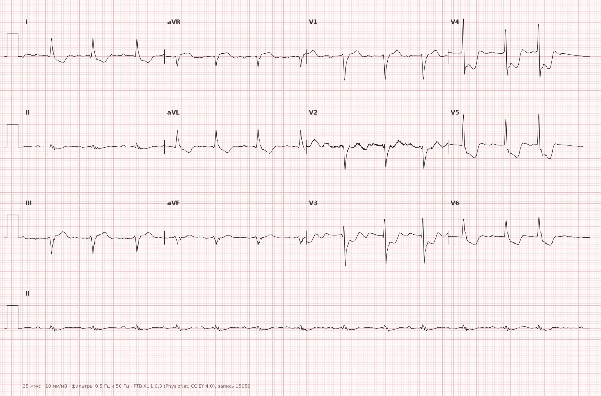
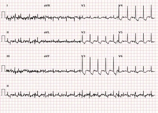
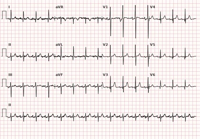
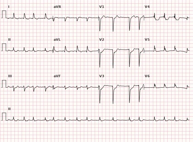
ECG from the open PTB-XL dataset (Wagner P. et al., Scientific Data, 2020; PhysioNet), CC BY 4.0 license. Rendering by WhiteCoat Pro.
Mechanism
When an artery supplying the posterior-basal segments of the LV is occluded (most commonly the circumflex branch of the left coronary artery, less commonly the posterior descending branch of the right coronary artery), acute injury of this zone develops. The ST-segment vector in acute injury is directed toward the injury zone — toward the posterior wall; therefore, leads V7–V9, located directly over it, record the direct pattern: ST-elevation and often, at the established necrosis stage, a pathological Q wave. Leads V1–V3 are located on the opposite side of the chest and register reciprocal (mirror) changes: instead of ST elevation — depression, instead of Q — a tall broad R wave, instead of a negative T wave — a positive one. This is the exact same electrical injury pattern viewed from two opposite points.
Causes
- Atherothrombotic occlusion of the circumflex artery (a common cause of isolated posterior MI)
- Occlusion of the posterior descending artery (in a right-dominant coronary circulation)
- Multivessel disease involving the circumflex artery
- Thrombosis in the setting of STEMI extending to the inferior and lateral walls (inferoposterior, posterolateral MI)
Differential diagnosis
- Diffuse subendocardial ischemia: horizontal or downsloping ST depression in multiple lead groups (not just V1–V3), often without a tall R in V1–V2 and without ST elevation in V7–V9, frequently combined with ST elevation in aVR in multivessel disease or left main stem stenosis
- Normal variant of tall R in V1–V2: without ST dynamics and without clinical symptoms, not accompanied by ST depression
- Right ventricular hypertrophy: tall R in V1 combined with right axis deviation and signs of right ventricular strain, rather than reciprocal ST depression
- Right bundle branch block: rsR' in V1, QRS ≥ 120 ms in complete block and 110–119 ms in incomplete block; secondary ST-T changes in V1–V3 should be assessed taking into account morphology, clinical presentation, and dynamics, as RBBB and posterior MI can coexist
- Wellens' syndrome: biphasic or deeply inverted T wave in V2–V3 without ST depression and without a tall R wave, indicating critical stenosis of the left anterior descending artery rather than posterior MI
Clinical significance and management
Characteristic ST depression in V1–V3 in a patient with ischemic chest pain is a sign suspicious for posterior coronary occlusion and one of the recognized STEMI equivalents (STEMI-equivalent/OMI), even if formal ST-elevation criteria in standard 12 leads are absent. The diagnosis of MI is established based on clinical data, ECG dynamics, and troponin rise/fall in the presence of at least one ischemia sign; coronary angiography identifies the occlusion or culprit artery but is not mandatory for the diagnosis itself. When posterior occlusion is suspected, the physician must immediately record additional leads V7–V9, but this should not delay the activation of reperfusion therapy: in a patient with ongoing ischemic symptoms and characteristic ST depression in V1–V3, the decision for urgent coronary angiography/PCI is made based on the clinical picture, without waiting for either the V7–V9 results or troponin. The choice between primary PCI and fibrinolysis (if timely PCI is unavailable) depends on symptom onset time, contraindications, and expected delay to the procedure — following the same principles as for anterior or inferior STEMI. It is important to actively search for concomitant inferior and lateral wall involvement, as well as right ventricular involvement.
Common interpretation errors
- Interpretation of ST depression in V1–V3 as "anterior wall ischemia" without recording V7–V9
- Ignoring the tall R and positive T waves in V1–V2 as significant reciprocal signs
- Underestimating isolated ST depression when the standard leads appear normal — missing a STEMI equivalent
- Applying lowered ST-elevation thresholds for V7–V9 (using criteria for V2–V3 instead of a separate threshold of ≥ 0.5 mm in two contiguous leads)
- Waiting for V7–V9 results or troponin as a prerequisite for activating reperfusion therapy in the presence of clear clinical symptoms and characteristic ST depression in V1–V3
- Failing to account for the frequent coexistence of inferior or lateral MI, which may shift the focus of attention to only a single wall
"Tall R and ST depression in V1–V3 — look for the mirror in the back: record V7–V9"
1. Which finding in leads V1–V3 is characteristic of posterior myocardial infarction?
This is a mirror reflection of direct posterior wall injury, which is directly visible in V7–V9.
2. What ST-elevation threshold in leads V7–V9 is typically used to diagnose posterior STEMI?
Due to the smaller diagnostic amplitude of posterior leads, the ST-elevation threshold is lower than that for anterior chest leads and requires confirmation in two contiguous leads.
3. Which artery most commonly causes an isolated posterior myocardial infarction?
The circumflex artery supplies the posterobasal segments of the LV and is particularly often responsible for an isolated posterior MI.
Frequently asked questions
How to distinguish posterior MI from conventional subendocardial ischemia?
In posterior MI, ST depression in V1–V3 is combined with a tall, broad R wave and often a positive T wave, and is confirmed by ST elevation in V7–V9. In diffuse subendocardial ischemia, such reciprocal signs are absent, changes are more widespread, and are often combined with ST elevation in aVR.
Is posterior myocardial infarction dangerous?
Yes, ST depression in V1–V3 confirmed in V7–V9 is a recognized STEMI equivalent in clinical significance. It is often combined with inferior or lateral wall involvement and requires emergency reperfusion within the same timeframe as classic STEMI.
Is it always necessary to record V7–V9?
Yes, if unexplained ST depression with a tall R wave is detected in V1–V3 in a patient with ischemic symptoms. Additional leads help confirm direct posterior wall injury, but recording them should not delay the decision for reperfusion in a clear clinical presentation.
Which artery is usually affected in posterior MI?
Both the circumflex branch of the left coronary artery and the posterior interventricular branch of the right coronary artery are possible; the choice depends on the type of coronary dominance. The circumflex artery is particularly often responsible for isolated posterior MI, while the posterior interventricular branch from the right coronary artery is responsible for inferoposterior MI in right dominance.
Why is posterior MI often combined with other localizations?
The blood supply zone of the posterior wall is anatomically closely connected with the inferior and lateral walls of the LV, so occlusion of the same artery often produces combined changes—inferoposterior or posterolateral infarction. Isolated posterior wall involvement is less common but possible, especially with circumflex artery occlusion.
ECG Course
Interpretation from basics to myocardial infarction — learn to read any ECG using a systematic algorithm.
- Video lessons and theory for every topic
- Simulator case studies under every lesson
- Unlimited ECG simulator practice
- Study guide: "All ECGs in 25 minutes"
ECG Strip Simulator
ECG, radiology, histology, pathomorphology, pharmacology — thousands of flashcards with detailed explanations.
- Real ECG strips, not textbook drawings
- Instant answers and explanations
- Streaks and progress tracking in your dashboard
Related terms
More in the section "Ischemia and Myocardial Infarction"
Sources
- Goldberger's Clinical Electrocardiography, latest edition
- Surawicz, Knilans. Chou's Electrocardiography in Clinical Practice
- AHA/ACCF/HRS Recommendations for Standardization of ECG, 2009
- Orlov V.N. Guide to Electrocardiography
Educational material for medical students and physicians. Does not replace clinical guidelines and physical examination; the ECG must always be interpreted in conjunction with the clinical picture.