ECG criteria
- ST elevation at the J point in two contiguous leads: in V2–V3 — ≥2.5 mm in men under 40 years old, ≥2 mm in men aged 40 and older, ≥1.5 mm in women; in all other leads — ≥1 mm (2009 AHA/ACCF/HRS criteria)
- Any Q wave in V2–V3 of duration ≥0.02 s or a QS complex in V2–V3; or Q ≥0.03 s and depth ≥1 mm or QS in I, II, aVL, aVF, V4–V6 (Universal Definition criteria); these features indicate a pathological Q wave, but do not by themselves prove acute infarction
- Poor R-wave progression from V1 to V3 is a non-specific finding; diagnosing a prior MI requires comparison with previous ECGs, clinical presentation, imaging, and other signs of scarring
- Reciprocal ST depression in the inferior leads (II, III, aVF) is not mandatory and does not always occur
- Time course: acute phase — ST elevation and possible hyperacute T waves; subacute phase — Q-wave formation and T-wave inversion; healed scar phase — persistent Q wave with normalization of ST-T
- Correlation with clinical presentation and troponin to differentiate STEMI from other causes of ST elevation in V1–V3; interpretation of pathological Q waves together with the clinical picture and dynamics
What it looks like on ECG
Zone of interest — leads V1–V3, occasionally extending to V4. In the acute phase, hyperacute broad T waves and subsequent or nearly simultaneous ST-segment elevation may occur; the sequence is variable, and hyperacute T waves may be absent or transient, with ST elevation often becoming the first noticeable sign. Pathological Q/QS waves may appear in the acute phase or later, reflecting changes in depolarization propagation and not independently determining the transmurality of necrosis.
Important feature: a QS complex in V1 is sometimes normal, whereas a QS complex in V2 and especially in V1–V2 requires checking electrode placement, comparison with previous ECGs, and evaluation of other signs. A pathological Q wave in these leads is considered to be a change compared to the baseline ECG, the appearance of notches, increasing width or depth, and most importantly — association with ST-T elevation/dynamics and the clinical picture.
| Lead | What to look for | Typical finding |
|---|---|---|
| V1–V2 | ST, Q | ST elevation, QS/Q |
| V3 (±V4) | ST, Q, R | ST elevation, reduced R, sometimes Q |
| II, III, aVF | reciprocity | minor ST depression possible, not mandatory |
| V7–V9 | rule out posterior MI | normally without ST elevation |
In isolated anteroseptal involvement, the Q wave in I, aVL, V5–V6 is usually absent — distinguishing it from extensive anterior or anterolateral infarction.
Real ECGs: 8 examples
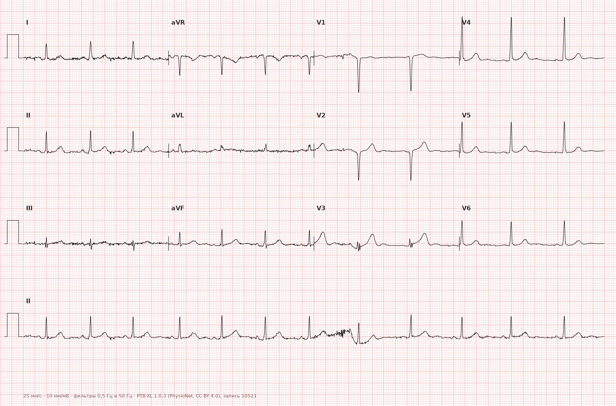
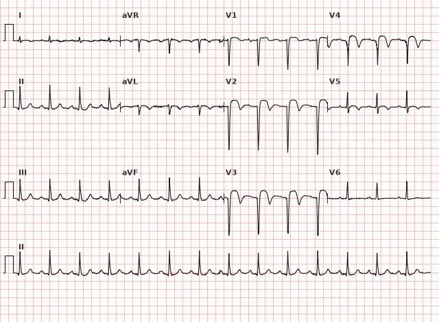
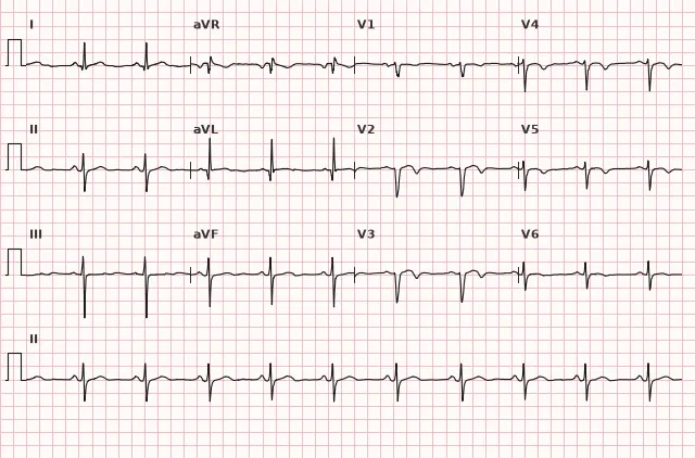
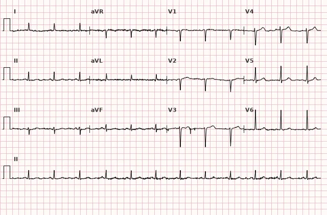
ECG from the open PTB-XL dataset (Wagner P. et al., Scientific Data, 2020; PhysioNet), CC BY 4.0 license. Rendering by WhiteCoat Pro.
Mechanism
The interventricular septum and adjacent anterior LV wall are supplied by perforating septal branches and proximal anterior septal branches of the LAD. Occlusion of this LAD segment leads to ischemia and necrosis of the septum, which is reflected on the ECG by ST elevation and Q-wave formation precisely in the leads electrically directed toward the septum — V1–V3. Because the septum contains the proximal parts of the right and left bundle branches, extensive septal necrosis can cause intraventricular conduction disturbances (right bundle branch block, rarely bifascicular blocks).
Causes
- Atherothrombotic occlusion or critical stenosis of the proximal/middle LAD involving septal branches
- Atherosclerotic plaque rupture with thrombosis (most common mechanism of STEMI)
- Coronary spasm in the LAD zone (variant angina)
- Coronary artery dissection (including spontaneous, SCAD)
- Embolism in the LAD (rare)
- Iatrogenic occlusion of a septal branch during coronary interventions
Differential diagnosis
- Normal variant of QS in V1: QS in V1 is sometimes normal, without ST-T dynamics, without clinical symptoms, stable on previous ECGs; QS in V2 and wider requires checking electrode placement and ruling out pathology
- Left bundle branch block: wide QRS, typical morphology in V1 and V5–V6, masks and mimics a Q wave; infarction is evaluated using Sgarbossa criteria (modified — Smith)
- Brugada syndrome (type 1 pattern): coved ST-segment/J-point elevation typically ≥2 mm in at least one of leads V1–V2 recorded in standard (4th intercostal space) or high (2nd–3rd intercostal space) position, followed by a negative T wave; requires clinical and electrocardiographic evaluation — an isolated Brugada-like pattern without appropriate context does not equal clinical syndrome
- Hypertrophic cardiomyopathy with septal hypertrophy: deep narrow Q waves combined with high R waves, without ST-T dynamics
- Myocarditis: diffuse ST-T changes without strict confinement to a single coronary artery territory, often accompanied by changes in inferior and lateral leads
- Early repolarization: ST elevation with a notch at the J point, concave, without reciprocal depression and without Q-wave formation
Clinical significance and management
New ST elevation in V1–V3(V4) in two contiguous leads with clinical ischemia is a STEMI requiring emergency reperfusion (primary PCI within the recommended time window, if unavailable — fibrinolysis according to current criteria and timeframes). One must actively look for extension to the lateral wall (I, aVL, V5–V6) and the right ventricular/conduction system risk zone, and take into account the appearance of new bundle branch blocks as a marker of extensive involvement.
ST elevation in aVR with diffuse ST depression indicates high-risk ischemia and potential left main/multivessel disease, but does not establish an anatomical diagnosis; an immediate invasive strategy is indicated when very high-risk criteria are met. In the absence of ST elevation but with suspected ACS (e.g., Wellens syndrome-like pattern in precordial leads), a different strategy applies — early NSTE-ACS invasive strategy, rather than emergency primary PCI as in STEMI.
Prior anteroseptal infarction (persistent Q wave without acute dynamics) requires assessment of LV ejection fraction and LV thrombus, risk of anterior wall/apical aneurysm, and prescribing antithrombotic and other secondary prevention measures as indicated. Anticoagulation is prescribed for documented LV thrombus, atrial fibrillation, or another established indication; antiarrhythmic drugs are used for specific arrhythmias and taking risk into account; in reduced LVEF, indications for ICD implantation for primary prevention of sudden death are evaluated after optimal medical therapy and in accordance with recommended waiting times.
Common interpretation errors
- Mistaking physiological QS in V1 for a pathological Q wave without evaluating dynamics and clinical context; forgetting that QS in V2 and wider requires verification
- Interpreting a pathological Q wave as a sign of acute STEMI by itself — without new ST elevation or an occlusion equivalent and clinical context, it is only an indication of possible necrosis/scar
- Failing to compare the current ECG with previous ones — missing the new appearance of a Q wave or ST elevation
- Ignoring left bundle branch block as a potential masking factor for infarction
- Failing to assess lead contiguity — a STEMI diagnosis requires ST elevation in two or more contiguous leads, not in a single random lead
- Forgetting gender and age differences in ST elevation thresholds in V2–V3 when interpreting borderline values
1. In which leads is ST elevation expected in anteroseptal infarction?
The septum and adjacent anterior wall project to leads V1–V3, sometimes extending to V4.
2. Which artery is most commonly responsible for anteroseptal infarction?
Septal perforating branches most commonly arise from the LAD, supplying the interventricular septum, but the ECG does not reliably identify the exact segment and affected branch.
3. What distinguishes a physiological QS in V1–V2 from a pathological Q wave in myocardial infarction?
A stable QS in V1 can be a normal variant; pathological Q/QS waves are evaluated using morphological criteria and comparison with prior ECGs, while acute ST-T dynamics and clinical presentation are used to determine the acuity of the infarction, though they are not mandatory for establishing the presence of a pathological Q wave.
Frequently asked questions
How to distinguish anteroseptal myocardial infarction from a normal QS variant in V1–V2?
QS in V1 is sometimes normal, whereas QS in V2 and especially across V1–V2 requires checking electrode placement, comparison with previous ECGs, and assessment of other features. In acute infarction, Q-wave changes are accompanied by new ST elevation, its dynamic evolution, and a corresponding clinical picture, which is confirmed by comparison with prior ECGs.
Which artery is typically affected in anteroseptal myocardial infarction?
The most frequent cause is LAD occlusion, but the ECG cannot reliably determine the exact segment and affected branch. The term "anteroseptal MI" is a traditional electrocardiographic localization; modern guidelines more frequently use "anterior MI / STEMI", as zone boundaries and anatomical correlations are variable.
Is anteroseptal myocardial infarction dangerous due to conduction blocks?
Yes, the septum contains the proximal divisions of the bundle of His branches; therefore, extensive septal damage can lead to right bundle branch block, bifascicular block, and even high-degree AV block, which worsens the prognosis.
Is emergency PCI required in the presence of anteroseptal ST elevation?
Yes, if the new ST elevation in V1–V3 meets STEMI criteria and there is a compatible clinical presentation, emergency reperfusion is indicated—primary PCI within recommended timeframes or fibrinolysis if PCI is unavailable.
Why do ST-elevation thresholds in V2–V3 vary by sex and age?
This is due to physiological differences in ST-amplitude thresholds in these leads among men and women of different ages; the 2009 AHA/ACCF/HRS recommendations propose stratified thresholds (e.g., higher in men under 40 years of age) to reduce the number of false-positive STEMI diagnoses.
ECG Course
Interpretation from basics to myocardial infarction — learn to read any ECG using a systematic algorithm.
- Video lessons and theory for every topic
- Simulator case studies under every lesson
- Unlimited ECG simulator practice
- Study guide: "All ECGs in 25 minutes"
ECG Strip Simulator
ECG, radiology, histology, pathomorphology, pharmacology — thousands of flashcards with detailed explanations.
- Real ECG strips, not textbook drawings
- Instant answers and explanations
- Streaks and progress tracking in your dashboard
Related terms
More in the section "Ischemia and Myocardial Infarction"
Sources
- Goldberger's Clinical Electrocardiography, current edition
- AHA/ACCF/HRS Recommendations for the Standardization and Interpretation of the Electrocardiogram, 2009
- Surawicz B., Knilans T. Chou's Electrocardiography in Clinical Practice
- Orlov V.N. Guide to Electrocardiography
Educational material for medical students and physicians. Does not replace clinical guidelines and physical examination; the ECG must always be interpreted in conjunction with the clinical picture.