ECG criteria
- New ST elevation at the J point of ≥1 mm in two contiguous leads from I and aVL — isolated lateral localization often has low amplitude; this is a standard ECG criterion for STEMI, but not a criterion to rule out occlusion
- New ST elevation at the J point of ≥1 mm in two contiguous leads V5–V6
- ST elevation in only one high lateral lead (isolated in I or in aVL) formally does not meet the "two contiguous leads" threshold, but in the presence of reciprocal depression in III and ACS symptoms, it must be interpreted as a possible acute coronary occlusion rather than a reason to exclude the diagnosis
- Reciprocal ST depression, most commonly in III and aVF, sometimes in II — supports the lateral component, but does not prove isolated lateral localization and does not rule out posterior or combined infarction
- Pathological Q wave in two contiguous leads from I, II, aVL, aVF, V4–V6: Q ≥0.03 s and depth ≥1 mm, or QS — a sign of current or prior infarction, but does not prove transmurality based on a single ECG
- Often combined with inferior or anterior infarction (inferolateral, anterolateral) — isolated lateral is less common
- Negative T wave in I, aVL, V5–V6 — a possible but nonspecific dynamic sign of ischemia or infarction evolution
- ST depression in V1–V3 in the appropriate clinical setting requires recording V7–V9 to rule out posterior infarction; ST elevation in aVR with diffuse ST depression is a nonspecific marker of severe ischemia, multivessel disease, or left main coronary artery disease, rather than a localization criterion for LCx occlusion
What it looks like on ECG
The lateral wall of the left ventricle is reflected by leads I, aVL (high lateral limb leads) and V5–V6 (low lateral precordial leads). In acute lateral STEMI, clinicians look for a new ST elevation at the J point of ≥1 mm in two contiguous leads from this group — this is the standard diagnostic threshold for STEMI. It is important to remember: it was created for classifying classic STEMI and is not a criterion for ruling out acute occlusion. Occlusion of the diagonal branch or circumflex artery frequently causes changes in only one lead — aVL or I — especially in combination with reciprocal depression in III and typical ACS symptoms. The absence of a second contiguous lead with ST elevation does not rule out occlusive infarction and does not eliminate the need for immediate expert evaluation, repeat ECG recordings, and consideration of emergency coronary angiography without waiting for formal criteria to be met.
The amplitude of ST elevation in lateral MI is often small, and sometimes a diagnostic elevation is absent altogether — this is due to the characteristics of the resulting injury vector and anatomy, rather than a sign of necessarily small necrotic area: lateral MI can be extensive, especially in proximal circumflex artery occlusion or occlusion of a large diagonal or marginal branch.
Reciprocal ST depression in the inferior leads is frequently observed, predominantly in III and aVF: lead aVL is the most opposed to the inferior leads along the axis, especially III. Such depression supports the lateral component of injury, but its absence does not rule out lateral MI, and its presence does not prove isolated lateral localization — changes may reflect combined inferolateral-posterior ischemia.
During the evolution of infarction, a pathological Q wave and T-wave inversion may appear in I, aVL, V5–V6, but this is not mandatory and is not linear. A diagnosis of prior infarction is not made based on a single ECG — clinical presentation, prior records, and, if necessary, imaging are required.
| Leads | What to look for |
|---|---|
| I, aVL | ST elevation, possible Q wave, possible T inversion |
| V5–V6 | ST elevation, possible Q wave, possible T inversion |
| III, aVF | possible reciprocal ST depression |
The lateral zone is frequently affected together with the inferior wall (inferolateral infarction, occlusion of a dominant LCx or RCA with lateral branches) or the anterior wall (anterolateral infarction — primarily occlusion of the LAD with diagonal branches; the circumflex artery is mainly associated with lateral, inferolateral, or posterolateral involvement). Isolated lateral STEMI is not the most frequent variant and is easily missed due to the low amplitude of changes.
Real ECGs: 8 examples
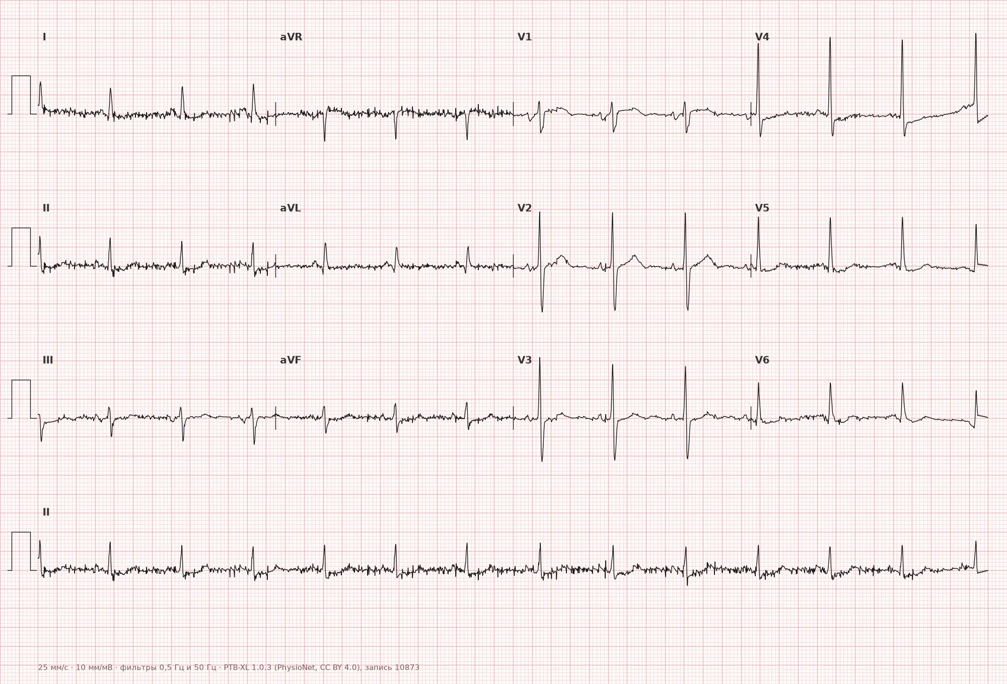
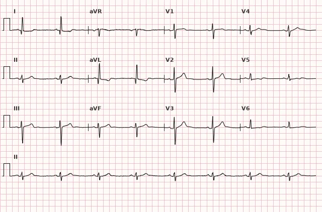
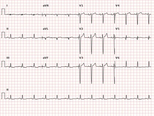
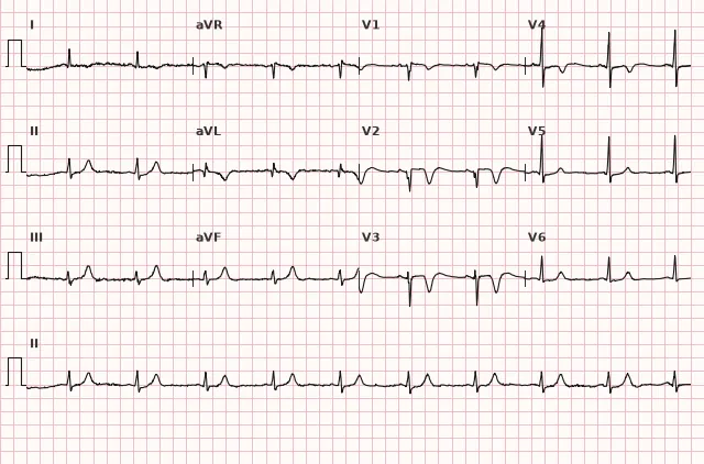
ECG from the open PTB-XL dataset (Wagner P. et al., Scientific Data, 2020; PhysioNet), CC BY 4.0 license. Rendering by WhiteCoat Pro.
Mechanism
Occlusion of the circumflex artery or its branches (obtuse marginal branch), or diagonal branches of the left anterior descending artery, halts blood flow to the lateral wall of the left ventricle. Transmural ischemia shifts the ST-segment vector toward the injured zone — toward I, aVL, V5–V6. The oppositely directed vector may form reciprocal ST depression in the inferior leads, but this is an optional finding. Infarction alters the initial vectors of lateral wall depolarization, which may manifest as a pathological Q wave in the same leads; its presence or absence does not allow judging the transmurality of necrosis from a single ECG.
Causes
- Atherothrombotic occlusion of the circumflex artery (LCx)
- Occlusion of the diagonal branch of the left anterior descending artery (LAD)
- Occlusion of the obtuse marginal branch (ramus marginalis)
- Part of combined involvement in inferolateral or anterolateral infarction
- Coronary artery embolism, spontaneous coronary artery dissection (SCAD) — atherosclerosis is less common
Differential diagnosis
- Early repolarization: ST elevation in I, aVL without reciprocal depression in III, concave ST, stable pattern on repeat ECGs
- Left ventricular hypertrophy with strain: ST elevation is usually in the right precordial leads, while leads I, aVL show ST depression and asymmetric T inversion, high QRS voltage
- Pericarditis: diffuse concave ST elevation and PR-segment depression in many leads, not limited to the basin of a single artery; reciprocal ST depression is usually absent, except in aVR and frequently V1; the T wave in the acute phase generally remains positive, therefore a "concordant T" is not used as an independent diagnostic sign
- Normal variant of the Q wave in I, aVL in some healthy individuals — low amplitude, no dynamics, and no ST elevation
- Left bundle branch block: ST-T changes are secondary; Sgarbossa criteria and modified Sgarbossa criteria (Smith-modified Sgarbossa) are evaluated; a new or presumably new LBBB does not confirm STEMI on its own and is interpreted together with the clinical picture and signs of acute occlusion.
Clinical significance and management
In the presence of ischemic symptoms and a new ST elevation of ≥1 mm in two contiguous leads from I, aVL and/or V5–V6, the patient is managed according to the STEMI algorithm with immediate reperfusion activation (primary PCI within the target time window, or thrombolysis per standard protocol if unavailable). The formal threshold of "two contiguous leads" is a convenient criterion for standardizing the diagnosis of STEMI, but not a criterion that can rule out acute coronary occlusion. ST elevation in only one high lateral lead (isolated aVL or I), especially with reciprocal depression in III and typical clinical presentation, requires the same vigilance: immediate expert evaluation, repeat serial ECGs, extended leads, and consideration of emergency coronary angiography and reperfusion without waiting for troponin results and without waiting for a second lead with elevation to appear.
Due to the low amplitude of changes, the physician must purposefully examine I, aVL, V5–V6 and not limit the search to classic high elevations only. When the ECG is non-diagnostic but ischemia symptoms persist, repeat recordings, additional leads (V7–V9, right precordial), and serial troponins are performed — without delaying emergency care if the probability of occlusion remains high.
When combined with inferior infarction (inferolateral) or anterior infarction (anterolateral), the extent of involvement and risk of complications are higher. Reciprocal ST depression in III, aVF supports the lateral component when accompanied by corresponding ST elevation in contiguous lateral leads, but on its own does not confirm localization and requires evaluation of posterior leads V7–V9 and clinical-angiographic correlation.
Common interpretation errors
- Ruling out STEMI and acute coronary occlusion solely based on ST elevation being present in only a single lead (only I or only aVL), without taking into account the clinical presentation and reciprocal changes
- Mistaking reciprocal depression in III, aVF for isolated inferior ischemia
- Missing lateral STEMI due to low amplitude of changes and focus of attention on anterior/inferior leads
- Judging infarct size by the amplitude of ST elevation
- Failing to compare with previous ECGs when a normal variant or prior changes are suspected
- Forgetting to check V5–V6 separately from I, aVL — changes may not be present in all lateral leads simultaneously
1. What does ST-segment elevation in only one high lateral lead (e.g., aVL only) in the setting of ACS presentation and reciprocal depression in lead III mean?
The formal "two contiguous leads" threshold is not a criterion to rule out occlusion; isolated changes in aVL with reciprocal depression and ACS symptoms warrant high clinical suspicion and immediate evaluation.
2. Which reciprocal change may accompany acute lateral STEMI?
Lead aVL is most directly opposed to the inferior leads, especially lead III; therefore, ST-segment elevation in lateral leads may be accompanied by reciprocal depression in III and aVF, although this is not a mandatory finding.
3. Which artery is most commonly responsible for an isolated lateral myocardial infarction?
The lateral wall of the LV is supplied by the circumflex artery (including the obtuse marginal branch) and the diagonal branches of the left anterior descending artery.
Frequently asked questions
Which leads reflect the lateral wall of the heart on the ECG?
High lateral leads I and aVL, and low lateral chest leads V5–V6. These are the main leads for assessing the lateral wall, but the diagnosis of ischemia or infarction is established based on the combination of clinical presentation, ECG dynamics, and troponin, rather than changes in these leads alone.
Can occlusive myocardial infarction be ruled out if ST-segment elevation is present in only one lead?
No. The criterion "ST-segment elevation in two contiguous leads" is a standard way to classify STEMI, not a rule to rule out acute occlusion. Occlusion of the diagonal branch or circumflex artery frequently causes changes only in aVL or I, especially with reciprocal depression in III and ACS symptoms, and requires immediate expert evaluation and repeat ECGs.
How does isolated lateral infarction differ from inferolateral infarction?
In isolated lateral myocardial infarction, ST elevation is present in two contiguous leads among I, aVL, V5–V6 without primary ST elevation in II, III, aVF. In inferolateral infarction, ST elevation is present simultaneously in inferior and lateral leads, indicating involvement of a dominant artery with branches to both walls.
Is lateral myocardial infarction dangerous?
Like any STEMI, it requires emergency reperfusion. ST amplitude cannot reliably determine infarct size; lateral MI can be extensive with proximal circumflex artery occlusion. When combined with involvement of other walls, the risk of complications increases.
How to distinguish lateral infarction from early repolarization in I, aVL?
In early repolarization, the ST segment is concave, stable on serial ECGs, and there is usually no reciprocal depression in III and aVF. In acute lateral infarction, there is ST-segment evolution, ACS clinical presentation, and often reciprocal changes.
ECG Course
Interpretation from basics to myocardial infarction — learn to read any ECG using a systematic algorithm.
- Video lessons and theory for every topic
- Simulator case studies under every lesson
- Unlimited ECG simulator practice
- Study guide: "All ECGs in 25 minutes"
ECG Strip Simulator
ECG, radiology, histology, pathomorphology, pharmacology — thousands of flashcards with detailed explanations.
- Real ECG strips, not textbook drawings
- Instant answers and explanations
- Streaks and progress tracking in your dashboard
Related terms
More in the section "Ischemia and Myocardial Infarction"
Sources
- Hampton J. The ECG in Practice
- Surawicz B., Knilans T. Chou's Electrocardiography in Clinical Practice
- Orlov V.N. Guide to Electrocardiography
- ESC Guidelines for the management of acute coronary syndromes
Educational material for medical students and physicians. Does not replace clinical guidelines and physical examination; the ECG must always be interpreted in conjunction with the clinical picture.