ECG criteria
- New J-point ST elevation in two contiguous anatomically adjacent leads: ≥ 1 mm — except in V2–V3, where separate age- and sex-specific thresholds apply
- In men ≥40 years: ST elevation in V2–V3 ≥ 2 mm; in men <40 years: ≥ 2.5 mm
- In women: ST elevation in V2–V3 ≥ 1.5 mm regardless of age
- In other leads (V1, V4–V6, I, aVL): ST elevation ≥ 1 mm in two contiguous leads
- ST-segment morphology (concave, straight, convex upward) and hyperacute tall T waves are supportive features that do not replace the magnitude of ST elevation criterion
- Reciprocal ST depression in the inferior leads (II, III, aVF) in extensive anterior MI, not mandatory for diagnosis
- Over time — pathological Q wave: in I, II, aVL, aVF, V4–V6 — Q ≥ 0.03 s and depth ≥ 1 mm (or QS) in two contiguous leads; in V2–V3 — any Q wave with a duration ≥ 0.02 s or a QS complex is already pathological (one of these two leads is sufficient); the appearance of Q is not mandatory for the diagnosis of acute STEMI
- Assess the extent of the lesion by the number of involved leads: V1–V2 (septal) vs V1–V6 + I, aVL (extensive anterior) — as a guide to the ischemic zone, without a definitive conclusion on the level of LAD occlusion
What it looks like on ECG
Anterior STEMI reflects ischemia of the anterior wall and interventricular septum, typically supplied by the LAD. The key finding is a new J-point ST-segment elevation in two contiguous precordial leads (± I, aVL); its distribution correlates with the extent of the lesion:
| Zone | Leads | Approximate basin |
|---|---|---|
| Septal | V1–V2 | septal perforators |
| Anterior | V3–V4 | LAD basin |
| Anterolateral | V5–V6, I, aVL | LAD, often with diagonal branches |
| Extensive anterior | V1–V6, I, aVL | large LAD basin |
This is a guide to the area of ischemia, not an exact anatomical localization of the occlusion level or a specific branch — this is unreliable on a standard ECG; the exact level of the lesion is determined by clinical data and coronary angiography. In the hyperacute phase, tall hyperacute T waves in V2–V4 may occur even before overt ST elevation. A new right bundle branch block, especially when combined with ischemic symptoms, is associated with extensive anterior ischemia and a poor prognosis, and requires immediate evaluation for urgent reperfusion strategy; it is not a precise marker of the LAD occlusion level and does not replace ST analysis as the primary diagnostic criterion. Reciprocal ST depression in the inferior leads is not always present, but its presence in anterior MI indicates a larger area of damage. ST elevation in aVR in the setting of diffuse ST depression in other leads is a sign of high-risk extensive subendocardial ischemia (found in multivessel disease, left main coronary artery occlusion, but also in tachycardia, anemia, hypotension, and other conditions of oxygen supply-demand imbalance); it requires urgent clinical evaluation, but does not prove left main disease by itself. Over hours to days, a pathological Q wave develops, and the T wave becomes inverted (reperfusion or part of the natural evolution of necrosis).
Real ECGs: 8 examples
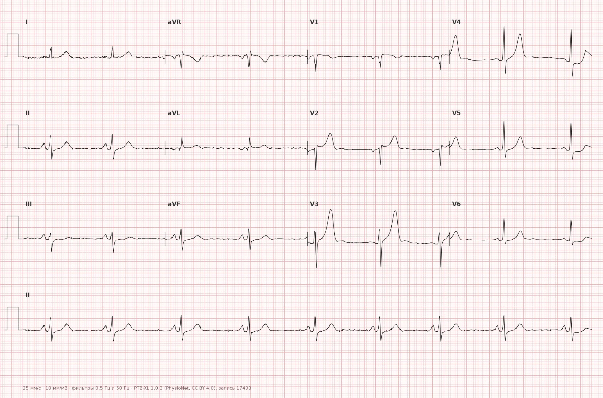
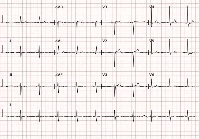
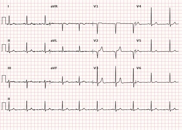
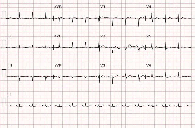
ECG from the open PTB-XL dataset (Wagner P. et al., Scientific Data, 2020; PhysioNet), CC BY 4.0 license. Rendering by WhiteCoat Pro.
Mechanism
Acute thrombotic occlusion of the LAD (or another culprit artery) halts blood flow to the anterior wall and LV septum. The subendocardium suffers first, then the ischemia becomes transmural — on the ECG, this is expressed as ST elevation (the injury current is directed from the endocardium to the epicardium, with the vector shifting toward electrodes overlying the injured area). The larger the ischemic myocardial mass, the broader the involvement of precordial leads by ST elevation. A decrease in ST elevation during treatment primarily reflects reduced ischemia or successful reperfusion, rather than myocardial death per se. A pathological Q wave increases the likelihood of irreversible necrosis, but may appear as early as the acute phase, is not equivalent to a completed scar, and regresses in some cases.
Causes
- Atherothrombotic occlusion of the LAD (main cause of anterior STEMI)
- Left main coronary artery disease or disease of its other branches, multivessel disease
- Coronary artery dissection (spontaneous, in young women, postpartum)
- Coronary spasm (Prinzmetal's variant) with or without thrombosis
- Embolism to the LAD (endocarditis, atrial fibrillation with thrombus, paradoxical embolism)
- Aortic dissection extending to the left main ostium (rare)
- Severe coronary vasospasm associated with cocaine use
Differential diagnosis
- Early repolarization syndrome — concave ST elevation, J-point notch, without reciprocal depression and time evolution
- Pericarditis — diffuse ST elevation in multiple leads without reciprocity (except aVR), often PR depression
- Takotsubo cardiomyopathy — clinical presentation and ECG resemble anterior MI, but coronary arteries are normal, diagnosed by coronary angiography and echocardiography
- Hypertrophic cardiomyopathy (apical) — deep T-wave inversions in precordial leads without acute ST elevation
- De Winter waves — tall, symmetrical, pointed T waves with upsloping ST depression approximately 1–3 mm from the J point, typically in V2–V6; ST elevation in aVR may occur but is not a diagnostic component of the pattern; considered an occlusive MI pattern (STEMI equivalent) reflecting acute occlusion or critical proximal LAD stenosis, requiring immediate reperfusion evaluation
- Wellens' syndrome — deep inverted or biphasic T waves in V2–V3 after a pain episode, a sign of critical LAD stenosis without ongoing occlusion; primary PCI windows of "<120 min" do not apply, the strategy is an early invasive NSTE-ACS strategy
Clinical significance and management
Anterior STEMI, especially extensive (V1–V6, I, aVL), is associated with the largest mass of damaged myocardium among all STEMI locations and a high risk of LV systolic dysfunction, cardiogenic shock, conduction disturbances (bundle branch block, AV block), and ventricular arrhythmias. When ST elevation meets criteria, immediate activation of the primary PCI pathway is indicated: the first medical contact-to-device time goal is generally ≤ 90 minutes for direct presentation to a PCI-capable center and ≤ 120 minutes for inter-hospital transfer. If the expected delay to primary PCI exceeds 120 minutes from diagnosis, with symptom onset < 12 hours and no contraindications, immediate fibrinolysis is considered (preferably within 10 minutes of diagnosis) followed by a pharmacoinvasive strategy; acute aortic dissection and other contraindications must be ruled out before fibrinolysis. The time of the first ECG must be recorded, repeat ECGs performed if symptoms worsen, axis and QRS width monitored (appearance of bundle branch block is an unfavorable sign), and hemodynamics controlled due to the risk of acute left ventricular failure.
Common interpretation errors
- Using a single ST-elevation threshold for V2–V3 without regard to sex and age
- Failing to recognize hyperacute T waves and de Winter waves as a potential equivalent of LAD occlusion in the absence of overt ST elevation
- Making a definitive conclusion about the level of LAD occlusion or a specific branch based solely on ST morphology or a new bundle branch block
- Confusing early repolarization and pericarditis with true anterior STEMI due to inadequate assessment of dynamics and reciprocity
- Failing to repeat ECGs in the setting of changing clinical status, thereby missing ST-T evolution
1. What J-point ST-elevation threshold in leads V2–V3 is considered diagnostic in men aged 40 years and older?
According to ECG standardization guidelines, for men ≥40 years the threshold in V2–V3 is ≥ 2 mm, whereas for men <40 years it is 2.5 mm, and for women 1.5 mm.
2. Which coronary artery most frequently causes anterior myocardial infarction?
The anterior wall and interventricular septum are typically supplied by the left anterior descending (LAD) artery; its occlusion produces a classic pattern of anterior STEMI, although other LMCA lesion patterns are possible.
3. Which ECG pattern corresponds to an extensive anterior STEMI with a large area of myocardial involvement?
Involvement of all precordial leads V1–V6 along with I and aVL reflects a large area of anterior wall ischemia; reciprocal depression in the inferior leads increases the suspicion of an extensive lesion, although the precise level of occlusion cannot be determined by ECG alone.
Frequently asked questions
Which leads are examined in anterior myocardial infarction?
The main leads are the precordial leads V1–V6, with the lateral leads I and aVL added in extensive involvement. Reciprocal changes are looked for in the inferior leads II, III, aVF.
How does an anterior MI differ from an anteroseptal MI?
Anteroseptal MI is limited to ST-elevation in V1–V2 (septal perforators), whereas an anterior or extensive anterior MI involves V3–V6 and frequently I, aVL. This indicates a larger ischemic area, although the exact level of LAD occlusion cannot be reliably determined from the ECG.
Is anterior myocardial infarction more dangerous than other locations?
On average, yes: the LAD supplies a major portion of the LV, which is why anterior STEMI is more frequently accompanied by a significant decrease in ejection fraction, cardiogenic shock, and conduction disturbances compared to inferior MI.
Can an anterior MI occur without overt ST-elevation?
Yes, with subtotal or acute LAD occlusion, occlusion equivalents such as de Winter waves or hyperacute T waves in the precordial leads may occur, while Wellens' syndrome reflects critical stenosis without active occlusion at the exact time of the ECG recording.
Is the appearance of a Q wave mandatory for diagnosing an anterior infarction?
No, a pathological Q wave develops over time and is not a prerequisite for diagnosing acute STEMI. The main ECG criterion for the acute phase is new ST-elevation in two contiguous leads, but the definitive diagnosis of myocardial infarction is based on clinical presentation, ECG, and cardiac troponin dynamics; in a classic presentation, reperfusion is not delayed while waiting for troponin results.
ECG Course
Interpretation from basics to myocardial infarction — learn to read any ECG using a systematic algorithm.
- Video lessons and theory for every topic
- Simulator case studies under every lesson
- Unlimited ECG simulator practice
- Study guide: "All ECGs in 25 minutes"
ECG Strip Simulator
ECG, radiology, histology, pathomorphology, pharmacology — thousands of flashcards with detailed explanations.
- Real ECG strips, not textbook drawings
- Instant answers and explanations
- Streaks and progress tracking in your dashboard
Related terms
More in the section "Ischemia and Myocardial Infarction"
Sources
- AHA/ACCF/HRS Recommendations for the Standardization and Interpretation of the Electrocardiogram, 2009
- Goldberger's Clinical Electrocardiography, textbook
- ESC Guidelines for the management of acute coronary syndromes, 2023
- Orlov V.N. Guide to Electrocardiography
Educational material for medical students and physicians. Does not replace clinical guidelines and physical examination; the ECG must always be interpreted in conjunction with the clinical picture.