ECG criteria
- A typical pattern, not a diagnostic criterion: local persistent ST elevation in the leads corresponding to the prior infarction zone (most commonly V1–V4, less frequently II, III, aVF) is an indication for imaging, not an independent confirmation of an aneurysm.
- Stability of the findings on serial tracings over a prolonged period (weeks to months) without a fixed mandatory duration threshold — consistency is what matters, not a specific timeframe.
- Frequently accompanied by a pathological Q wave or QS complex — criteria for prior myocardial infarction according to AHA/ACCF/HRS: in V2–V3 — a Q wave with a duration >0.02 s or a QS complex; in I, II, aVL, aVF, V4–V6 — a Q wave with a duration ≥0.03 s and a depth ≥1 mm or a QS complex in any two contiguous leads (these are criteria for a pathological Q wave / prior MI, not standalone criteria for an anatomical aneurysm; the Q wave may also appear in leads that do not coincide with the zone of ST elevation).
- The T wave is frequently inverted or flattened, but without ischemic dynamics on serial tracings.
- Absence of clinical and biochemical dynamics of acute MI (no crescendo chest pain, no rise or fall of troponin typical of an acute event).
- Confirmation by echocardiography, cardiac MRI, or CT: local out pouching of the LV wall with akinesia or dyskinesia, and diastolic wall thinning — an obligatory diagnostic step.
What it looks like on ECG
The typical presentation is ST-segment elevation in the leads corresponding to the zone of a prior myocardial infarction, most commonly anteroseptal and anterior (V1–V4), less frequently inferior (II, III, aVF). The elevation is typically local and stable on serial tracings, but its amplitude and morphology are non-specific; it must be evaluated in comparison with prior ECGs and the clinical context. In the same or adjacent leads, a pathological Q wave or QS complex (absence of R wave) is often present, although its absence does not rule out an aneurysm. The T wave is frequently inverted or low-amplitude, but its morphology does not change from tracing to tracing — this consistency over weeks and months is referred to as a "frozen ECG".
| Sign | Acute STEMI | Persistent ST elevation (potential aneurysm) |
|---|---|---|
| ST elevation | rises/falls over hours to days | stable for weeks to months |
| Q wave | may appear in the first hours to days, but not mandatory | if present, already formed and stable |
| Clinical Practice | symptoms, troponin dynamics | usually an asymptomatic phase or heart failure |
The diagnosis of acute STEMI is based on clinical presentation, ECG dynamics, and troponin levels, rather than the mandatory development of a Q wave. The absence of reciprocal changes may support a chronic presentation, but does not rule out an acute MI; symptoms, serial ECGs, troponin, and urgent clinical evaluation are decisive. Persistent ST elevation by itself is merely an ECG finding that requires imaging to confirm or exclude a true aneurysm.
Real ECGs: 8 examples
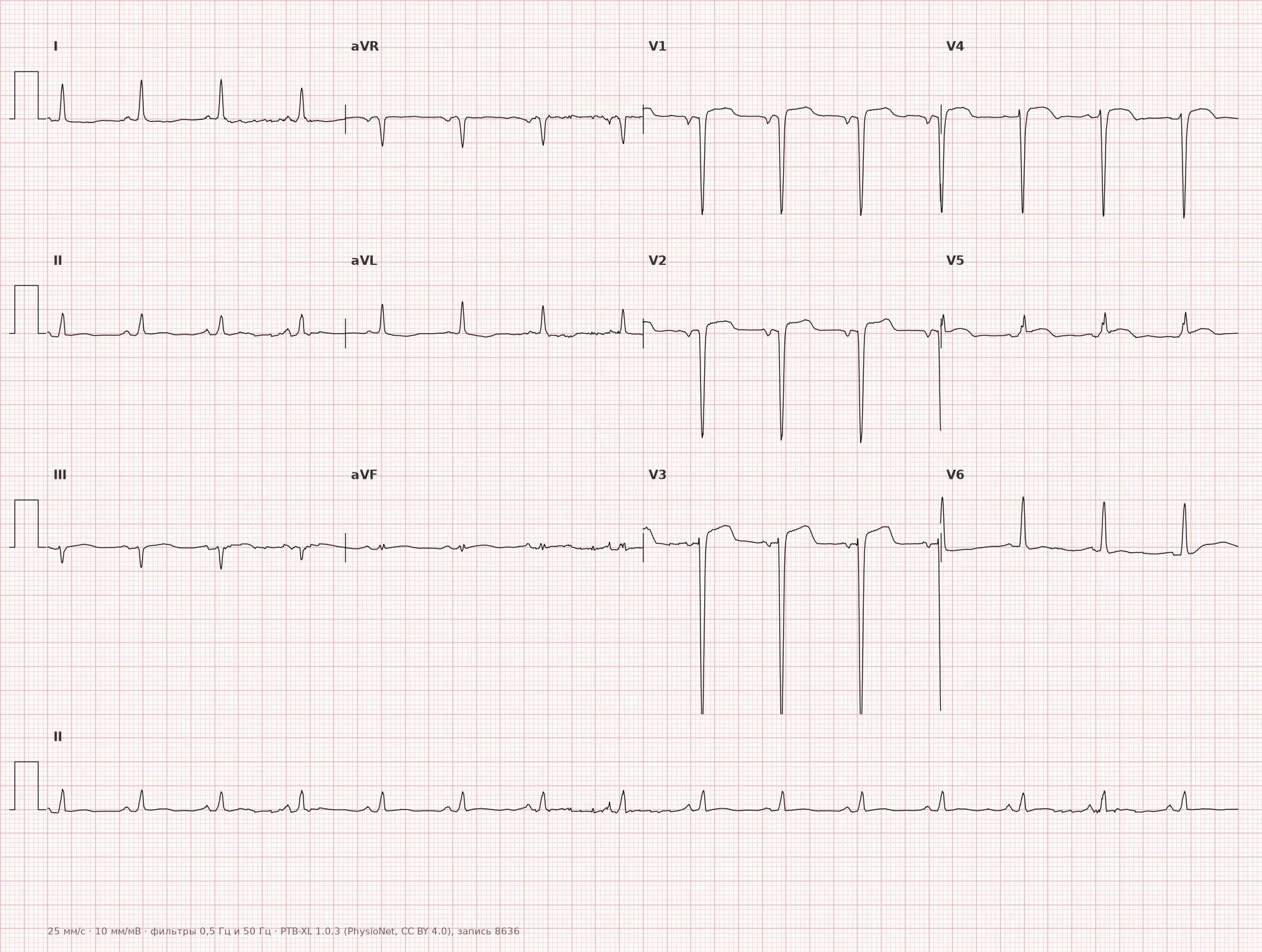
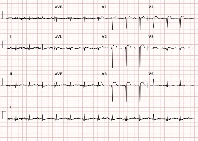
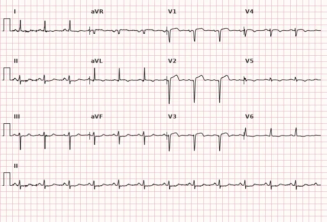
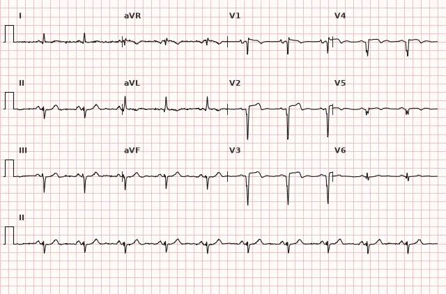
ECG from the open PTB-XL dataset (Wagner P. et al., Scientific Data, 2020; PhysioNet), CC BY 4.0 license. Rendering by WhiteCoat Pro.
Mechanism
Following a transmural myocardial infarction, necrotic myocardium is replaced by fibrous tissue. The scar does not contract and stretches under the pressure of blood during systole, forming a wall out pouching — an aneurysm. Persistent ST elevation is presumably associated with the post-infarction scar and persistent wall motion abnormality (akinesia or dyskinesia), but this ECG pattern is non-specific: it occurs even without a true anatomical aneurysm, and conversely, a confirmed aneurysm may present without notable ST elevation or a pathological Q wave. Therefore, the ECG does not rule out ischemia in the presence of new symptoms — an evaluation for acute coronary syndrome is necessary.
Causes
- Large focal transmural myocardial infarction, most commonly anterior (occlusion of the left anterior descending artery)
- Late or ineffective reperfusion therapy (late revascularization, failed thrombolysis)
- Extensive zone of necrosis with scar formation in the absence of adequate collateral blood flow
- Less commonly — post-infarction aneurysm of the inferior or posterior wall
Differential diagnosis
- Acute STEMI: evaluated based on symptoms, serial ECGs, and high-sensitivity troponin dynamics; absence of chest pain or initial troponin rise does not rule out acute myocardial infarction
- Reinfarction / recurrent ischemia in the same zone: new ST dynamics against the backdrop of an old "frozen" pattern, worsening chest pain, or new symptoms
- Pericarditis: diffuse concordant ST elevation in multiple leads, PR depression, no pathological Q wave
- Early repolarization syndrome: ST elevation in young individuals without a Q wave and without a history of myocardial infarction
- Brugada pattern type 1: ST elevation exclusively in V1–V2 with a characteristic coved morphology, without a Q wave or history of MI
- Pseudoaneurysm (false aneurysm): rupture of the ventricular wall with the formation of a cavity contained by the pericardium; carries a significantly higher risk of rupture and requires urgent echocardiography/CT/MRI and consultation with a cardiovascular surgeon
- Persistent ST elevation without a true aneurysm (the so-called "ECG aneurysm pattern" without echocardiographically confirmed wall out pouching) — requires imaging for differentiation
Clinical significance and management
LV aneurysm is a marker of extensive transmural injury and a risk factor for heart failure, ventricular arrhythmias (re-entry around the scar), and thromboembolism due to a mural thrombus. Wall rupture is rare in a true chronic post-infarction aneurysm, as it is typically a complication of the acute phase. The finding of persistent ST elevation on an ECG in a patient with a history of MI is an indication for echocardiography, cardiac MRI, or CT, which confirm the diagnosis (local out pouching, wall dyskinesia/akinesia, thinned wall in diastole); the ECG alone does not establish the diagnosis. When recent ischemia is suspected in the setting of an old aneurysm, repeat ECGs, troponin, and clinical evaluation are necessary to avoid missing a reinfarction. Subsequent management follows cardiology guidelines: heart failure control; if an LV thrombus is visualized, this is typically an indication for therapeutic anticoagulation for a defined period (with follow-up echocardiography or MRI), and the specific regimen and duration are determined individually considering bleeding risk and thrombus persistence; an aneurysm without a thrombus is not an automatic indication for anticoagulation. Implantation of an implantable cardioverter-defibrillator is decided primarily based on LV ejection fraction, documented ventricular arrhythmia, time elapsed since MI, and optimal medical therapy, rather than the presence of an aneurysm alone. Surgical reconstruction (aneurysm resection) is intended for carefully selected patients with a large aneurysm, symptomatic heart failure, refractory ventricular tachycardia, or embolic complications.
Common interpretation errors
- Mistaking persistent ST elevation for acute STEMI without comparing with prior tracings and medical history
- Failing to account for the stability of the findings over time — a key differentiating feature
- Missing new ST dynamics in the setting of an old aneurysm and failing to recognize reinfarction
- Diagnosing an aneurysm based solely on ECG without confirmation by an imaging modality (echocardiography, cardiac MRI)
- Confusing local ST elevation in V1–V4 with a diffuse pericarditis pattern
- Failing to differentiate between a true aneurysm and a pseudoaneurysm (false aneurysm); if a pseudoaneurysm is suspected, urgent echocardiography/CT/MRI and consultation with a cardiovascular surgeon are required
1. Which feature is most characteristic of a chronic left ventricular aneurysm on the ECG?
The key feature is the absence of dynamics in ST elevation on repeat tracings a long time after myocardial infarction, which distinguishes it from acute MI; however, this is merely a suspicious pattern that requires confirmation by imaging.
2. Which additional ECG feature frequently accompanies persistent ST elevation in post-infarction aneurysm?
A pathological Q wave or QS complex frequently accompanies a post-infarction scar, but its absence does not rule out an aneurysm, and its presence does not confirm it — imaging is required.
3. What should be done in the presence of persistent ST elevation in a patient with a history of myocardial infarction?
In a stable condition without signs of acute ACS, comparison with baseline ECGs and imaging confirms the chronic nature of the finding; if acute MI is suspected, reperfusion must not be delayed for imaging.
Frequently asked questions
How to differentiate LV aneurysm from acute myocardial infarction on ECG?
The key factors are temporal dynamics and clinical presentation. In acute STEMI, evaluation is based on symptoms, serial ECGs, and high-sensitivity troponin dynamics. In chronic aneurysm, the picture remains stable for weeks and months. Comparison with previous ECGs, clinical status, and biochemical markers plays a decisive role.
Is a left ventricular aneurysm dangerous?
True chronic aneurysm increases the risk of heart failure, ventricular arrhythmias due to re-entry around the scar, and thromboembolism if a thrombus is present in the aneurysm cavity. Wall rupture is rare in true aneurysms and is typically a complication of the acute phase. Pseudoaneurysm (false aneurysm) carries a significantly higher risk of rupture and requires urgent cardiac surgery evaluation.
Is it necessary to confirm the ECG finding with other methods?
Yes, persistent ST elevation after myocardial infarction is merely a nonspecific ECG pattern that raises suspicion but does not confirm the diagnosis. It can persist without an anatomical aneurysm, and a true aneurysm can occur without ST elevation or pathological Q waves. The diagnosis is confirmed by echocardiography, cardiac MRI, or CT.
Why does the ST segment not return to baseline over time?
Persistent ST elevation is presumably associated with a post-infarction scar and persistent regional wall motion abnormality. These changes are stable, unlike the dynamics of acute injury. However, this ECG pattern is nonspecific and does not rule out ischemia; if new symptoms appear, evaluation for acute coronary syndrome is necessary.
In which leads is an LV aneurysm most commonly visible?
Most frequently in V1–V4, as aneurysms typically form after anterior myocardial infarction in the anterior interventricular artery basin. Inferior and posterior aneurysms with changes in II, III, and aVF are less common.
ECG Course
Interpretation from basics to myocardial infarction — learn to read any ECG using a systematic algorithm.
- Video lessons and theory for every topic
- Simulator case studies under every lesson
- Unlimited ECG simulator practice
- Study guide: "All ECGs in 25 minutes"
ECG Strip Simulator
ECG, radiology, histology, pathomorphology, pharmacology — thousands of flashcards with detailed explanations.
- Real ECG strips, not textbook drawings
- Instant answers and explanations
- Streaks and progress tracking in your dashboard
Related terms
More in the section "Ischemia and Myocardial Infarction"
Sources
- Goldberger's Clinical Electrocardiography (guide)
- Surawicz, Knilans — Chou's Electrocardiography in Clinical Practice
- Orlov V.N. Guide to Electrocardiography
- AHA/ACCF/HRS Recommendations for the Standardization of the Electrocardiogram, 2009
Educational material for medical students and physicians. Does not replace clinical guidelines and physical examination; the ECG must always be interpreted in conjunction with the clinical picture.