ECG criteria
- ST-segment elevation ≥1 mm in two contiguous leads from the group II, III, aVF
- ST-segment elevation ≥1 mm in two contiguous leads from the group I, aVL, V5–V6 (lateral wall)
- In the presence of a compatible clinical picture (ischemic pain, typical dynamics), the combination of ST-segment elevation in both inferior and lateral leads allows establishing a working diagnosis of inferolateral STEMI and initiating reperfusion strategy without waiting for troponin results; the definitive diagnosis of MI according to the universal definition requires dynamic changes in myocardial injury biomarkers
- Reciprocal ST-segment depression in aVL and/or I with isolated inferior wall involvement without high lateral elevation (sensitive early sign)
- Pathological Q wave according to the universal definition of MI: ≥0.03 s and depth ≥0.1 mV (≥1 mm) in two contiguous leads; in V2–V3 — any Q wave with duration ≥0.02 s or QS complex indicates subacute/scar stage; evaluate taking into account the clinical context, as other causes of Q waves are possible
- Compare ST-segment elevation amplitude in III and II: III > II and elevation in V1 more often indicate RCA occlusion rather than LCx occlusion
- Acquire right-sided chest leads V3R–V4R and posterior leads V7–V9 to assess concomitant RV and posterior wall involvement
What it looks like on ECG
Inferolateral myocardial infarction combines two anatomically contiguous regions — the inferior wall, which is projected in II, III, aVF, and the lateral wall, reflected in I, aVL (high lateral) and V5–V6 (low lateral). In the acute stage of STEMI, ST-segment elevation appears in these leads, often merging with the T wave (hyperacute T waves at the onset of the process). The elevation shape is convex upward, straight, or concave; a concave shape alone does not exclude STEMI and does not automatically classify changes as early repolarization — the diagnosis is made based on thresholds in contiguous leads, reciprocal changes, dynamics, and clinical presentation.
In isolated inferior wall involvement without high lateral wall involvement, one of the earliest and most sensitive signs is reciprocal ST depression in aVL and/or I — it sometimes appears before ST elevation becomes apparent in II, III, aVF. If the high lateral wall is involved in the process, ST elevation, rather than depression, is expected in I and aVL.
The ratio of ST-segment elevation between leads helps identify the culprit artery: ST elevation in III greater than in II, plus ST elevation in V1 or depression in I, favors right coronary artery (RCA) occlusion; the reverse pattern (II ≥ III, unchanged or elevated in I) points to the circumflex artery (LCx). When RCA occlusion is proximal to the takeoff of right ventricular branches, one should look for ST elevation in V3R–V4R (RV infarction) and ST depression in V1–V3 with elevation in V7–V9 (concomitant posterior infarction).
As the infarction evolves, ST elevation decreases, the T wave becomes inverted in the same leads, and a pathological Q wave may form — these are signs of the subacute and scar stages. A scar-stage ECG may show only a pathological Q wave in II, III, aVF and/or I, aVL, V5–V6 without ST-T dynamics; in some patients, inferolateral MI presents as non-ST-segment elevation ACS, in which case localization cannot be reliably determined from a single ECG alone — it is refined using coronary angiography and imaging data.
Real ECGs: 8 examples
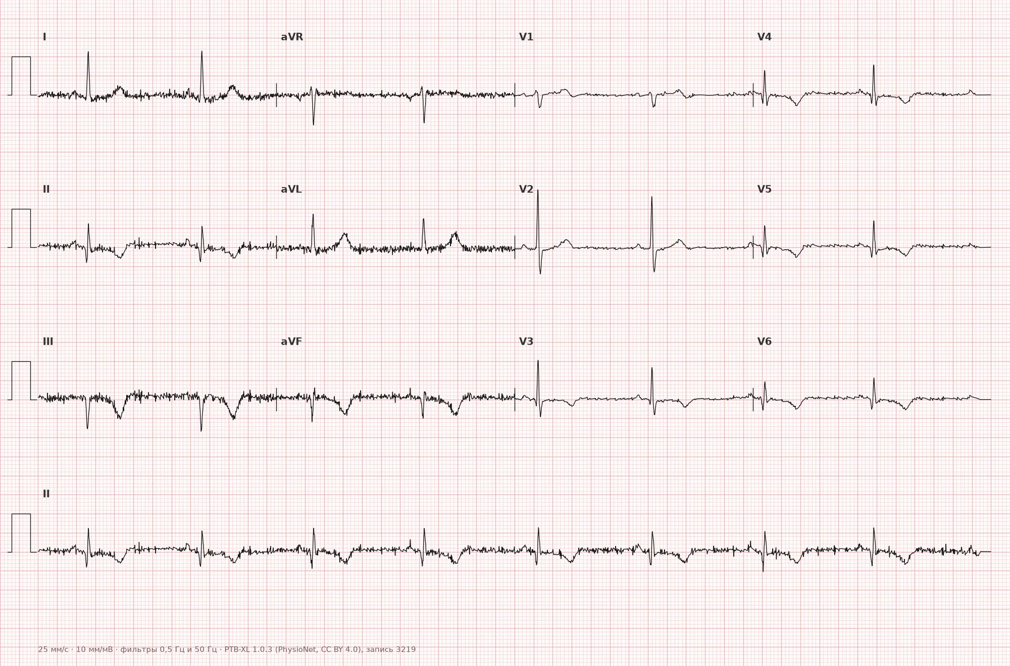
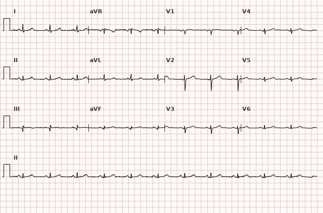
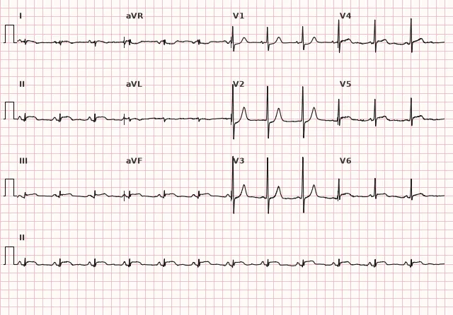
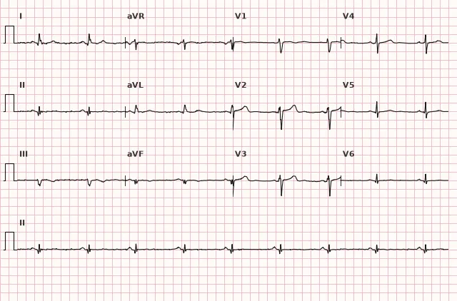
ECG from the open PTB-XL dataset (Wagner P. et al., Scientific Data, 2020; PhysioNet), CC BY 4.0 license. Rendering by WhiteCoat Pro.
Mechanism
The underlying mechanism is transmural myocardial ischemia of the inferior and lateral walls of the LV due to acute thrombotic occlusion of a coronary artery supplying both zones. Most often, this is a dominant right coronary artery, which in most individuals gives off the posterior descending artery to the inferior wall and may participate in supplying the lateral divisions, or the circumflex branch of the left coronary artery, which supplies the lateral wall and frequently the posterior part of the inferior wall in left-dominant circulation. The damaged epicardial layer loses its negative charge relative to the endocardium, and the injury vector is directed toward the electrode overlying the injury zone — hence ST elevation in the inferior and lateral leads; in leads electrically opposite to the injury zone, reciprocal ST depression may be recorded.
Causes
- Atherothrombotic occlusion of the right coronary artery (dominant)
- Atherothrombotic occlusion of the circumflex branch of the left coronary artery
- Embolic occlusion (cardioembolism, thrombosis in atrial fibrillation or endocarditis)
- Spontaneous coronary artery dissection (SCAD), more frequent in young and middle-aged women
- Coronary spasm (Prinzmetal variant angina) with a transient pattern of inferolateral ST elevation
- Multivessel disease involving the RCA/LCx basin
Differential diagnosis
- Isolated inferior MI — no ST elevation in I, aVL, V5–V6, only inferior leads; reciprocal ST depression in I/aVL is possible.
- Isolated lateral/high lateral MI — ST elevation in I, aVL (and/or V5–V6) without elevation indicating an inferior component; reciprocal ST depression is possible in inferior leads.
- Early repolarization — concave ST elevation, J-point notch, no dynamics on serial ECGs; the shape of ST alone is not a reliable criterion for differentiation.
- Pericarditis — diffuse concordant ST elevation in almost all leads, PR depression; localized reciprocal ST depression outside of aVR and sometimes V1 is not typical for a classical presentation; no pathological Q waves.
- Brugada syndrome — saddleback or coved ST elevation limited to the right chest leads V1–V3, not involving inferior and lateral leads; accompanied by characteristic QRS morphology rather than typical MI waves.
- Hyperkalemia — can cause pseudoinfarction ST elevation, including locally in inferior or other leads, but a tall peaked T wave, shortening or disappearance of the P wave, PR prolongation, and QRS widening are more typical; when suspected, ST elevation localization does not rule out electrolyte imbalance — potassium levels must be checked.
- Inferoposterior MI without lateral component — ST depression in V1–V3 without elevation in I, aVL, V5–V6; posterior leads V7–V9 are needed for confirmation.
Clinical significance and management
Inferolateral ST elevation meeting STEMI criteria requires immediate reperfusion. In patients with symptom onset within 12 hours, primary PCI is preferred if the expected time from STEMI diagnosis to recanalization (guidewire crossing) is ≤120 minutes. If this is unachievable, and in the absence of contraindications, fibrinolysis should be performed as early as possible followed by transfer to a PCI center; in late presentation (significant delay from symptom onset), the strategy may differ and requires individual evaluation.
In inferior STEMI, right-sided chest leads V3R–V4R (primarily V4R) should be recorded as early as possible, without delaying reperfusion, to detect signs of right ventricular involvement — ST elevation here alters fluid management tactics and increases the risk of hypotension with nitrates. It is important to remember that these changes can be transient and disappear upon late recording, so the absence of ST elevation in V3R–V4R does not reliably rule out RV infarction: if clinical suspicion persists (hypotension, jugular venous distension with clear lungs), the time of recording from symptom onset is considered and supplemented by imaging. Posterior leads V7–V9 should also be acquired if extension to the posterior wall is suspected.
With RCA occlusion, the risk of bradyarrhythmias is high: sinus bradycardia, AV blocks of various degrees (up to complete) due to ischemia of the AV node, which in most cases is supplied by a branch of the RCA. Rhythm monitoring and preparedness for temporary pacing are required. Extensive involvement with lateral wall recruitment (LCx basin) may be accompanied by a larger mass of ischemic myocardium and LV dysfunction; individual risk of mechanical complications (e.g., papillary muscle rupture with mitral regurgitation) and ventricular arrhythmias is assessed based on clinical status, imaging, and coronary anatomy, rather than solely on the number of leads with ST elevation.
Common interpretation errors
- Evaluating only II, III, aVF and missing concomitant ST elevation in I, aVL, V5–V6, leading to underestimation of the extent of damage.
- Failing to compare ST-elevation amplitude in II and III — missing an indication of the culprit artery.
- Failing to record V3R–V4R leads in inferior/inferolateral STEMI, missing signs of right ventricular involvement.
- Automatically interpreting ST depression in aVL as an independent finding without considering that it may be a reciprocal sign of an inferior MI without high lateral elevation.
- Confusing an isolated lateral MI with an inferolateral MI — diagnosing an inferolateral MI requires changes in both lead groups.
- Diagnosing a prior MI based on any Q wave with a duration of 0.02–0.03 s without regard to lead, amplitude, and clinical context.
1. Which leads show ST elevation in acute inferolateral STEMI?
Acute inferolateral STEMI is characterized by a combination of ST elevation in the inferior (II, III, aVF) and lateral (I, aVL, V5–V6) leads.
2. Which ECG sign in inferolateral MI indicates likely occlusion of the right coronary artery rather than the circumflex branch?
ST elevation in III higher than in II, along with elevation in V1 or depression in I, is typical for right coronary artery occlusion.
3. Which additional ECG examination should be performed in inferolateral STEMI to detect signs of right ventricular involvement?
Right precordial leads V3R–V4R allow detection of ST elevation characteristic of right ventricular involvement, although a normal pattern does not completely rule it out, especially with late recording.
Frequently asked questions
How to distinguish inferolateral infarction from isolated inferior infarction?
In inferolateral myocardial infarction, ST elevation is present not only in II, III, aVF, but additionally in I, aVL and/or V5–V6. If changes are limited to the inferior leads only, this is an isolated inferior MI, in which only reciprocal ST depression may be present in I/aVL.
Which artery most commonly causes inferolateral infarction?
Most often it is the dominant right coronary artery or the circumflex branch of the left coronary artery — both can supply blood to both the inferior and lateral walls depending on the type of coronary dominance.
Is inferolateral infarction more dangerous than isolated inferior infarction?
Widespread involvement of the lateral wall may be accompanied by a greater mass of ischemic myocardium and LV dysfunction, but the mere number of leads with ST elevation does not determine the extent of necrosis or the risk of mechanical complications. Individual risk is assessed based on the clinical picture, imaging, and the specific culprit artery.
Is it necessary to record additional leads in inferolateral MI?
Yes, right precordial leads V3R–V4R to detect signs of right ventricular involvement and posterior leads V7–V9 to assess extension to the posterior wall change management tactics. A normal pattern in V3R–V4R does not reliably rule out RV infarction, especially with late recording. Recording should not delay reperfusion.
Why do bradycardia and AV block occur in inferolateral infarction?
In most cases, the AV node is supplied by a branch of the right coronary artery, so its occlusion leads to nodal ischemia with bradyarrhythmias, up to complete AV block.
ECG Course
Interpretation from basics to myocardial infarction — learn to read any ECG using a systematic algorithm.
- Video lessons and theory for every topic
- Simulator case studies under every lesson
- Unlimited ECG simulator practice
- Study guide: "All ECGs in 25 minutes"
ECG Strip Simulator
ECG, radiology, histology, pathomorphology, pharmacology — thousands of flashcards with detailed explanations.
- Real ECG strips, not textbook drawings
- Instant answers and explanations
- Streaks and progress tracking in your dashboard
Related terms
More in the section "Ischemia and Myocardial Infarction"
Sources
- AHA/ACCF/HRS Recommendations for the Standardization and Interpretation of the Electrocardiogram, 2009
- ESC Guidelines for the management of acute coronary syndromes, 2023
- Hampton J.R. The ECG in Practice
- Orlov V.N. Guide to Electrocardiography
Educational material for medical students and physicians. Does not replace clinical guidelines and physical examination; the ECG must always be interpreted in conjunction with the clinical picture.