ECG criteria
- New ST-segment elevation at the J-point in at least 2 contiguous leads: anterior (V3–V4) and/or lateral (I, aVL, V5–V6)
- Diagnostic ST elevation requires ≥2 contiguous leads; isolated elevation in a single lead (e.g., in aVL only) does not meet this criterion
- ST threshold values in V2–V3 depend on sex and age: ≥2.5 mm in men under 40 years, ≥2.0 mm in men ≥40 years, ≥1.5 mm in women (in other chest leads and in I, aVL, V5–V6 — ≥1.0 mm)
- Reciprocal ST depression in the inferior leads II, III, aVF may be detected — this is not a mandatory sign
- Pathological Q wave: in any two contiguous leads from I, II, aVL, aVF, V4–V6 — Q ≥0.03 s and ≥1 mm in depth or a QS complex; in V2–V3 — Q >0.02 s or a QS complex. A Q wave is not mandatory for the diagnosis of acute MI and may be absent
- Typical but non-mandatory time evolution: hyperacute T waves → ST elevation → possible appearance of Q waves → possible T-wave inversion
- ST depression in aVL can be a mirror image of an inferior MI — for anterolateral MI, specifically ST elevation in I/aVL is significant, rather than isolated depression
- Assess ≥2 contiguous leads; the combination of anterior and lateral leads indicates an extensive anterolateral pattern, but is not a mandatory condition for diagnosing MI in general
What it looks like on ECG
Acute STEMI is diagnosed based on the clinical picture of ischemia and new ST elevation in ≥2 contiguous leads. In the anterolateral variant, elevation appears simultaneously in the anterior chest leads V3–V4 and in the lateral leads — I, aVL and/or V5–V6. This distinguishes it from isolated anterior or isolated lateral MI: two adjacent zones of the anterior LV wall are affected at once.
In the acute phase, tall pointed (hyperacute) T waves are sometimes visible, followed by upwardly convex ST elevation, in some cases merging with the T wave. This sequence is typical but not mandatory: hyperacute T waves may not be recorded, and ST elevation may be the first noticeable change. Reciprocal ST depression in the inferior leads II, III, aVF may be detected, but its absence does not exclude the diagnosis.
A pathological Q wave is not mandatory for the diagnosis of acute MI: it may appear later, and especially with timely reperfusion, it may not appear at all. If a Q wave forms, it typically does so in the same leads (I, aVL, V3–V6), while the R-wave amplitude decreases. As it evolves, the ST segment gradually returns to the isoelectric line, and the T wave may become inverted — the timing of this evolution is individual.
| Leads | What can be seen |
|---|---|
| V3–V4 | ST elevation, possible Q and decreased R (anterior zone) |
| I, aVL | ST elevation, possible Q (high lateral zone) |
| V5–V6 | ST elevation, possible Q (low lateral zone) |
| II, III, aVF | possible reciprocal ST depression |
Changes only in I and aVL represent a high-lateral pattern, not an anterolateral one. In the presence of ischemic symptoms and ST elevation meeting the criteria, the patient is managed via the STEMI/occlusion pathway without waiting for troponin; if elevation is present in only a single lead, urgent repeat ECG, comparison with a previous ECG, and expert evaluation are required. The typical anterior/anteroseptal area of changes is V1–V4, but an anterior MI can extend to V5–V6, I, and aVL — in this case, it is referred to as an anterolateral pattern.
Real ECGs: 8 examples
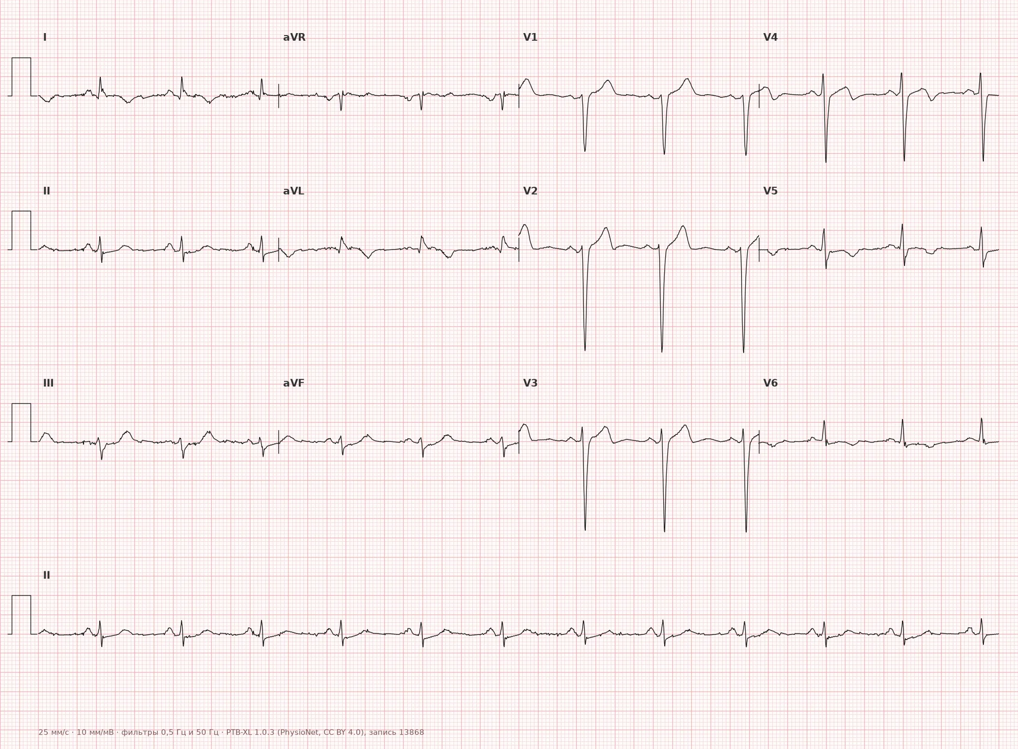
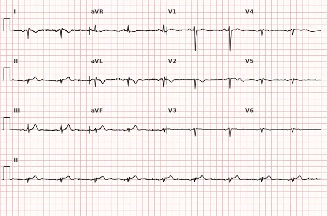
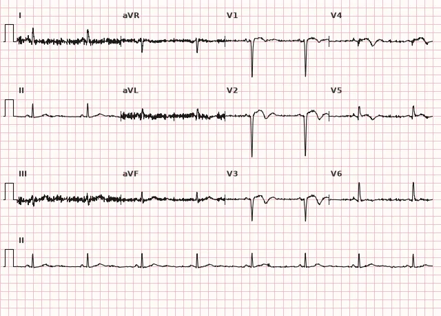
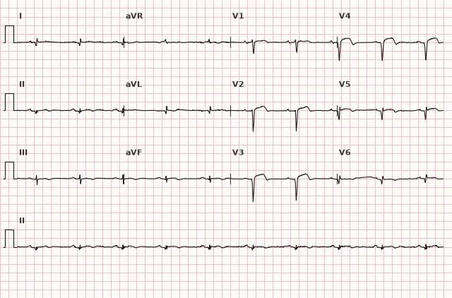
ECG from the open PTB-XL dataset (Wagner P. et al., Scientific Data, 2020; PhysioNet), CC BY 4.0 license. Rendering by WhiteCoat Pro.
Mechanism
The left anterior descending artery (LAD) supplies blood to the anterior wall and interventricular septum via septal branches, and the lateral wall often via diagonal branches. The circumflex artery (LCx) primarily supplies the lateral and posterolateral wall; isolated LCx occlusion usually produces a lateral and/or posterolateral pattern, rather than simultaneous anterior and lateral involvement. An anterolateral picture is more often associated with proximal or mid-LAD occlusion with a large diagonal branch, left main coronary artery disease, or combined multi-vessel disease; coronary anatomy is variable, so other combinations are also possible.
ST elevation reflects the current of injury — the spatial shift of the repolarization vector toward the ischemic/injured myocardial zone. This produces ST elevation in leads facing the focus and possible mirror-image ST depression in opposite leads. Cardiomyocyte death and the loss of their electrical activity can form a pathological Q wave, but ST elevation by itself is not a specific marker of transmural injury.
Causes
- Atherothrombotic occlusion of the LAD (especially proximal, with a large diagonal branch)
- Left main coronary artery disease or multi-vessel disease involving the LAD and LCx
- Circumflex artery occlusion (more often produces a lateral/posterolateral pattern, but anatomical variations are possible)
- Spontaneous coronary artery dissection (more common in young women)
- Rarely — coronary embolism (with a confirmed source: valve prostheses, endocarditis, intracardiac thrombus; atrial fibrillation per se does not prove coronary embolism)
- Pronounced coronary spasm (cocaine, vasospastic angina)
Differential diagnosis
- Isolated anterior or anteroseptal myocardial infarction — typical zone V1–V4, without involvement of I, aVL, V5–V6
- High lateral myocardial infarction — changes only in I, aVL, without chest leads
- Early repolarization syndrome — concave ST elevation, J-point notch, no reciprocal depression and Q-wave evolution
- Pericarditis — diffuse concordant ST elevation in almost all leads, PR depression, no local reciprocity and Q waves
- Left ventricular hypertrophy with strain — tall R wave in lateral leads with discordant ST-T, but without acute evolution and typical reciprocity
- Takotsubo cardiomyopathy — extensive ST elevation that in distribution may resemble anterior or anteroseptal/anterolateral MI; coronary angiography typically shows no obstructive disease/culprit lesion; concomitant coronary artery disease is possible. The diagnosis is confirmed by a combination of clinical data, coronary angiography, and cardiac imaging
Clinical significance and management
Anterolateral STEMI is one of the most dangerous variants: a large volume of the LV myocardium is affected, which increases the risk of acute left ventricular failure, cardiogenic shock, ventricular tachyarrhythmias, and mechanical complications (wall rupture, mitral regurgitation when the papillary muscle is involved).
In the presence of ST elevation meeting the criteria and a clinical picture of ischemia — immediate activation of the STEMI pathway. Primary PCI is preferred if achievable within recommended timeframes; fibrinolysis is considered when symptoms are generally ≤12 hours, contraindications are absent, and timely PCI is not feasible, followed by a pharmacoinvasive strategy and rescue PCI if thrombolysis fails.
Additionally, one must consider patterns without explicit ST elevation that are associated with acute coronary occlusion: hyperacute T waves (require evaluation in dynamics, non-specific on their own) and the de Winter pattern (regarded as a probable occlusion pattern, usually requiring urgent angiography). Diffuse ST depression with ST elevation in aVR is non-specific, does not prove left main or multi-vessel occlusion, and is not an independent STEMI equivalent — the urgency of invasive management in this case is determined by high-risk clinical criteria (hemodynamics, recurrent ischemia, ECG dynamics, biomarkers) rather than the pattern alone.
Post-reperfusion — rhythm monitoring (risk of ventricular arrhythmias), assessment of LV contractility via echocardiography, follow-up ECG monitoring to assess reperfusion efficacy and detect complications.
Common interpretation errors
- Attributing changes confined to I/aVL without chest leads to anterolateral MI — this is an isolated lateral focus
- Requiring a mandatory pathological Q wave for the diagnosis of acute MI — a Q wave may be absent, especially with early presentation and successful reperfusion
- Applying ST thresholds without accounting for sex and age in V2–V3
- Missing patterns associated with coronary occlusion without overt ST elevation (hyperacute T waves, de Winter pattern) due to the formal absence of ST elevation
- Interpreting ST depression with elevation in aVR as an absolute STEMI equivalent
1. In which leads is ST elevation expected in anterolateral myocardial infarction?
Anterolateral MI combines anterior (V3–V4) and lateral (I, aVL, V5–V6) localization.
2. Which sign can be reciprocal in anterolateral myocardial infarction?
Damage to the anterior and lateral wall may produce reciprocal ST depression in the inferior leads, although this sign is not mandatory.
3. Which statement about the pathological Q wave in acute anterolateral myocardial infarction is correct?
The diagnosis of acute STEMI is established based on clinical presentation and ST elevation; a pathological Q wave is an optional and often later sign.
Frequently asked questions
How does anterolateral infarction differ from anterior infarction?
In anterior infarction, the typical zone of changes is the anterior precordial leads V1–V4. In anterolateral infarction, lateral leads I, aVL, and/or V5–V6 are added to them, indicating a larger volume of affected myocardium.
Is anterolateral infarction dangerous?
Yes, this is one of the most extensive STEMI variants in terms of left ventricular affected area. The risk of acute heart failure, cardiogenic shock, and ventricular arrhythmias is elevated, which is why reperfusion is attempted as quickly as possible.
Which artery is typically affected?
Most commonly, the proximal or middle left anterior descending artery with a large diagonal branch, the left main coronary artery, or multivessel disease. Isolated occlusion of the circumflex artery usually produces a lateral or posterolateral rather than anterolateral pattern, although anatomical variants are possible.
Should posterior leads be checked in anterolateral infarction?
If there is ST depression in V1–V3 along with changes in I, aVL, V5–V6, it is worth recording leads V7–V9 to rule out a concomitant posterior infarction, since circumflex artery involvement can also affect the posterior wall.
Can anterolateral infarction present without ST elevation?
Yes, a non-ST-segment elevation ACS picture is possible with ST depression or negative T waves in the same leads. Patterns associated with acute coronary occlusion (hyperacute T waves, de Winter waves) are assessed separately — the urgency of their evaluation is determined by the probability of occlusion, symptoms, dynamics, and hemodynamics, rather than the formal presence of ST elevation.
ECG Course
Interpretation from basics to myocardial infarction — learn to read any ECG using a systematic algorithm.
- Video lessons and theory for every topic
- Simulator case studies under every lesson
- Unlimited ECG simulator practice
- Study guide: "All ECGs in 25 minutes"
ECG Strip Simulator
ECG, radiology, histology, pathomorphology, pharmacology — thousands of flashcards with detailed explanations.
- Real ECG strips, not textbook drawings
- Instant answers and explanations
- Streaks and progress tracking in your dashboard
Related terms
More in the section "Ischemia and Myocardial Infarction"
Sources
- AHA/ACCF/HRS Recommendations for the Standardization and Interpretation of the Electrocardiogram, 2009
- ESC Guidelines for the management of acute coronary syndromes, 2023
- Surawicz B., Knilans T. Chou's Electrocardiography in Clinical Practice
- Orlov V.N. Guide to Electrocardiography
Educational material for medical students and physicians. Does not replace clinical guidelines and physical examination; the ECG must always be interpreted in conjunction with the clinical picture.