Summary
Postpartum hemorrhage (PPH) remains the leading cause of maternal mortality worldwide, particularly in resource‐constrained and remote settings. The cornerstone of reducing PPH‐related morbidity and mortality lies in its early recognition, timely treatment, and adherence to evidence‐based protocols, all of which are heavily dependent on an accurate assessment of postpartum blood loss. Visual estimation, unfortunately, is still widely used, highly inaccurate, and often leads to underdiagnosis, resulting in a delayed or sometimes absent response to PPH. Objective quantification of blood loss, although not perfectly precise, provides a far more reliable estimate and is critical to triggering timely and effective interventions. This approach requires coordinated teamwork, leadership, institutional commitment, and a shift in clinical culture from subjective to standardized measurement practices. Scientific evidence strongly supports the integration of objective quantification into routine obstetric care. The use of calibrated drapes, cumulative measurement, and the combination of quantification with early warning systems have all proven to be effective in reducing PPH‐related complications and deaths. Practical barriers are faced in implementing objective measurement strategies at the facility and institutional level, including availability, supply costs, environmental concerns, national and institutional advocacy, policy change, leadership, appropriate training, and, more importantly, resistance to change. Low‐cost, reusable, and locally made devices can offer promising solutions for scaling up this intervention in resource‐limited environments. Sustained success will depend on engagement with health departments of the governments for framing policies, guidelines for implementation, procurement and regular supplies, inter‐institutional collaboration, and ongoing and refresher training for in‐service healthcare providers. In the longer term, it requires inclusion into the medical curriculum, local leadership, champions, behavioral interventions, recognition and rewards to individuals, and teams who promote long‐term adherence to best practice. Ultimately, although technical knowledge on PPH management is now well established, the real challenge lies in its consistent and context‐appropriate identification and application. This article discusses the different definitions of PPH. It highlights the importance of objective quantification of blood loss during and after childbirth and the various available methods for blood loss measurement and explores their implementation across different clinical settings.
Keywords: blood loss, evidence‐based practice, health resources, maternal mortality, measurement, multiprofessional working, postpartum hemorrhage, surgical, teamwork
1. INTRODUCTION
Each year, millions of women experience postpartum hemorrhage (PPH), resulting in approximately 70 000 maternal deaths globally, which is approximately 20% of all maternal deaths. Most of these occur in resource‐limited settings 1 and are almost entirely preventable. 2 Multiple effective interventions exist for the management of PPH, applicable even at basic levels of care provision; however, delays in recognizing and diagnosing the condition are common, leading to late or non‐utilization of these interventions. 3
There are various definitions of PPH, with the current definition proposed by the World Health Organization (WHO) being the most widely used. 4 WHO currently defines PPH as blood loss exceeding 500 mL after a vaginal delivery or 1000 mL after a cesarean birth 4 , 5 within the first 24 h postpartum (see Box 1).
Visual estimation of blood loss remains the most commonly practiced method for quantifying postpartum bleeding; however, it has been shown to be inaccurate, overestimating small volumes of blood loss 6 and underestimating losses when volumes are high, 7 thus delaying treatment 8 and increasing PPH‐related morbidity. 9 Further, when bleeding is quantified immediately after delivery, cumulative estimation during the first 24 h postpartum is often neglected, leading to missed identification of patients with excessive bleeding.
A review of the evidence in 2023 resulted in two recommendations (WHO recommendations for the prevention and treatment of postpartum hemorrhage).
First, for all women giving birth, routine objective measurement of postpartum blood loss is recommended to improve the detection and prompt treatment of PPH.
Second, a standardized and timely approach to the management of PPH, comprising an objective assessment of blood loss and use of a treatment bundle supported by an implementation strategy, is recommended.
The care bundle for the first‐line treatment of PPH in vaginal births should include rapid institution of uterine massage, administration of an oxytocic agent and tranexamic acid, intravenous fluids, examination of the genital tract, and escalation of care. 10
Our paper highlights the importance of objective quantification of blood loss during and after childbirth, the various methods available for blood loss measurement, and their application in different clinical settings. Graphic materials and videos are included, which we hope will facilitate the implementation of objective postpartum blood loss quantification strategies, particularly in resource‐limited settings.
2. DEFINITION OF POSTPARTUM HEMORRHAGE
There are several published definitions of PPH; however, at the time of writing this paper, no clear consensus exists regarding which is most appropriate, contextually relevant, and effective in initiating timely evidence‐based treatment protocols. A physiologically based definition, focused on hemodynamic and metabolic impact, is likely to correlate closely with clinical outcomes, however typical signs of hypovolemia do not usually appear until approximately 25% of the total blood volume has been lost. 11 In addition, determination of the exact volume corresponding to that percentage in each patient is challenging, making this definition unreliable for clinical use.
The American College of Obstetricians and Gynecologists (ACOG) defines PPH as a cumulative blood loss of greater than or equal to 1000 mL or blood loss accompanied by signs or symptoms of hypovolemia within 24 h after the birth process. 12 This definition acknowledges that the mode of delivery (vaginal or cesarean) should not alter the threshold for initiating therapeutic interventions. Furthermore, during a cesarean birth, the patient might receive neuraxial anesthesia, which is associated with a loss of sympathetic tone in certain vascular territories, thereby impairing some of the adaptive mechanisms to blood loss. This might result in a greater hemodynamic impact than what a patient might experience when losing the same volume during a vaginal birth. ACOG emphasizes that, regardless of the mode of delivery, blood loss exceeding 500 mL should raise concerns among the treating team, and depending on the clinical context (e.g. pre‐existing anemia or very low body mass index), it might indicate the need for therapeutic measures.
The current WHO definition includes a lower threshold of 500 mL within the first 24 h after vaginal birth (while aligning with the 1000 mL threshold for cesarean birth), 4 , 5 recognizing that in resource‐limited settings (where vaginal birth is the predominant mode of delivery), access to healthcare services is often delayed, and visual estimation of blood loss frequently underestimates the true volume lost. 13
The continued high prevalence of PPH as a leading cause of maternal morbidity and mortality worldwide, especially in resource‐limited settings, has recently led to expert consensus recommendations advocating for lower diagnostic and treatment thresholds for bleeding during both vaginal and cesarean births. 14 It has been proposed that during cesarean birth, the first response should be triggered once the woman has lost at least 500 mL of blood with ongoing bleeding or when clinical signs of hemodynamic instability appear, whichever occurs first. 14
Similarly, the initiation of a PPH treatment bundle could be considered in some women from as little as 300 mL of blood loss. 10 Indeed, a trigger criterion of 300 mL of quantified postpartum blood loss after vaginal birth plus one abnormal clinical observation (uterine tone, increased bleeding flow, or altered level of consciousness) or an abnormal vital sign was one of the criteria used to initiate interventions in the intervention group of the E‐MOTIVE study, in which a reduction in PPH‐related mortality was demonstrated following early detection and protocolized management. 15
Other definitions are also valid and might be useful when the treating team cannot accurately quantify blood loss or in specific clinical situations. These include a hematocrit drop of more than 10%, 16 any blood loss that results in hemodynamic compromise, or significant bleeding in patients with low tolerance to blood loss (e.g. those with severe pre‐existing anemia or cardiovascular conditions). 17
3. IMPORTANCE OF OBJECTIVE QUANTIFICATION OF POSTPARTUM BLOOD LOSS (Box 2)
If the assessment of postpartum blood loss is inaccurate, PPH might go unrecognized, potentially delaying or preventing the timely initiation of life‐saving interventions. In addition, underestimation might lead to suboptimal interventions, when the provider grades the hemorrhage as not severe. Timely recognition of PPH minimizes the risk of life‐threatening complications, such as hemorrhagic shock. Effective interventions, such as the administration of tranexamic acid, can lose their efficacy if administered too late, 18 likely due to the metabolic deterioration associated with prolonged inadequate oxygen delivery to tissues.
Early identification of abnormal bleeding is one of the key pillars of the intervention bundles proposed by the California Maternal Quality Care Collaborative 19 and endorsed by ACOG. 17 Therapeutic interventions used to control abnormal bleeding require time for proper execution, and often by the time they are applied, blood loss far exceeds the thresholds established for diagnosing PPH. It is evident that immediate recognition of abnormal bleeding is fundamental to the successful management of this condition. Changes in maternal vital signs or laboratory parameters often provide late or misleading information. 20 Visual estimation of blood loss after both vaginal and cesarean birth is notoriously inaccurate 21 and is thereby of limited clinical use. Accurate and effective assessment of ongoing blood loss, both during and after delivery, is therefore essential for reducing maternal morbidity and mortality.
Evidence has increasingly supported the objective quantification of postpartum blood loss as a fundamental component of safe birth management. The 2018 Cochrane review concluded that there was insufficient evidence to recommend one blood loss quantification strategy over another, as studies found no significant differences in severe morbidity, transfusion rates, or the use of uterotonics when comparing visual estimation with calibrated drapes. 22 Despite the lack of conclusive evidence favoring a specific method, overall, objective quantification techniques are generally considered to offer more accurate estimates than subjective visual assessments. 23
Morbidity among women with severe PPH might be reduced when quantitative blood loss estimation is integrated into maternal safety protocols. 22 Regional experiences in high‐resource countries 24 as well as in resource‐limited settings 25 reflect a shift in the weighting of published evidence, now far more clearly recommending a diagnostic approach that combines a calibrated drape for objective blood loss measurement with clinical observations. 26
4. METHODS FOR QUANTIFYING POSTPARTUM BLOOD LOSS
Table 1 describes the different methods for quantifying postpartum bleeding. A commonly used method to assess blood loss during the third stage of labor is visual estimation, typically performed by a trained health worker. 26 Visual estimation is inaccurate; obstetric caregivers tend to underestimate blood loss by approximately 30%–50%, particularly at higher volumes of blood loss. 7 This occurs even when the measurement is performed in specialized centers and by personnel with extensive training. 27 Other factors contributing to the inaccuracy of visual estimation of blood loss include the speed and duration of blood flow and the influence of mode of birth on the health worker's perceptions.
Methods for quantifying postpartum bleeding.
| Method | Description | Advantages | Disadvantages | |
|---|---|---|---|---|
| Methods based on direct collection | ||||
| Methods based on gravimetry | The use of a scale to weigh absorbent material before and after use. The formula is used: (weight after—weight before)/1.05 = blood volume (in mL; 1.05 g/mL is the density of blood) | High accuracy | Time‐consuming; requires preparation of equipment knowing the “dry weights” of all materials | |
| Volumetric measurement: collection of blood in containers to measure its volume | Blood suction collectors | Method routinely used during cesarean birth. Some collection bags placed under the patient's pelvis can be connected to a negative pressure blood collector | Relatively high accuracy | Attention must be paid to the inclusion of other fluids in the collection containers (urine, cleaning solutions, amniotic fluid) |
| Blood collection bags for vaginal birth | Under‐buttock plastic bags that collect blood loss through the vagina | Low cost; can be calibrated and allow real‐time assessment of blood loss volume; can be handmade in each hospital using plastic bags, with marked lines indicating volumes of 300 mL and 500 mL; additional volume marks can be added to facilitate quantification; non‐calibrated bags must be weighed to calculate the fluid volume inside | Lithotomy position required; additional non‐clinical workload for the service provider to prepare the bags with marked lines | |
| Other commercial blood collection containers | Other low‐cost, reusable containers have been designed with pre‐established volumes that facilitate immediate identification of patients with PPH | Low cost; reusable; lower environmental impact; useful for births in positions other than lithotomy | Not accessible in all countries; unknown even to many doctors | |
| Other non‐reusable collector devices | Visual analog estimation based on the preset characteristics of the device | Not accessible in all countries | ||
| Artisanal methods for quantifying bleeding | In settings such as home birth or intercultural birth, traditionally accepted fabrics are used by the community during vaginal delivery. These fabrics have a standard size (approximately 100 × 155 cm) and absorb a relatively consistent volume of fluid. Among the names given to this fabric pieces are: “kanga,” “lappa,” and “sarong” | A useful strategy in resource‐limited settings or when the community has low acceptance of medical recommendations; once saturated with blood, these fabrics act as an alarm to trigger hospital transfer or the administration of available and approved medication by traditional healers or midwives | Low accuracy | |
| Visual methods | ||||
| Visual estimation | Visual assessment of blood loss volume; a visual reference method is used based on images of known volumes of blood on different surfaces (e.g. gauze or sheets) | Quick; widely used in the world | Low accuracy | |
| Colorimetry cards | Cards with red gradients to compare pad saturation and estimate loss volume | — | Low accuracy; training and graphic material are required to remember the proposed equivalences | |
| Methods based on clinical and paraclinical assessment | ||||
| Clinical assessment | Evaluation based on clinical signs and symptoms | No additional equipment required | Subjective and less accurate; clinical signs of hypovolemia are late | |
| Hemoglobin test | Comparison of hemoglobin levels before and after delivery | High accuracy in determining blood loss | Requires blood analysis and laboratory resources; physiologic hemoconcentration during early stages of bleeding may lead to false negative | |
| Advances technological methods | ||||
| Spectrophotometry | It allows the measurement of hemoglobin in the collected fluids and to make an accurate quantification of the actual blood volume (discounting other fluids such as amniotic fluid) | Accurate measurement under predetermined conditions | High cost; availability in low‐ to medium‐resource settings is an issue | |
| Applications and sensors | Use of artificial intelligence and sensors to more accurately estimate postpartum bleeding in real time | Automatic calculation; immediate results | High cost; availability in low‐ to medium‐resource settings is an issue | |
Abbreviation: PPH, postpartum hemorrhage.
Although strategies have been reported to improve the performance of visual blood loss estimation, including staff training and the use of cognitive aids to approximate blood loss, based on the appearance of blood‐soaked surgical materials, the observed improvement is only temporary. 28
There are other approaches to assess postpartum blood loss after birth more objectively. Implementation of quantitative assessment of blood loss requires the following: (i) use of direct measurement of blood loss (quantitative blood loss); and (ii) clear protocols for collecting and reporting a cumulative record of blood loss after delivery. 19
Considering the differences between vaginal and cesarean births, we will describe both situations separately.
4.1. Vaginal birth
In direct blood collection, a vessel (such as a plastic collecting bag or a tray) is used to collect all blood lost during the third stage of labor 29 (Figure 1a). To avoid overestimating blood loss due to other fluids (urine, perineal wash solutions, amniotic fluid) that might enter the collection container and considering that in most cases bleeding occurs after placental delivery, placing the device under the maternal perineum immediately before placental removal is recommended. In the case of a collection bag, it can be positioned underneath the woman's buttocks, folded to prevent the collection of other fluids, and then unfolded after delivery of the infant (Video S1).
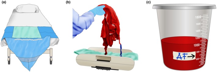
Components of the objective quantification of postpartum bleeding. (a) Graduated under‐buttock plastic bags. (b) Weight of blood‐soaked materials. (c) Fluid level in the suction device after draining the amniotic fluid (AF).
Graduated collection bags with marked volume indicators (e.g. at 300, 500, and 1000 mL) enable the clinical team to monitor blood loss in real time and to easily recognize when predefined thresholds are exceeded.
Although plastic bags are relatively inexpensive and can even be locally handmade upon hospital request (Figure 2), maintaining consistent availability in resource‐limited settings might still pose a challenge. A calibrated drape (Dhaka/Dk drape) made with thin, transparent, low‐cost, polythene shopping bags and readily available in the market (capacity 12–15 kg) appears to be effective and safe. The drape might be disposed of, as for other contaminated medical waste. The calculated cost of making the drape is only approximately US$0.05. 30
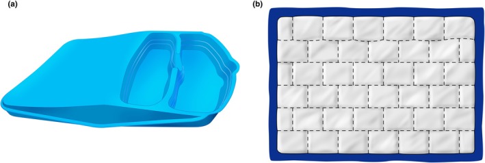
Other devices for postpartum blood collection. (a) MaternaWell Tray. A low‐cost, reusable plastic container designed in South Africa with visual cues corresponding to volumes of 300, 500, and 1000 mL. (b) Signaling a postpartum hemorrhage emergency (SAPHE) mat, designed so that each square absorbs up to 50 mL of blood, allowing for an approximate calculation of the volume lost.
Alternatively, non‐graduated plastic drapes can be used. After the bleeding stops, the bag and its contents can be weighed. The team can estimate blood loss using a 1:1 equivalence between grams and milliliters (Video S1). Although there might be economic concerns about adding another item to the standard birth supply kit, as well as environmental concerns related to increased plastic use, these can be addressed through well‐defined disposal protocols. Moreover, the relatively low additional cost is minimal compared to the devastating impact of a PPH‐related maternal death. As an alternative, various models of reusable plastic collection devices are also available with visual cues corresponding to 300‐, 500‐, and 1000‐mL volumes (Figure 3a).

Collection bag in place with the bed tilted. In patients who have experienced postpartum hemorrhage, once they are positioned in the supine position, the collection under the buttock bag can be maintained by Inclining the bed (reverse Trendelenburg position). It is recommended to keep the bag in place for at least 1 h after bleeding control.
These devices are similarly placed under the buttocks after the infant is born, allowing for easy identification of abnormal postpartum bleeding, 31 with high rates of approval among birth attendants and birthing women themselves. 32 In cases where the team prefers to keep the collection bag in place for an extended period after birth, simple strategies have been reported to ensure the woman's comfort, such as adjusting the bed position when the bag is in use (Figure 4).
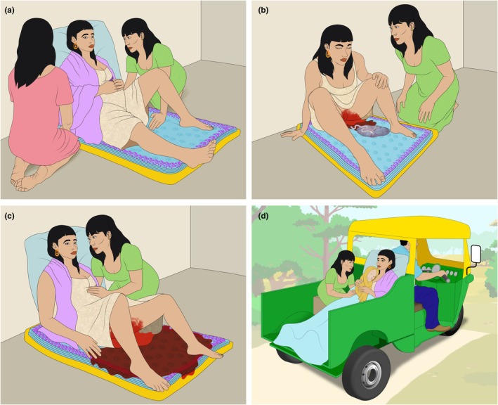
Traditional practices to estimate blood loss during delivery (“Kanga”‐based blood loss estimation). In some communities where home birth is common, traditional healers or midwives use standardized pieces of fabric with an approximate absorption capacity of 500 mL to estimate postpartum blood loss. (a) The laboring woman is positioned over the folded “Kanga,” which absorbs (b) postpartum bleeding. (c) When the entire Kanga is soaked with blood, an alert is triggered. (d) If bleeding continues, the patient must be transferred to the nearest hospital. This approach should be discussed between the hospital covering the patient's residential area and the community members responsible for assisting home births. In such settings, where no other options are available, interventions such as oral misoprostol administration for the active management of the third stage of labor and abnormal postpartum bleeding, manual aortic compression, and hypothermia prevention can be part of the initial management of patients with postpartum hemorrhage (PPH).
In addition to collecting blood in a container, the attending team should quantify the volume of blood soaked into the surgical clothing used. Surgical sponges, gauze, diapers, sheets, and other blood‐soaked items should all be weighed (Figure 1b). The dry weight of these items must be known and subtracted from the total weight obtained for each case evaluation (Table 2).
Dry weight of surgical clothing and personal hygiene implements used during or after delivery at Fundacion Valle de Lili, Cali, Colombia.
| Element | Dry weight (g) a |
|---|---|
| Surgical sponge | 21 |
| Gauze | 5 |
| Standard surgical drape | 52 |
| Patient gown | 150 |
| Adult diaper | 110 |
| Postpartum sanitary pad | 10 |
In each hospital, the materials and their sizes vary, and consequently, the volume of absorbed blood differs. Each team must weigh their materials and create a list of “dry weights,” which should be clearly visible to all team members.
In resource‐limited settings, other strategies are valid, addressing local needs and incorporating practices accepted by the local community. 33 For example, during home births, traditional birth attendants use the number of blood‐soaked cloth items (“kangas” in Tanzania and “sarongs” in Asia) as a threshold measure, after which they administer misoprostol or refer the woman to a health facility. 34 It has been demonstrated that these cloth items have an average blood absorption capacity of approximately 500 mL (Figure 5). 35
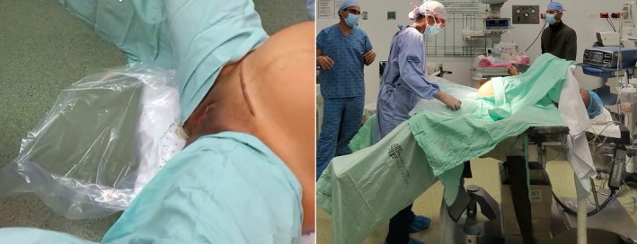
Low lithotomy positioning during cesarean birth for placenta accreta spectrum, allowing the collection bag to be placed under the perineum. Left: The plastic collection bag (graduated or non‐graduated) is placed beneath the patient's pelvis before applying sterile drapes. Lateral stirrups are used to support the lower limbs. Right: The low lithotomy position with the hips abducted, allowing a surgical assistant to be positioned between the thighs and providing a clear view of the perineum through a sterile transparent plastic surgical drape. During the procedure, an assistant periodically checks for vaginal bleeding and monitors the volume of blood accumulated in the collection bag placed beneath the surgical drape.
Other devices have also been developed to collect blood lost vaginally, featuring predefined characteristics that enable a rapid visual estimation of blood loss. An example is the “Signaling a Postpartum Hemorrhage Emergency (SAPHE) mat,” which is composed of superabsorbent polymer, nylon, pellon, and SurgiMat dressings. The mat is designed so that each square absorbs up to 50 mL of blood. 36 Counting the number of blood‐filled squares allows for an approximate calculation of the volume lost (Figure 3b).
4.2. Cesarean birth
The traditional positioning of a woman during a cesarean birth limits the use of a collection bag under the perineum. In certain situations, such as a cesarean for placenta accreta spectrum (PAS), the use of low lithotomy positioning is recommended, allowing the collection bag to be placed under the perineum (Figure 6). In most cases, however, cesarean births are performed without vaginal bleeding; therefore, blood loss can be estimated more objectively by summing up the weight of the blood‐soaked surgical items (subtracting the dry weight, as in a vaginal birth) and the content of suction devices.
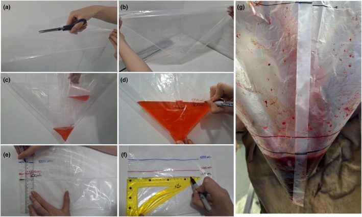
Hand‐made collection bag. By using large, transparent plastic bags, a graduated collection bag can be constructed to quantify postpartum bleeding. (a) One side of the bag is cut and (b) the bag is folded so that it acquires a funnel shape with the closed part facing down. (c) A predefined volume (300 mL, 500 mL, 1000 mL, and other volumes as the local team prefer) is poured in to determine the placement of (d) the alert mark using an indelible marker. (e, f) This marked bag serves as a template to transfer the markings onto other bags without the need to use liquid in each one. (e) For example, a ruler can be used to transfer the markings obtained from the first bag, where the predefined liquid volume was deposited, onto (f) other bags. Additional markings can be added to guide the team in assessing blood loss progression, such as at 300 mL to alert the team (e, f) and at 1000 mL to indicate a higher‐volume hemorrhage. (g) Bags are used during vaginal birth to collect postpartum bleeding. Further calibrations can be made to measure the amount of actual blood loss.
To avoid overestimating blood loss due to amniotic fluid, it is useful for the surgical team to note the fluid level in the suction device after draining the amniotic fluid. At that point, a mark can be placed on the suction device (Figure 1c), and the volume collected after that moment should be considered for quantifying blood loss (Video S2).
There are also other, more complex methods for the measurement of blood loss. However, these can be difficult to perform and are not always available in routine clinical practice (Table 1). This includes hemoglobin concentration change in venous blood sampling comparing pre‐ and postpartum evaluation, spectrophotometry, 37 dye dilution technique, 38 contrast‐enhanced ultrasound, 39 nuclear medicine imaging with the use of radiopharmaceuticals. 29 New technologies, such as digital analysis of photos of soaked pads and linens, have also been tested. 29 , 40 Unfortunately, methods that document blood loss retrospectively are useful for statistical purposes but not for the real‐time blood loss tracking required to trigger effective clinical intervention.
4.3. First 24 h after birth blood loss quantification
Current definitions of PPH include not only the volume of blood lost, but also the time frame in which the loss occurs. 11 Accurate identification of PPH requires objective quantification of blood loss, where possible, over a 24‐h period, not just during the immediate postpartum phase. It is therefore recommended that blood loss be documented in medical records similarly to the way urine output is routinely charted 41 (Figure 7).
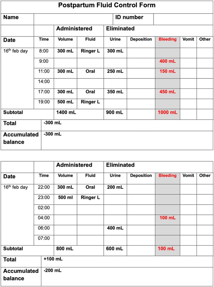
Blood quantification as urine output measurement. Objective quantification of postpartum bleeding should include the documentation of measurements using a format specifically designed to facilitate cumulative tracking over the first 24 h postpartum, similar to the way urinary output is monitored. The definition of postpartum hemorrhage includes the total volume of blood lost during the first 24 h after childbirth.
In addition to measuring objective blood loss after birth, immediate postpartum monitoring should, where possible, include the weight of blood‐soaked clothing during the first 24 h. Diapers, sanitary pads, or sheets soaked with blood should be weighed and the attending midwife should subtract the dry weight of these items. The final weight should be converted to milliliters and recorded in the medical record. For example, a patient with a blood loss of 400 mL during vaginal birth is not immediately diagnosed with PPH. However, if a clot weighing 150 g (equivalent to 150 mL) is expelled through the vagina 2 h later and a blood‐soaked diaper weighing 600 g (150 g dry weight of the diaper and 450 g = 450 mL of blood) is removed from that patient 8 h after birth, the diagnosis of PPH can be established because the volume of blood loss exceeds 500 mL (400 mL during delivery + 150 mL 2 h later and 450 mL at 8 h postpartum = 1000 mL lost in the first 24 h after vaginal birth) and immediate treatment must be administered (Figure 7).
As the most widely accepted current definition of PPH is blood loss exceeding 500 or 1000 mL within the first 24 h postpartum, we recommend an objective and cumulative quantification of blood loss at least during this period, unless, or until, the WHO definition changes.
To date, there is insufficient evidence to strongly recommend a specific timeframe for continuing blood loss assessment after diagnosing PPH. However, it appears reasonable to continue ongoing blood loss monitoring as long as active bleeding persists, or, if the patient remains hemodynamically unstable after blood loss exceeding 500 mL, 11 unless, or until, clear guidance regarding timing is available.
Other methods for identifying patients with abnormal postpartum bleeding
Despite best efforts to quantify postpartum blood loss, the volume of estimated blood loss remains unreliable in many cases. Therefore, attention should also be directed toward the patient's overall clinical status. This is particularly important in the postoperative period. 23 Recently, some guidelines have incorporated the shock index (SI; heart rate/systolic blood pressure) and obstetric early warning systems into their recommendations for assessing hemorrhage (Figure 8, Box 3). 42
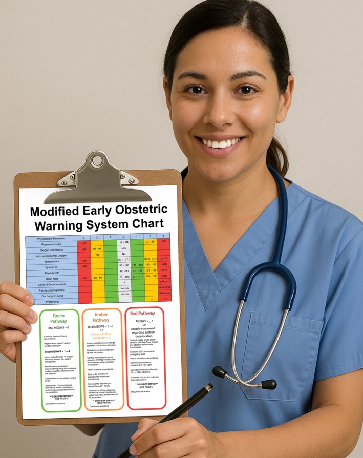
Obstetric early warning system chart. Several models of early warning systems in obstetrics exist, many of which have been scientifically validated. Their routine implementation in obstetric emergency services facilitates the identification of patients at risk of clinical deterioration. These systems are based on the monitoring of physiological parameters and the detection of abnormalities, each assigned a specific score. The cumulative score across all parameters determines the risk of deterioration and guides the need for immediate or delayed interventions.
Any amount of obstetric bleeding that is associated with clinical signs of hypovolemia should prompt a PPH evaluation and consideration of interventional protocols. The SI, along with the “rule of 30,” represents an important set of tools to assist clinicians in emergency situations by estimating blood loss and assessing the degree of hemodynamic instability.
Shock index
To date, the normal obstetric SI has been defined as 0.7–0.9, compared to 0.5–0.7 in the non‐pregnant population, considering that the hemodynamic changes of pregnancy might delay the recognition of hypovolemia. 11 , 43 To compensate for blood loss, the heart rate increases before a drop in systolic blood pressure, leading to an increase in SI. An SI of 0.9 or higher is associated with increased mortality, and an SI greater than 1 raises the likelihood of requiring a blood transfusion. 44 FIGO considers the shock index a potential marker of PPH severity and a warning sign of hemodynamic instability when its value exceeds 0.9. 43
Rule of 30
The rule of 30 estimates an approximate blood loss of 30% of normal circulating volume (70 mL/kg in adults, 100 mL/kg during pregnancy). It is defined by a 30% decrease in hematocrit, a 30% reduction in hemoglobin (approximately 3 g/dL), a 30 mmHg drop in systolic blood pressure, and an increase in heart rate by 30 beats per minute. 43
5. IMPLEMENTATION OF OBJECTIVE MEASUREMENT OF POSTPARTUM BLOOD LOSS
Objective strategies for quantifying postpartum bleeding aim to estimate blood loss as accurately as possible, recognizing that absolute accuracy is not possible. Implementing objective measurement requires a coordinated team effort and a cultural shift at an individual and institutional level. This means moving away from a reliance on visual estimation and involving all stakeholders in obstetric care, both in the delivery room and the operating theater. It is essential that the entire team is familiar with and consistently follows established procedures. Table 3 presents process maps for blood loss quantification during vaginal and cesarean births, respectively (Box 4).
Process maps for blood loss quantification during vaginal and cesarean birth.
| Vaginal birth | Cesarean birth |
|---|---|
| Gather your team and design a sustainable strategy accepted by all members | |
|
Determine the dry weight of clothing and other items that may become soaked with blood to improve the accuracy of blood loss estimation Create a standardized reference list and pictorial guide with the dry weights of commonly used materials (e.g. surgical drapes, gauze, pads, and kangas) and their estimated blood absorption capacity Ensure this list/guide remains visible to all team members in critical areas such as labor and delivery rooms, operating theaters, and emergency departments | |
| Train the team on using this method consistently and integrating it into routine PPH management protocols | |
|
Begin blood loss quantification immediately after the birth of the baby and the expulsion of amniotic fluid, before placental delivery | Aspirate and quantify all fluids in the suction device before placental delivery. Mark the suction canister and notify the team of the fluid level |
| When the bleeding is considered controlled and before adding irrigation fluid, quantify the volume in the suction device and inform the team of the result | |
| Weigh all blood‐soaked materials and clots. Subtract the dry weight of the materials (using the reference list created for each hospital, as weights might vary between institutions) and calculate the blood content using the equivalence 1 g = 1 mL | |
| At the end of the procedure, sum up the estimated blood loss obtained by weighing materials with the volume collected in the vaginal collection bag or the suction device in cesarean births | |
| Record the quantified blood loss in the fluid intake and output chart to allow subsequent shift personnel to account for additional blood loss during the first 24 h of postpartum monitoring | |
| Regularly review and update the strategy to ensure its effectiveness and alignment with best practices | |
Abbreviation: PPH, postpartum hemorrhage.
5.1. Useful strategies for implementation
The most successful experiences in reducing morbidity and mortality associated with PPH are based on the organization of interdisciplinary teams that implement protocols focused on immediate diagnosis and treatment. 19 , 24 , 25
One of the most widely accepted models is the use of intervention bundles (Figure 9), which recognize that an appropriate response to a patient with PPH requires prior preparation of the team and hospital, as well as early identification of abnormal bleeding. These three activities (Readiness, Recognition, Response) can only be progressively improved if they are complemented by continuous audit of results, team feedback, and quality improvement in care (Reporting/System learning). 10 , 19 Recently, a fifth component has been added to this bundle—Respectful, Equitable & Supportive Care—which encourages open, empathetic, and transparent communication among members of the multidisciplinary team, including the patient and their support network. 45
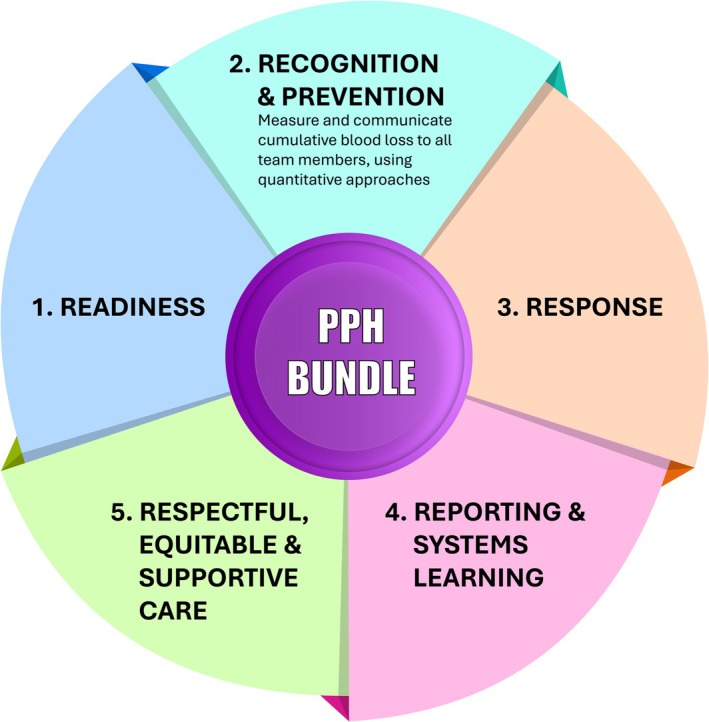
PPH intervention bundle. The organization of activities aimed at improving PPH care includes five established domains, ranging from the preparedness of healthcare systems and professionals (Readiness), through Prevention and early Recognition of the condition, to an optimal, evidence‐based Response. Following the management of the emergency, activities related to Reporting and Systems learning support continuous quality improvement for future cases. The integration of Respectful, equitable, and supportive care policies is essential to ensure a humanized approach to care. One key component of the Recognition domain is the objective quantitative and cumulative measurement of blood loss during the first 24 h postpartum, along with clear communication of the results to the entire care team. Abbreviation: PPH, postpartum hemorrhage.
Training and continuous updating of the team is essential for implementation. Simulation training for PPH, including virtual simulation, significantly improves competency of health service providers by improving knowledge, skill, and attitude, resulting in improvement of clinical performance.
Enhancing team spirit and team response times in a safe and controlled environment for healthcare providers to practice identification and management of PPH helps develop confidence in handling life‐threatening emergencies. It is a cost‐effective way to provide high‐quality training to a large number of healthcare providers, compared to traditional methods, and can be used to continuously update the existing and new workforce. Virtual obstetric simulation training is widely considered to be feasible, acceptable, and effective. 46 , 47 , 48
Debriefing after a PPH is a crucial process for learning, performance improvement, and reducing potential psychological trauma of the staff team. It involves a structured discussion immediately after an event, to analyze what went well, and what could be improved, which helps improve management of future cases. It is also used as a checklist during emergencies, including near‐miss cases. 49 , 50
Although the evidence‐based technical knowledge required to manage a case of PPH has been well‐documented in the scientific literature for many years, the real challenge lies in adapting this knowledge to diverse settings, ensuring provider adherence, and sustaining its application over time, despite considerable challenges. Nearly 10 years ago, the implementation of bundles in regional hospital systems and individual hospitals reported reductions in severe maternal morbidity among patients who experienced PPH after bundle implementation. 25 , 51 , 52 More recently, the application of a similar intervention bundle in Africa confirmed its effectiveness in reducing maternal deaths due to PPH. 25
The PPH emergency response using the bundle approach has clinical components. These are the first response bundle (uterine message, oxytocic, intravenous fluid, and tranexamic acid along with supportive measure) and refractory bundle (compressions, intrauterine tamponade, anti‐shock garments, necessary surgery, blood/blood product transfusion, and proper referral). Non‐clinical components include facility readiness, network integration, teamwork and communication, respectful maternity care, data, monitoring, quality improvement, and leadership. Professional member societies can play a significant role in implementation, advocacy, and scale‐up. 53
It is impossible to determine whether these improvements are due to specific practices within the bundle or to the implementation of the bundle as a whole; however, early identification of PPH patients is very likely to be a key factor in improving outcomes, particularly when implementing other interventions requires additional time.
5.2. Clinical impact of blood loss quantification
In May 2023, the findings of the E‐MOTIVE trial were published. 25 This was a cluster randomized trial on early detection and a PPH treatment care bundle involving 80 hospitals. The intervention consisted of a calibrated drape for early PPH detection, followed by a first‐response PPH treatment bundle. The intervention also included the use of implementation strategies (audit and feedback, health worker training, PPH trolley, carry case, and change champions). The control group received postpartum blood loss collection in uncalibrated drapes (without alert or action lines) to quantify blood loss for the study, and their PPH treatment interventions were administered according to usual care practices.
In the intervention arm, early detection of PPH included the following: a calibrated blood collection drape was used to collect all postpartum blood and to prompt health workers to treat PPH when excessive blood loss occurred. The drape was applied for 1 h for all women after vaginal birth. For women with excessive bleeding, the drape remained in place for an additional 2 h from the point of excessive bleeding detection. Health workers received training on the use of a blood loss monitoring chart to document blood volume loss, blood flow, and uterine tone every 15 min in the first hour postpartum. Blood pressure and pulse were checked and documented at least once in the first hour postpartum.
Postpartum hemorrhage was detected in 93.1% of patients in the intervention group compared to 51.1% in the usual‐care group (rate ratio = 1.58; 95% confidence interval [CI] = 1.41–1.76), and the treatment bundle was used in 91.2% and 19.4%, respectively (rate ratio = 4.94; 95% CI = 3.88–6.28). The study concluded that early detection of PPH and the use of a bundled treatment approach led to a lower risk of severe PPH, laparotomy for bleeding, or death from bleeding than usual care among patients having a vaginal birth.
The E‐MOTIVE trial illustrates how the organized use of strategies, previously described as effective in PPH treatment, can improve clinical outcomes in resource‐limited settings. Interventions such as uterine compression, administration of uterotonic medications, and intravenous fluids are well known to all healthcare professionals involved in childbirth care. Moreover, this study crucially highlights the importance of immediate detection of abnormal bleeding (in this case, using a calibrated drape) as the trigger for these interventions.
5.3. The role of scientific societies and academic groups
Adherence to the recommendation for objective blood loss measurement also depends on non‐behavioral interventions, 54 which were emphasized in the E‐MOTIVE trial. 25
In all these successful experiences incorporating objective quantification of blood loss, collaboration between hospitals and/or academic groups played a critical role. Hospitals that began using blood quantification devices received training in these processes and mid‐term follow‐up from a central support group. The identification and empowerment of local champions, who sustained initial momentum and mobilized their teams over time, was a determining factor.
One model of implementation can be seen in the “Leadership Development Initiative: Removing barriers to Effective Access and Coverage of maternal Healthcare (LDI:REACH),” a FIGO‐delivered program that facilitates leadership development and quality improvement in clinical practice, aiming to effect change in healthcare systems. Its long‐term goal is to improve maternal and newborn health outcomes through increased coverage of key clinical interventions, rapidly implemented at scale, through the leadership of National Member Societies, all conducted through the lens of gender diversity and equity.
FIGO's member societies are critical to improving maternal and newborn health outcomes within their respective countries. They actively participate in national legislative policy guideline development, providing technical assistance, capacity building, educating communities, and conducting implementation research.
Many member societies not only draw on obstetricians and gynecologists but also include key health professionals, such as midwives and general practitioners. One of the four clinical areas FIGO LDI:REACH has focused on, is E‐MOTIVE for PPH. 55
It is not always easy to change ingrained behaviors among healthcare workers. Medium‐ to long‐term interventions, such as reminders, local leadership promotion, positive feedback, and indicator measurement, are essential. 25 , 54 , 56 Surgical checklists have been shown to promote the implementation of safe practices. 57 One example of a checklist to promote implementation of blood loss measurement during third stage care is the mnemonic “BOND”: Baby skin‐to‐skin; Oxytocin; iNitiate blood loss monitoring; and Delay cord clamping. 58
Routine objective quantification of blood loss during and after childbirth represents a structural change in obstetric care, addressing the challenges, including manufacturing and standardization costs, supply, transportation, and training on blood collection device usage. One study examined the cost of introducing quantitative postpartum blood loss assessment compared to visual estimation, considering the direct costs of resources such as the calibrated drape, transportation, staffing, additional training, periodic refresher training, equipment, supervision, and monitoring, 59 concluding that the measure is associated with significant savings of resources.
5.4. Barriers to implementation
Concerns have been raised regarding the environmental impact of plastic drapes, as well as the safe disposal of these materials in areas with a high prevalence of human immunodeficiency virus. These concerns are conflicting: discarding the collection device after a single use and avoiding washing and reuse reduces the bio‐sanitary risk, whereas reusing it decreases the environmental impact. In response, cheaper, reusable, environmentally friendly, 31 and easier‐to‐use options (Figure 3a) have been developed, as well as “in‐hospital” handmade alternatives 30 (Figure 2) that can help mitigate cost‐related challenges. The concern of environmental hazards can be mitigated by biodegradable transparent materials made from jute and corn flour. 30 It is the responsibility of professional societies and government organizations to facilitate access to these devices in resource‐limited settings.
Interestingly, according to one report, the most significant barrier to adopting objective blood loss quantification as a management standard for all births is the resistance to change among healthcare professionals. 60
There is now clear and sufficient evidence to strongly recommend this practice 25 and to incorporate it into the educational content provided to all healthcare professionals involved in maternity care. It is essential to recognize that the widespread adoption of evidence‐based interventions can take more than a decade. 61 The decision to accelerate the implementation of objective blood loss quantification in all births will require the use of innovative, validated strategies that serve as change accelerators. Behavioral interventions and sustained inter‐institutional collaboration at all levels have been associated with protocol adherence and reduced maternal mortality due to PPH.
Recently, the incorporation of virtual communication and mentorship programs, where resource‐limited hospitals are supported by more experienced institutions, has demonstrated positive outcomes in improving obstetric emergency management. 62
Although a single specific strategy for objective postpartum blood loss quantification has not yet been defined, the minimum requirements for an accessible measurement device in resource‐limited settings (where most PPH‐associated morbidity occurs) have been outlined (Videos S1–, S3), highlighting that this strategy must be low cost, easy to use and interpret in different scenarios, and ideally environmentally friendly. 63
For now, each hospital must define which strategy (e.g. graduated drape, non‐graduated drape, or reusable plastic container) is most applicable to its local context, facilitating easy, sustainable implementation and long‐term adherence to this early PPH detection strategy.
We hope that the graphic content of this article will be helpful for the implementation of objective blood loss measurement methods during vaginal and cesarean births.
6. CONCLUSION
There is an urgent need to implement objective quantification of postpartum blood loss as a standard practice in maternity care. Early detection, combined with timely therapeutic interventions, is critical to reducing the global burden of PPH and associated maternal morbidity and mortality. Clinical trials, such as E‐MOTIVE, have demonstrated the effectiveness of bundled approaches that include calibrated blood loss measurement, provider training, and structured response protocols.
To ensure sustainability and widespread adoption, these practices must be formally integrated into training curricula for all levels of maternal healthcare providers, incorporated into institutional guidelines, and supported by national health policies. The shift from visual estimation to objective quantification represents a fundamental change in clinical culture and will require coordinated behavioral, structural, and policy‐level interventions to overcome resistance, address resource constraints, and adapt tools to diverse care environments.
Global implementation must prioritize context‐appropriate strategies, including the use of reusable or biodegradable drapes, virtual simulation training, and inter‐institutional support networks. Only through a systemic and collaborative approach can we achieve lasting improvements in the timely recognition and effective treatment of postpartum hemorrhage worldwide.
AUTHOR CONTRIBUTIONS
Article conceptualization: FB, AJN‐C, DS. Literature review or critical analysis of the topic: FB, AJN‐C, DS. Manuscript writing—original draft: AJN‐C. Critical review and editing of the manuscript: FB, AJN‐C, DS, JH, JP‐J, AB, MAS, JMB‐L, JB‐K, AEU, AW. Viewing or preparing graphs/figures: FB, AJN‐C, DS. All authors read and approved the final manuscript.
FUNDING STATEMENT
No funding was received.
CONFLICT OF INTEREST STATEMENT
The authors have no conflicts of interest to declare.
Supporting information
ACKNOWLEDGMENTS
We thank Fabian Cabrera, graphic designer and professor of the Universidad del Valle, Cali, Colombia, for developing the graphics. We also thank Cristian Nieto, graphic designer, for preparing the videos that accompany this article. We extend our sincere thanks to the Centro de Simulacion y Entrenamiento Continuo (CESEC), the Clinical Research Center of the Fundacion Valle de Lili, and the Obstetrical and Gynecological Society of Bangladesh (OGSB) for their support in the development of the graphic material accompanying this paper. We especially thank our patients who consented to the recording of surgeries.
Begum F, Nieto‐Calvache AJ, Schlembach D, et al. FIGO recommendations on objective measurement of blood loss after birth for early detection of postpartum hemorrhage. Int J Gynecol Obstet. 2025;171:933‐950. doi: 10.1002/ijgo.70523
Ferdousi Begum and Albaro J. Nieto‐Calvache are joint first authors.
DATA AVAILABILITY STATEMENT
Data sharing is not applicable to this article as no new data were created or analyzed in this study.
REFERENCES
Associated Data
Supplementary Materials
Data Availability Statement
Data sharing is not applicable to this article as no new data were created or analyzed in this study.
References
- Say L, Chou D, Gemmill A, et al. Global causes of maternal death: a WHO systematic analysis. Lancet Glob Health. 2014;2(6):e323‐e333.
- Berg CJ, Harper MA, Atkinson SM, et al. Preventability of pregnancy‐related deaths: results of a state‐wide review. Obstet Gynecol. 2005;106:1228‐1234.
- Seacrist MJ, VanOtterloo LR, Morton CH, Main EK. Quality improvement opportunities identified through case review of pregnancy‐related deaths from obstetric hemorrhage. J Obstet Gynecol Neonatal Nurs. 2019;48:288‐299.
- World Health Organization . The prevention and management of postpartum haemorrhage. Report of a Technical Working Group, Geneva, 3–6 July 1989. Geneva: World Health Organization 1990.
- Pahlavan P, Nezhat C, Nezhat C. Hemorrhage in obstetrics and gynecology. Curr Opin Obstet Gynecol. 2001;13(4):419‐424.
- Patel A, Goudar SS, Geller SE, et al. Drape estimation vs. visual assessment for estimating postpartum hemorrhage. Int J Gynaecol Obstet. 2006;93:220‐224.
- Natrella M, Di Naro E, Loverro M, et al. The more you lose the more you miss: accuracy of postpartum blood loss visual estimation. A systematic review of the literature. J Matern Fetal Neonatal Med. 2018;31:106‐115.
- Rath WH. Postpartum hemorrhage‐‐update on problems of definitions and diagnosis. Acta Obstet Gynecol Scand. 2011;90(5):421‐428.
- Howard TF, Grobman WA. The relationship between timing of postpartum hemorrhage interventions and adverse outcomes. Am J Obstet Gynecol. 2015;213(2):239.e1‐239.e3.
- World Health Organization . WHO recommendations on the assessment of postpartum blood loss and use of a treatment bundle for postpartum haemorrhage [Internet]. Geneva: WHO; Accessed May 7, 2025. https://www.who.int/publications/i/item/9789240080931 (ISBN 978‐92‐4‐008539‐8 (electronic version). 2023;ix)
- Pacagnella RC, Souza JP, Durocher J, et al. A systematic review of the relationship between blood loss and clinical signs. PLoS One. 2013;8(3):e57594.
- Menard MK, Main EK, Currigan SM. Executive summary of the reVITALize initiative: standardizing obstetric data definitions. Obstet Gynecol. 2014;124(1):150‐153.
- Lertbunnaphong T, Lapthanapat N, Leetheeragul J, Hakularb P, Ownon A. Postpartum blood loss: visual estimation versus objective quantification with a novel birthing drape. Singapore Med J. 2016;57(6):325‐328.
- Pingray V, Williams CR, Al‐Beity FMA, et al. Strategies for optimising early detection and obstetric first response management of postpartum haemorrhage at caesarean birth: a modified Delphi‐based international expert consensus. BMJ Open. 2024;14(5):e079713. doi: 10.1136/bmjopen-2023-079713
- Gallos I, Devall A, Martin J, et al. Randomized trial of early detection and treatment of postpartum hemorrhage. N Engl J Med. 2023;389(1):11‐21. doi: 10.1056/NEJMoa2303966
- Combs CA, Murphy EL, Laros RK Jr. Factors associated with postpartum hemorrhage with vaginal birth. Obstet Gynecol. 1991;77:69‐76.
- Committee on Practice Bulletins‐Obstetrics . Practice bulletin No. 183: postpartum hemorrhage. Obstet Gynecol. 2017;130(4):e168‐e186. doi: 10.1097/AOG.0000000000002351
- WOMAN Trial Collaborators . Effect of early tranexamic acid administration on mortality, hysterectomy, and other morbidities in women with post‐partum haemorrhage (WOMAN): an international, randomised, double‐blind, placebo‐controlled trial. Lancet. 2017;389(10084):2105‐2116.
- Main EK, Goffman D, Scavone BM, et al. National Partnership for maternal safety: consensus bundle on obstetric hemorrhage. Obstet Gynecol. 2015;126:155‐162.
- Doctorvaladan SV, Jelks AT, Hsieh EW, Thurer RL, Zakowski MI, Lagrew DC. Accuracy of blood loss measurement during cesarean delivery. AJP Rep. 2017;7(2):e93‐e100.
- Bose P, Regan F, Paterson‐Brown S. Improving the accuracy of estimated blood loss at obstetric haemorrhage using clinical reconstructions. BJOG. 2006;113(8):919‐924.
- Diaz V, Abalos E, Carroli G. Methods for blood loss estimation after vaginal birth. Cochrane Database Syst Rev. 2018;9(9):CD010980.
- American College of Obstetricians and Gynecologists , Committee on Obstetric Practice . Quantitative blood loss in obstetric hemorrhage: ACOG Committee opinion summary, number 794. Obstet Gynecol. 2019;134(6):1368‐1369.
- Main EK. Reduction of severe maternal morbidity from hemorrhage using a state perinatal quality collaborative. Am J Obstet Gynecol. 2017;216(3):298.e1‐298.e11.
- Gallos ID, Papadopoulou A, Tsiami A, et al. Randomized trial of early detection and treatment of postpartum hemorrhage. N Engl J Med. 2023;389(12):1124‐1133.
- Yunas I, Gallos ID, Devall AJ, et al. Tests for diagnosis of postpartum haemorrhage at vaginal birth. Cochrane Database Syst Rev. 2025;1(1):CD016134. doi: 10.1002/14651858.CD016134
- Zea‐Prado F, Espino‐Y‐Sosa JS, Sarria‐Ortiz D, Muñoz‐Córdoba L, Nieto‐Calvache AJ. Postpartum bleeding visual underestimation: a persistent problem. Int J Gynaecol Obstet. 2024;164(2):803‐804. doi: 10.1002/ijgo.15155
- Toledo P, Eosakul ST, Goetz K, Wong CA, Grobman WA. Decay in blood loss estimation skills after web‐based didactic training. Simul Healthc. 2012;7(1):18‐21. doi: 10.1097/SIH.0b013e318230604f
- World Health Organization . Web Annex A. Methods of assessing postpartum blood loss for the detection of postpartum haemorrhage: evidence‐to‐decision framework. In: WHO recommendations on the assessment of postpartum blood loss and use of a treatment bundle for postpartum haemorrhage. Geneva: WHO; 2023. Accessed Apr 11, 2025. https://iris.who.int/
- Begum F. Innovation of calibrated dk drape for quantitative measurement of blood loss during and immediately after vaginal delivery. Bangladesh J Obstet Gynaecol. 2024;39(2):65‐73.
- Maternova Inc . MaternaWell—Comprehensive Supplement for Pregnancy. Maternova; Accessed Apr 11, 2025. https://maternova.net/es/products/maternawell
- Two postpartum blood collection devices: the Brass‐V drape and MaternaWell tray—as experienced by birth attendants and birthing women—a questionnaire‐based randomised study. Obstet Gynecol Int. 2024;2024:6605833. doi: 10.1155/2024/6605833
- bij de Vaate A, Coleman R, Manneh H, Walraven G. Knowledge, attitudes and practices of trained traditional birth attendants in The Gambia in the prevention, recognition and management of postpartum haemorrhage. Midwifery. 2002;18(1):3‐11. doi: 10.1054/midw.2001.0289
- Medical Aid Films . How to manage bleeding after birth (no care within 4 hours). 2014. Accessed Apr 11, 2025. https://www.youtube.com/watch?v=xU7da4SO5Gs
- Prata N, Mbaruku G, Campbell M. Using the kanga to measure postpartum blood loss. Int J Gynaecol Obstet. 2005;89(1):49‐50. doi: 10.1016/j.ijgo.2005.01.019
- Wilcox LL, Ramprasad C, Gutierrez A, Oden M, Richards‐Kortum R, Gandhi M. Accuracy in estimation of blood loss using the SAPHE (signaling a postpartum hemorrhage emergency) mat. Obstet Gynecol. 2015;125(Suppl 1):25S. doi: 10.1097/AOG.0000000000000753
- Larsson C, Saltvedt S, Wiklund I, Pahlen S, Andolf E. Estimation of blood loss after cesarean section and vaginal delivery has low validity with a tendency to exaggeration. Acta Obstet Gynecol Scand. 2006;85(12):1448‐1452. doi: 10.1080/00016340600985032
- Quinlivan WL, Brock JA. Blood volume changes and blood loss associated with labor. I. Correlation of changes in blood volume measured by I‐131 albumin and Evans blue dye, with measured blood loss. Am J Obstet Gynecol. 1970;106(6):843‐849. doi: 10.1016/0002-9378(70)90477-1
- Imai K, Kotani T, Tsuda H, Nakano T, Hirakawa A, Kikkawa F. A novel approach to detecting postpartum hemorrhage using contrast‐enhanced ultrasound. Ultrasound Med Biol. 2017;43(3):615‐620. doi: 10.1016/j.ultrasmedbio.2016.11.008
- OR Manager . New technology tracks blood loss, reduces transfusions. OR Manager; Accessed Apr 11, 2025. https://www.ormanager.com/ai‐series/new‐technology‐tracks‐blood‐loss‐reduces‐transfusions/
- Gabel KT, Weeber TA. Measuring and communicating blood loss during obstetric hemorrhage. J Obstet Gynecol Neonatal Nurs. 2012;41(4):551‐558. doi: 10.1111/j.1552-6909.2012.01375.x
- Cole MF. A modified early obstetric warning system. Br J Midwifery. 2014;22(12):862‐868. doi: 10.12968/bjom.2014.22.12.862
- Escobar MF, Nassar AH, Theron G, et al. FIGO recommendations on the management of postpartum hemorrhage 2022. Int J Gynaecol Obstet. 2022;157(Suppl 1):3‐50. doi: 10.1002/ijgo.14184
- Oglak SC, Obut M, Tahaoglu AE, Demirel NU, Kahveci B, Bagli I. A prospective cohort study of shock index as a reliable marker to predict the patient's need for blood transfusion due to postpartum hemorrhage. Pak J Med Sci. 2021;37(3):863‐868. doi: 10.12669/pjms.37.3.3875
- Safer Births. About us. SaferBirths.org; Accessed Apr 11, 2025. https://saferbirth.org/about‐us/
- Nelissen E, Ersdal H, Mduma E, et al. Clinical performance and patient outcome after simulation‐based training in prevention and management of postpartum haemorrhage: an educational intervention study in a low‐resource setting. BMC Pregnancy Childbirth. 2017;17:301. doi: 10.1186/s12884-017-1481-7
- Minor KC, Bianco K, Mayo JA, et al. Virtual simulation training for postpartum hemorrhage in low‐to‐moderate‐volume hospitals in the US. AJOG Glob Rep. 2024;4:100357.
- Almutairi WM, Almutaraiy SM, Al‐Zahrani A, et al. Transforming postpartum care: the efficacy of simulation training in hemorrhage management among nurses. Health. 2025;13(5):549. doi: 10.3390/healthcare13050549
- Joseph NT, Worrell NH, Collins J, et al. Implementation of a post partum hemorrhage safety bundle at an urban safety‐net hospital. Am J Perinatol Rep. 2020;10:e255‐e261.
- Corbett N, Hurko P, Vallee TJ. Debriefing as a strategic tool for performance improvement. JOGNN. 2012;41(2):572‐579.
- Shields LE, Wiesner S, Fulton J, Pelletreau B. Comprehensive maternal hemorrhage protocols reduce the use of blood products and improve patient safety. Am J Obstet Gynecol. 2015;212:272‐280. doi: 10.1016/j.ajog.2014.08.004
- Einerson BD, Miller ES, Grobman WA. Does a postpartum hemorrhage patient safety program result in sustained changes in management and outcomes? Am J Obstet Gynecol. 2015;212:140‐144.e1. doi: 10.1016/j.ajog.2014.09.014
- Begum F, Haththotuwa R, Thapa K, Chowdhury S, Dewan F. Engaging National Professional Society to combat postpartum hemorrhage using bundle approach—a FIGO‐SAFOG‐OGSB‐MGH project: experience of implementation in Bangladesh. Bangladesh J Obstet Gynaecol. 2022;37(2):98‐108.
- Althabe F, Buekens P, Bergel E, et al. A behavioral intervention to improve obstetrical care. N Engl J Med. 2008;358(18):1929‐1940. doi: 10.1056/NEJMsa071456
- FIGO . Leadership development initiative: removing barriers to effective access and coverage of maternal healthcare (LDI:REACH). https://www.figo.org/what‐we‐do/figo‐programmes/leadership‐development‐initiative‐removing‐barriers‐effective‐access‐and
- García‐Elorrio E, Aleman A, Cafferata ML, et al. A multifaceted intervention to increase prophylactic oxytocin use during the third stage of labor and to reduce routine episiotomies in Nicaragua. Int J Gynaecol Obstet. 2014;127(1):31‐34. doi: 10.1016/j.ijgo.2014.04.019
- Eco S, Petcka NL, Li K, Hechenbleikner EM. Quality improvement in rural and low‐resource settings. Am Surg. 2025;91(5):723‐731. doi: 10.1177/00031348251331296
- Rubgega F, Nkuba MN, Hanson S, et al. Rapid response, E‐MOTIVE and suction tamponade for postpartum haemorrhage (ARREST PPH): a quality improvement project. Presented at: 42nd Conference on Priorities in Perinatal Care in Southern Africa; 2024 Mar 12–15; Goudini Spa, South Africa.
- Katz D, Wang R, O'Neil L, et al. The association between the introduction of quantitative assessment of postpartum blood loss and institutional changes in clinical practice: an observational study. Int J Obstet Anesth. 2020;42:4‐10. doi: 10.1016/j.ijoa.2019.05.006
- Duzyj CM, Boyle C, Mahoney K, Johnson AR, Ogot G, Ayers C. The postpartum hemorrhage patient safety bundle implementation at a single institution: successes, failures, and lessons learned. Am J Perinatol. 2021;38(12):1281‐1288. doi: 10.1055/s-0040-1712452
- Rubin R. It takes an average of 17 years for evidence to change practice—the burgeoning field of implementation science seeks to speed things up. JAMA. 2023;329(Apr 25):1333‐1336. doi: 10.1001/jama.2023.4387
- Escobar MF, Echavarría MP, Carvajal JA, et al. Hospital padrino: a collaborative strategy model to tackle maternal mortality: a mixed methods study in a middle‐income region. The Lancet Regional Health. 2024;31:100705. doi: 10.1016/j.lana.2024.100705
- Concept Foundation . Postpartum Blood Loss Measurement for Vaginal Births: Target Product Profile. 2024. Accessed Apr 11, 2025. https://www.conceptfoundation.org/wp‐content/uploads/2024/11/TPP‐Postpartum‐Blood‐loss‐Measurement‐for‐Vaginal‐Births_AIM‐Final.pdf
Conditions covered
Other guidelines on this topic
Reproduced for free educational use under the licence shown above. WhiteCoat Pro is not affiliated with or endorsed by the authors, the publisher or the issuing organisation. Always check the original document for the current version.