ECG criteria
- Right axis deviation: electrical axis angle typically > +120°; some criteria use > +90° or ≥ +100°, but the diagnosis can only be made with typical QRS morphology and the exclusion of other causes of right axis deviation
- rS-type QRS in leads I and aVL (small r wave, deep S wave)
- qR-type QRS in III and aVF, frequently also in II; R wave in III is usually larger than in II
- Intrinsicoid deflection time (R wave peak) in aVF ≥ 45 ms
- QRS duration is usually < 120 ms; with QRS ≥ 120 ms, a classic diagnosis of isolated LPFB is less likely — one should look for concomitant bundle branch block or another cause of QRS widening and describe the identified conduction abnormality in full
- Other causes of right axis deviation are excluded: right ventricular hypertrophy/strain, lateral myocardial infarction, vertical heart position in young and asthenic patients, pulmonary embolism, dextrocardia, limb lead misplacement
What it looks like on ECG
The posterior fascicle of the left bundle branch is thicker and shorter than the anterior fascicle and typically receives a dual blood supply from the anterior interventricular and posterior descending coronary arteries (anatomic variations are possible; the posterior descending artery may arise from the right or circumflex artery), which is why isolated posterior fascicular block is less common than anterior fascicular block.
Key changes:
| Leads | QRS morphology | What to look for |
|---|---|---|
| I, aVL | rS | small r, deep S |
| III, aVF | qR | small q wave, tall R wave, frequently R in III > R in II |
| II | frequently qR | may be less prominent than in III/aVF |
| Precordial Leads | typically no specific changes | evaluated to assess for concomitant bundle branch block, hypertrophy, and other etiologies |
The electrical axis is deviated to the right, typically beyond +120°; some criteria set the threshold at > +90° or ≥ +100°, but an isolated axis deviation in the +90° to +120° range often reflects a normal vertical heart axis and does not support the diagnosis on its own. QRS duration is generally normal (< 120 ms), unlike complete bundle branch block, because activation still reaches the myocardium via the anterior fascicle and Purkinje network, albeit with a delay and an altered vector direction. When QRS duration is ≥ 120 ms, a classic diagnosis of isolated LPFB is less likely — one must search for a coexisting bundle branch block or another cause of QRS widening. The intrinsicoid deflection time in lead aVF is prolonged (≥ 45 ms), reflecting delayed propagation of excitation along the posteroinferior wall of the left ventricle.
The ST segment and T wave are typically unremarkable unless concomitant ischemia or hypertrophy is present.
Real ECGs: 8 examples
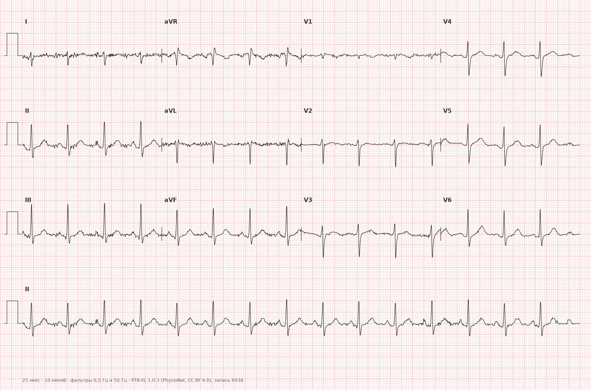
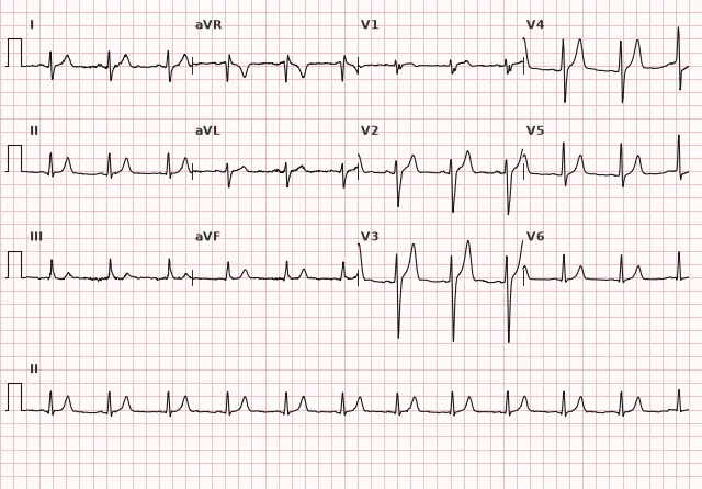
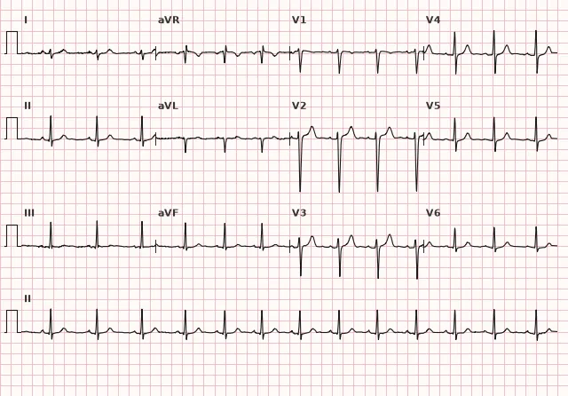
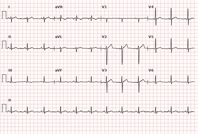
ECG from the open PTB-XL dataset (Wagner P. et al., Scientific Data, 2020; PhysioNet), CC BY 4.0 license. Rendering by WhiteCoat Pro.
Mechanism
The left posterior fascicle conducts excitation to the posteroinferior and inferior walls of the left ventricle. When it is blocked, initial depolarization proceeds via the anterior fascicle: the initial vector is directed superiorly and to the left, which forms an initial q wave in the inferior leads (II, III, aVF) and a small r wave in the lateral leads (I, aVL). Subsequently, excitation reaches the posteroinferior wall by an alternative pathway through the myocardium, and late activation of this region creates a vector directed to the right and inferiorly — hence the tall terminal R wave in III/aVF and the deep terminal S wave in I/aVL. The net QRS vector shifts to the right, producing right axis deviation.
Causes
- Coronary artery disease and myocardial infarction, particularly extensive or complicated by other conduction disturbances; both inferior and anterior infarctions are possible
- Degenerative conduction system fibrosis (Lenègre-Lev disease)
- Cardiomyopathies
- Myocarditis
- Aortic valve calcification and cardiac fibrous skeleton calcification
- Cardiac surgery
- Isolated LPFB in structurally normal hearts is rare, unlike anterior fascicular block
Differential diagnosis
- Right ventricular hypertrophy/strain — RVH may present with R>S or a tall R wave in V1, right axis deviation, ST-T changes in V1–V3, and signs of right atrial overload; the diagnosis is established based on the overall ECG and clinical findings (pulmonary pathology)
- Lateral myocardial infarction — pathological Q wave in leads I and aVL combined with ST-T changes, evolutionary changes on serial ECGs
- Vertical electrical axis in young, thin individuals — the axis angle typically does not exceed +90° to +120°, and the QRS complex lacks the characteristic qR pattern in III/aVF with prolonged intrinsicoid deflection time
- Pulmonary embolism — sinus tachycardia, S1Q3T3 pattern, right bundle branch block, and signs of right ventricular strain may be present, but the ECG is nonspecific; the diagnosis is established based on clinical probability and imaging rather than a single ECG pattern
- Dextrocardia — negative P and QRS waves in lead I, reverse R-wave progression across the precordial leads
- Limb lead misplacement — evaluate P and QRS waves in leads I, II, aVR, aVL and their mutual concordance; normally, the QRS complex in aVR is negative, but this is not an absolute rule and serves only as a general landmark. When in doubt, repeat the ECG recording after verifying lead placement
Clinical significance and management
Specific treatment for the fascicular block itself is generally not required; however, isolated LPFB is uncommon and necessitates evaluation for structural heart disease, myocardial ischemia, and other conduction abnormalities. Prognosis is determined by the underlying condition rather than the fascicular block per se.
The physician should:
- search for concomitant pathology: coronary artery disease, cardiomyopathy, arterial hypertension with target organ damage
- assess for coexisting right bundle branch block — this combination constitutes a bifascicular block with a risk of progression to complete AV block, which requires closer monitoring and, in the presence of syncopal episodes, further workup (up to electrophysiological testing and consideration of cardiac pacing)
- if newly developed in the setting of acute coronary syndrome, interpret as a sign of extensive conduction system involvement and manage according to ACS protocols
- for an incidental, asymptomatic finding — clinical evaluation, assessment for structural heart disease (including echocardiography when indicated), and ECG follow-up
Common interpretation errors
- Diagnosing LPFB without ruling out right ventricular hypertrophy and lateral myocardial infarction — a mandatory step in differential diagnosis
- Mistaking a normal vertical electrical axis in young patients for LPFB without analyzing QRS morphology and intrinsicoid deflection time
- Failing to check correct limb lead placement before interpreting right axis deviation
- Missing coexisting right bundle branch block (bifascicular block) — crucial for prognosis
- Assuming an isolated finding is entirely benign without clinical evaluation and assessment for underlying structural heart disease
1. Which QRS pattern in leads III and aVF is characteristic of left posterior fascicular block?
An initial small q wave and a tall terminal R wave (qR) in leads III and aVF (frequently also in II) reflect a late activation vector directed toward the posteroinferior wall of the left ventricle.
2. Which condition must be ruled out before diagnosing LPFB in the presence of right axis deviation?
Right axis deviation can be caused by right ventricular hypertrophy, lateral myocardial infarction, or a vertical heart position—these causes must be ruled out before diagnosing left posterior fascicular block.
3. What is the clinical significance of combined left posterior fascicular block and right bundle branch block?
Involvement of the right bundle branch and the left posterior fasciculus leaves only the left anterior fasciculus functional—such a bifascicular block increases the risk of complete AV block.
Frequently asked questions
How to distinguish left posterior fascicular block from right ventricular hypertrophy?
Right ventricular hypertrophy may present with R>S or a tall R wave in V1, right axis deviation, changes in V1–V3, and signs of right heart strain; the diagnosis is established based on a combination of ECG and clinical data. In left posterior fascicular block, precordial leads are typically unchanged, and right axis deviation is explained by the QRS morphology in I, II, III, aVF and prolonged intrinsicoid deflection time in aVF.
Is left posterior fascicular block dangerous?
Specific treatment for the block itself is usually not required, but isolated left posterior fascicular block is rare and warrants clinical evaluation to search for structural heart disease, ischemia, or other conduction disturbances—the prognosis is determined by the underlying condition. The risk increases when combined with right bundle branch block (bifascicular block) or when it occurs acutely in the setting of myocardial infarction, which requires closer monitoring and ruling out the progression to complete AV block.
Why is left posterior fascicular block less common than left anterior fascicular block?
The left posterior fasciculus is thicker than the anterior fasciculus and typically has a dual blood supply from the anterior interventricular and posterior descending coronary artery basins, making it more resistant to ischemic and degenerative injury. Anatomical variations in blood supply are possible.
Is additional workup necessary for an incidental finding of left posterior fascicular block?
An asymptomatic incidental finding requires clinical evaluation and the exclusion of structural heart disease (echocardiography when indicated), followed by follow-up monitoring. If syncope, dyspnea, or concomitant blocks are present, the diagnostic workup should be expanded.
Can left posterior fascicular block be diagnosed in the presence of a widened QRS?
Classic isolated posterior fascicular block typically yields a QRS duration < 120 ms. With QRS ≥ 120 ms, this diagnosis is less likely—one should search for a concomitant block of another fasciculus or bundle branch, or another cause of QRS widening (nonspecific intraventricular conduction delay, hyperkalemia, drug effect, pre-excitation, ventricular rhythm, etc.) and describe the identified abnormality in full.
ECG Course
Interpretation from basics to myocardial infarction — learn to read any ECG using a systematic algorithm.
- Video lessons and theory for every topic
- Simulator case studies under every lesson
- Unlimited ECG simulator practice
- Study guide: "All ECGs in 25 minutes"
ECG Strip Simulator
ECG, radiology, histology, pathomorphology, pharmacology — thousands of flashcards with detailed explanations.
- Real ECG strips, not textbook drawings
- Instant answers and explanations
- Streaks and progress tracking in your dashboard
Related terms
More in the section "Bundle Branch and Fascicular Blocks"
Sources
- Goldberger's Clinical Electrocardiography, 2021
- Surawicz B., Knilans T. Chou's Electrocardiography in Clinical Practice, 2008
- Orlov V.N. Guide to Electrocardiography
- AHA/ACCF/HRS Recommendations for the Standardization of ECG, 2009
Educational material for medical students and physicians. Does not replace clinical guidelines and physical examination; the ECG must always be interpreted in conjunction with the clinical picture.