ECG criteria
- QRS duration of 110–119 ms according to the 2009 AHA/ACCF/HRS criteria for adults is greater than normal (typically < 110 ms) but less than the criterion for complete RBBB (≥ 120 ms).
- In lead V1 (less commonly V2), an rSr', rsr', or similar morphology complex with a terminal R' wave; the amplitude of R' is not an independent criterion.
- In leads I and V6, a terminal S wave.
- The intrinsicoid deflection time in V1 (from QRS onset to the peak of R') may be slightly prolonged, but by itself does not serve as a criterion to differentiate incomplete from complete block; the defining parameters are QRS duration and morphology.
- ST–T changes in iRBBB may be absent or less pronounced than in complete RBBB; secondary discordant changes are possible, especially in V1–V3. Pronounced or primary ST–T changes require separate evaluation.
- The rhythm is sinus in the majority of cases; the conduction disturbance is limited solely to QRS morphology.
What it looks like on ECG
The main changes are visible in the right precordial leads V1–V2 and in leads with a leftward vector orientation (I, aVL, V5–V6).
| Lead | Typical picture |
|---|---|
| V1–V2 | rSr', rsr', or similar morphology — the initial r usually reflects early septal activation, a small S wave, followed by a terminal R' wave (late right ventricular activation); the height of R' is not a diagnostic criterion. |
| I, aVL | normal or slightly widened terminal S wave |
| V5–V6 | the terminal S wave is present as a supporting morphological sign |
According to the 2009 AHA/ACCF/HRS criteria in adults, the QRS in incomplete RBBB is 110–119 ms with typical morphology. A QRS ≥ 120 ms with corresponding morphology (rsR'/rSR' in V1–V2 and a broad S in I and V6) corresponds to complete RBBB; QRS duration alone does not confirm the diagnosis of complete block. In other classifications, as well as in children and adolescents, lower QRS thresholds down to 100 ms may be used for incomplete RBBB. With a QRS of 100–109 ms, it is more correct to describe the morphology in detail (e.g., rsr' in V1) and take into account age and clinical context without making an automatic diagnosis. The electrical axis of the heart is usually not deviated. ST–T changes in V1–V2 in iRBBB are usually absent or minimal, unlike complete block, where discordant ST depression and a negative T wave are more common.
Real ECGs: 8 examples
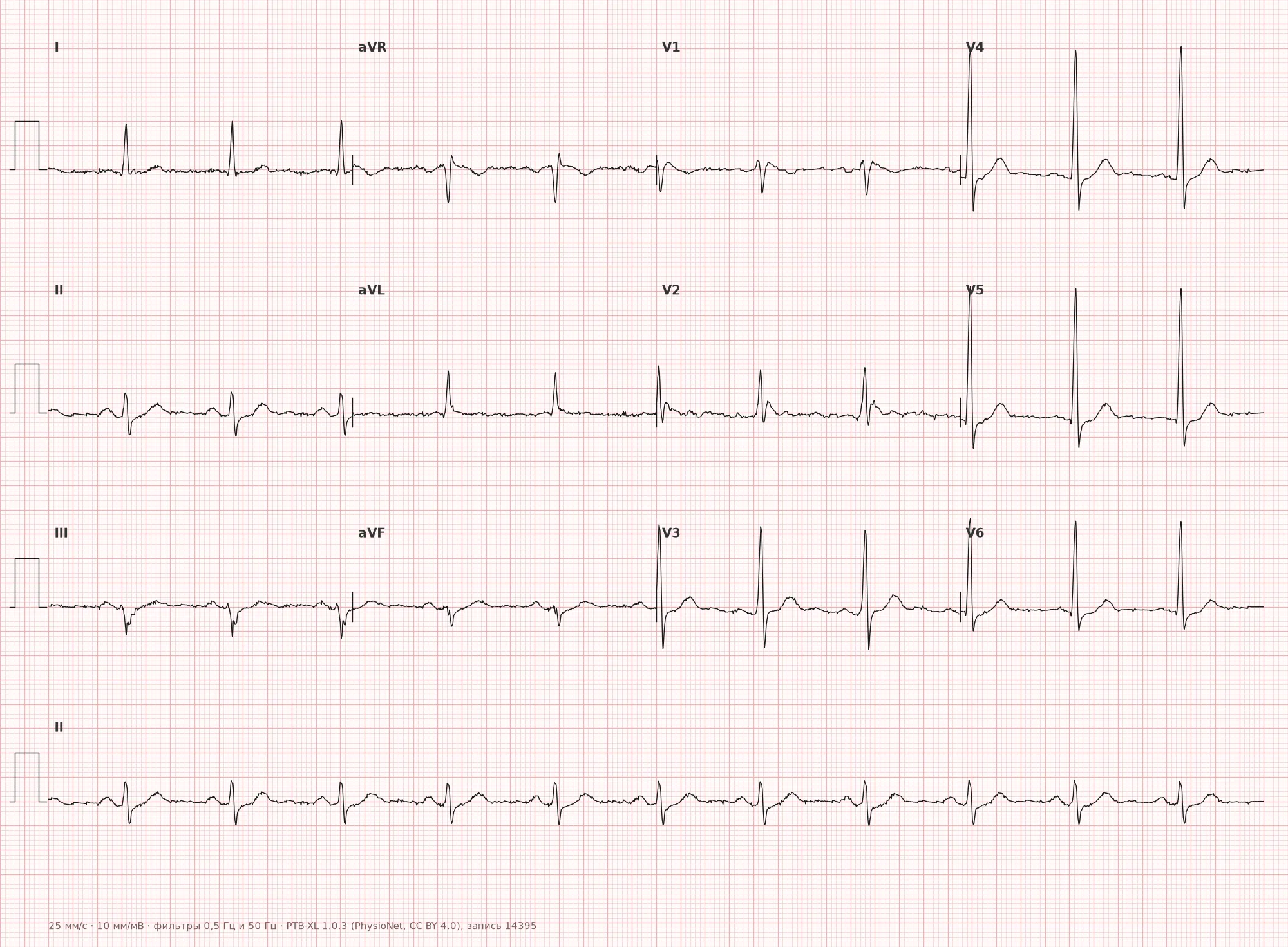
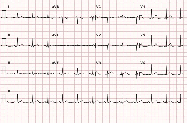
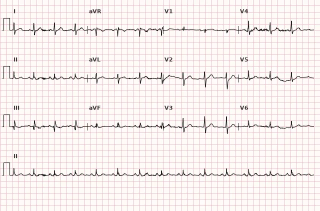
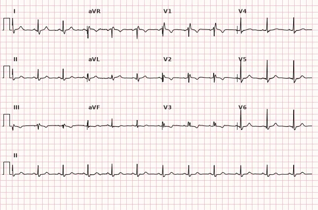
ECG from the open PTB-XL dataset (Wagner P. et al., Scientific Data, 2020; PhysioNet), CC BY 4.0 license. Rendering by WhiteCoat Pro.
Mechanism
The right bundle branch receives its primary blood supply from the septal branches of the left anterior descending artery, although anastomoses and an additional contribution from the right coronary artery are possible—the blood supply to the His–Purkinje system is variable. Damage to the conduction system, its age-related remodeling, or right heart overload can slow conduction through the right bundle branch; the mechanism of iRBBB should not be linked strictly to the presence of a single blood supply source. In iRBBB, conduction is delayed but not completely interrupted: the right ventricle depolarizes later than the left, and the impulse still travels through the specialized conduction system, albeit with a delay. This produces a terminal R' wave in V1—reflecting delayed activation of the right ventricle—but without the severe delay and pronounced QRS widening characteristic of complete block.
Causes
- A normal variant, especially in young individuals, athletes, and asthenic body types (cardiac rotation/stretching, high diaphragm position).
- Right ventricular volume overload: atrial septal defect, tricuspid or pulmonary regurgitation.
- Pulmonary hypertension, chronic cor pulmonale, COPD.
- Congenital heart disease with right-sided overload.
- Initial/mild form of degenerative conduction system disease (Lenègre–Lev disease).
- Sometimes as an incidental finding without structural heart disease.
Differential diagnosis
- Complete RBBB — characterized by a QRS duration ≥ 120 ms and, as a rule, a more prominent, wide terminal R' with discordant ST-T changes in V1–V2.
- Brugada syndrome, type 1 pattern — distinguished by a coved-type ST elevation ≥ 2 mm in ≥ 1 right precordial lead (standard V1–V2 or high positions in the 2nd–4th intercostal spaces) followed by a negative T wave, rather than an isolated rSr' without significant ST changes.
- Right ventricular hypertrophy — distinguished by signs of overload: tall R wave in V1, right axis deviation, confirmed by echocardiography.
- Normal variant of incomplete r' in healthy individuals — benign rSr' is typically accompanied by a QRS < 110 ms and the absence of other ECG abnormalities; with a QRS of 110–119 ms and typical morphology in adults, incomplete RBBB should be considered, and definitive differentiation sometimes requires clinical context and serial tracings.
Clinical significance and management
In most individuals without structural heart disease, iRBBB is a benign finding that does not require further workup in the absence of symptoms and other ECG abnormalities. When iRBBB is newly detected, the physician should review the medical history (congenital heart disease, pulmonary pathology), assess for signs of right heart overload (right axis deviation, right atrial enlargement), and, if structural heart disease is suspected, refer the patient for echocardiography. Isolated iRBBB is not considered an indication for specific antiarrhythmic therapy or device implantation; management is determined by the underlying condition, if present.
Common interpretation errors
- Confusing it with complete RBBB without measuring the actual QRS duration in ms.
- Interpreting any rSr' in V1 as pathology without considering that it is a frequent normal variant.
- Failing to check for signs of concomitant right ventricular hypertrophy or overload when clinically indicated.
- Mistaking improper placement of the V1–V2 electrodes (too high) for iRBBB.
- Confusing it with the Brugada pattern due to insufficient attention to the ST segment morphology.
1. What QRS complex duration is typical for incomplete right bundle branch block in adults according to the 2009 AHA/ACCF/HRS criteria?
110–119 ms is an intermediate value between normal (< 110 ms) and the complete block criterion (≥ 120 ms).
2. In which lead is the main diagnostic sign of iRBBB sought?
Lead V1 (less commonly V2) shows the characteristic rSr' or rsr' complex, reflecting delayed depolarization of the right ventricle.
3. What distinguishes incomplete RBBB from the Brugada type 1 pattern in V1?
The Brugada type 1 pattern features a specific coved ST elevation ≥ 2 mm followed by a negative T wave, which is absent in isolated iRBBB.
Frequently asked questions
Is incomplete right bundle branch block dangerous?
In most cases, iRBBB is not dangerous on its own and is often a normal variant, especially in young people and athletes. Its clinical significance depends on whether there is concomitant structural heart or lung disease.
How to distinguish incomplete from complete RBBB?
The main criterion is QRS duration: in incomplete block in adults, it is usually 110–119 ms, while in complete block, it is 120 ms or more with corresponding morphology. The shape of the complex in V1 (rSr', rsr') is similar, but in complete block, the secondary R' is usually wider, and discordant ST-T changes are more common.
Is additional workup necessary for incomplete RBBB?
For an isolated finding without complaints or other ECG changes, additional workup is usually not required. If congenital heart disease, pulmonary hypertension, or other structural pathology is suspected, echocardiography is indicated.
Can incomplete RBBB occur in a healthy person?
Yes, this is one of the most common normal variants on the ECG, especially in young asthenic individuals and athletes, related to the anatomical position of the heart and chest.
Does incomplete bundle branch block progress to complete block?
Isolated incomplete RBBB in a healthy individual typically does not progress. The development of complete block over time warrants an investigation for a new structural or degenerative conduction system disease (e.g., progressive cor pulmonale) rather than being viewed as a natural evolution of an isolated finding.
ECG Course
Interpretation from basics to myocardial infarction — learn to read any ECG using a systematic algorithm.
- Video lessons and theory for every topic
- Simulator case studies under every lesson
- Unlimited ECG simulator practice
- Study guide: "All ECGs in 25 minutes"
ECG Strip Simulator
ECG, radiology, histology, pathomorphology, pharmacology — thousands of flashcards with detailed explanations.
- Real ECG strips, not textbook drawings
- Instant answers and explanations
- Streaks and progress tracking in your dashboard
Related terms
More in the section "Bundle Branch and Fascicular Blocks"
Sources
- Goldberger's Clinical Electrocardiography, latest edition
- Surawicz B., Knilans T. Chou's Electrocardiography in Clinical Practice
- AHA/ACCF/HRS Recommendations for the Standardization of ECG, 2009
- Orlov V.N. Guide to Electrocardiography
Educational material for medical students and physicians. Does not replace clinical guidelines and physical examination; the ECG must always be interpreted in conjunction with the clinical picture.