ECG criteria
- QRS duration of 110–119 ms according to AHA/ACCF/HRS 2009 for adults (at 25 mm/s, this is 2.75–3 mm); some classifications set the lower limit starting from 100 ms — source discrepancy
- In V1 (less commonly V2), QS or rS predominates, a small r and deep broad S are possible; morphology is variable and does not confirm the diagnosis on its own
- In I, aVL, V5–V6 — broad, often notched or slurred R wave without a Q wave; the finding may not be fully expressed
- Intrinsicoid deflection time in V5–V6 is moderately prolonged; a value ≥ 60 ms is typical for complete LBBB, but in incomplete block this parameter is variable and is not an exclusion criterion
- Absence of a Q wave in I, aVL, V5–V6 (septal activation is altered as in complete block, but this feature is not mandatory in all cases)
- ST segment and T wave may be discordant with the QRS complex, the degree of expression is variable and not guaranteed to be milder than in complete block
- The electrical axis is usually normal, with a possible moderate leftward deviation
- The diagnosis is established based on a combination of QRS duration and morphology, rather than a single isolated feature
What it looks like on ECG
Incomplete LBBB occupies the intermediate zone between normal intraventricular conduction and complete left bundle branch block (LBBB). It is not a strictly defined single pattern, but rather an electrocardiographic category where the key parameter is QRS duration. According to AHA/ACCF/HRS 2009 guidelines for adults, incomplete left bundle branch block is defined by a QRS duration of 110–119 ms and corresponding morphology. Certain clinical classifications cite a broader range starting from 100 ms — this discrepancy arises from different sources, and when analyzing a specific ECG, it is important to clarify which criterion is applied. With a QRS duration of 110–119 ms, features of left ventricular/left bundle activation may be present, but they do not have to be a "diminished copy" of complete LBBB — the morphology is often incomplete and variable, and the diagnosis requires evaluating the features in combination rather than relying on a single element.
In lead V1 (sometimes V2), a QS or rS pattern is most commonly observed: a small or absent initial r wave, and a deep, relatively wide S wave. The morphology in V1 is variable on its own and does not confirm the diagnosis — the decisive factor remains the combination of QRS duration with typical left ventricular morphology in leads I, aVL, V5–V6. In the left lateral precordial leads V5–V6 and limb leads I, aVL, the R wave is often broad, with a notch or slurring on the peak or descending limb — a consequence of delayed activation of the left ventricular free wall, although this feature may be weakly expressed or absent.
The Q wave in I, aVL, V5–V6 is usually absent because the interventricular septum activates from right to left, similar to complete block, though this is not an absolute mandatory finding in the incomplete form. The intrinsicoid deflection time (the interval from the onset of QRS to the peak of the R wave) in V5–V6 often exceeds 60 ms in complete LBBB — this is one of the morphological signs of complete block, but in the incomplete form, this parameter is variable and does not have to remain below this threshold, so it should not be used as a mandatory criterion for incomplete LBBB.
| Parameter | Normal | Incomplete LBBB | Complete LBBB |
|---|---|---|---|
| QRS | < 110 ms | 110–119 ms (according to AHA/ACCF/HRS 2009; in some classifications, starting from 100 ms) | ≥ 120 ms |
| V1 | rS or QS | QS or rS, morphology is variable | broad QS or rS with notching |
| V5–V6, I, aVL | q, R, s | broad R, notching possible | broad, notched R, slurring |
| Q wave in I, aVL, V5–V6 | may be present | usually absent | absent |
The ST segment and T wave may be discordant with the QRS complex (ST depression and T-wave inversion in leads with a dominant R wave); the severity of these changes is variable and not always milder than in overt block.
Real ECGs: 8 examples
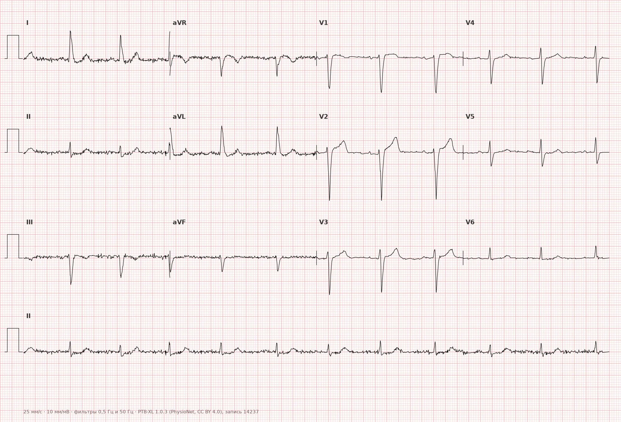
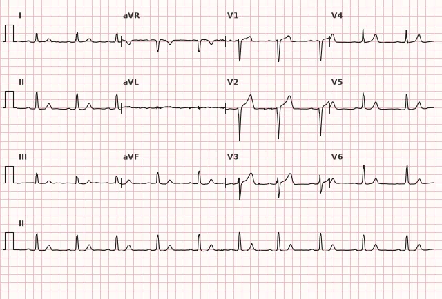
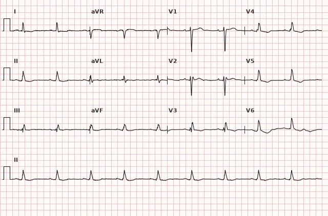
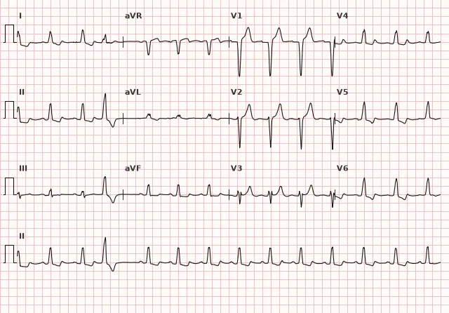
ECG from the open PTB-XL dataset (Wagner P. et al., Scientific Data, 2020; PhysioNet), CC BY 4.0 license. Rendering by WhiteCoat Pro.
Mechanism
Normally, the ventricles are activated via the complex His-Purkinje fiber system, and both walls depolarize almost synchronously. Incomplete left bundle branch block is a category representing delayed or partially impaired conduction through the left bundle branch; it does not have a single mandatory mechanism. Possible mechanisms include slowed conduction through the main trunk or branches of the left bundle, block of a portion of the fibers (fascicular level) without complete interruption of conduction, and partial recruitment of the right-sided His-Purkinje system with transseptal spread of the wave to the left ventricle. In different cases, the contribution of these mechanisms varies. The net effect is a moderate prolongation of the QRS and a shift in the direction of the resultant depolarization vector, creating a complex morphology that partially resembles complete block, but without the complete loss of conduction through the bundle branch. The degree and nature of the conduction disturbance determine whether the QRS remains within the 110–119 ms range (incomplete block) or reaches 120 ms or more with full morphology (complete block).
Causes
- Isolated finding without apparent structural heart disease (more common in the elderly, but requires evaluation rather than automatic dismissal as normal)
- Early stage of degenerative fibrosis of the conduction system (Lenègre-Lev disease)
- Coronary artery disease
- Arterial hypertension with left ventricular hypertrophy
- Cardiomyopathies (dilated, hypertrophic)
- Aortic valve disease
- Myocarditis
- Dynamic or intermittent conduction block morphology in conduction system disease, including in the setting of progressive left bundle branch pathology
Differential diagnosis
- Complete LBBB — QRS duration ≥ 120 ms combined with typical morphology: broad/notched R in I, aVL, V5–V6, absence of normal q waves in I, V5–V6, predominantly negative QRS in V1–V3; a QRS duration of ≥ 120 ms alone is insufficient for the diagnosis of complete block, as such a complex may correspond to a nonspecific intraventricular delay, hyperkalemia, ventricular rhythm, and other conditions — the specific combined morphology is required
- Left ventricular hypertrophy — QRS duration alone does not reliably differentiate LVH from incomplete LBBB (in LVH, the QRS can reach 110–119 ms and be associated with intraventricular delay); the diagnosis of LVH relies on a combination of voltage criteria (Sokolow-Lyon index, Cornell criteria) and repolarization abnormalities rather than the width of the complex
- Nonspecific intraventricular conduction delay — QRS duration is increased, but the morphology does not match either the left or right bundle branch pattern
- Left anterior fascicular block — changes primarily affect the electrical axis (marked leftward deviation) and QRS morphology in the limb leads, while the precordial leads appear different
- Normal QRS variant in young individuals — with a QRS width of about 100 ms and without typical morphology in V1 and V5–V6, this represents a borderline normal variant rather than a block
Clinical significance and management
Isolated incomplete LBBB in an individual without signs of organic heart disease may be a benign finding, but it should not be routinely labeled as a normal variant, especially in the elderly: such morphology can reflect structural heart disease, ischemia, cardiomyopathy, or conduction system disease. The role of the functional diagnostics physician is not to make a diagnosis based on a single tracing, but to evaluate the finding in context: patient age, history of coronary artery disease, arterial hypertension, heart failure, and previous ECGs.
In newly detected incomplete LBBB, it is advisable to compare it with archival ECGs (if available). For newly detected or unclear incomplete LBBB, echocardiography should reasonably be considered, especially in the presence of symptoms, risk factors, physical examination abnormalities, or concomitant ECG changes; for a long-standing, stable, isolated finding in an asymptomatic patient, the necessity of the test is determined by the clinical context rather than performed automatically.
Persistent progression of the QRS duration to 120 ms or more on repeat ECGs may indicate progressive conduction system disease, but reversible and rate-dependent causes of QRS widening must first be excluded—such as rate-related aberration, drug effects, electrolyte disturbances, and measurement variability. QRS widening alone does not prove the progression of structural heart disease. The urgency of response is determined by symptoms, hemodynamics, and suspicion of acute coronary syndrome, rather than merely crossing the 120 ms QRS threshold; a new bundle branch block morphology in the setting of ischemic symptoms warrants urgent evaluation regardless of whether the QRS has reached 120 ms.
If acute coronary syndrome is suspected, prompt clinical evaluation, serial ECGs, and high-sensitivity troponin are required. Sgarbossa criteria and their modified version (Smith) were historically developed and validated for assessing ischemia in complete LBBB and paced rhythms; their validated thresholds primarily apply to wide QRS complexes, and applying them to incomplete block requires caution. Block morphology can obscure ST-T interpretation, so diagnosing ischemia in the setting of incomplete LBBB should not rely on a mechanical application of general ST criteria—decisions must be based on clinical presentation, ECG dynamics, troponin, and imaging when necessary.
Common interpretation errors
- Diagnosing complete left bundle branch block based solely on a QRS duration ≥ 120 ms without checking for typical morphology in V1–V3 and I, aVL, V5–V6
- Ignoring the finding as "insignificant" without assessing the clinical context, age, symptoms, and changes compared to prior ECGs
- Confusing it with left ventricular hypertrophy without checking voltage and repolarization criteria
- Mechanically applying Sgarbossa or Smith criteria to incomplete block when assessing ischemia without considering clinical presentation and serial changes
- Treating any QRS widening up to 120 ms on a repeat ECG as definitive progression of conduction system disease without ruling out rate-dependent aberration, medications, and electrolyte disturbances
- Failing to account for the fact that QRS morphology in V1–V2 during incomplete block may be uninformative if chest electrodes are misplaced
1. Which QRS duration range corresponds to incomplete left bundle branch block in adults according to the 2009 AHA/ACCF/HRS recommendations?
According to the 2009 AHA/ACCF/HRS criteria for adults, a QRS duration of 110–119 ms distinguishes incomplete block from normal (< 110 ms) and from complete block (120 ms and greater).
2. Which QRS morphology in lead V1 is most common in incomplete LBBB?
In incomplete left bundle branch block, lead V1 most commonly shows a QS or rS pattern, similar to complete block, though morphology is variable and does not confirm the diagnosis on its own.
3. Is a QRS duration ≥ 120 ms sufficient to diagnose complete left bundle branch block?
A QRS duration ≥ 120 ms can occur in nonspecific intraventricular conduction delay, hyperkalemia, ventricular rhythms, and other conditions; complete LBBB requires a combination of typical morphological features.
Frequently asked questions
How does incomplete left bundle branch block differ from complete LBBB?
The main criterion is QRS duration: 110–119 ms for incomplete block versus 120 ms or more for complete block. However, duration alone is insufficient—typical morphology is also required: broad/notched R waves in I, aVL, V5–V6, absence of normal q waves in these leads, and predominantly negative QRS in V1–V3. In complete block, notches and slurring are more prominent, and the intrinsicoid deflection time in V5–V6 frequently exceeds 60 ms.
Is incomplete left bundle branch block dangerous?
The finding alone does not determine prognosis, but it requires evaluation of the clinical context and exclusion of structural heart disease, especially when the block is newly detected. Its significance depends on the underlying cause—if coronary artery disease, hypertrophy, or cardiomyopathy is responsible, the prognosis is driven by the primary condition.
Is further workup necessary when incomplete LBBB is detected?
For a newly detected or unclear finding, echocardiography is reasonable to consider, particularly in the presence of symptoms, cardiovascular risk factors, physical examination abnormalities, or concomitant ECG changes. For a long-standing, stable, isolated finding in an asymptomatic individual without risk factors, the necessity of the test is determined by the clinical context, and serial monitoring may be appropriate.
Can incomplete block progress to complete block?
Yes, this can occur with progressive conduction system disease, such as Lenègre–Lev disease or advancing coronary artery disease. Progression does not occur in everyone and may take years; when assessing serial changes, it is important to rule out reversible causes of QRS widening, such as rate-dependent aberration, medications, or electrolyte abnormalities.
Can myocardial ischemia be evaluated in the presence of incomplete LBBB?
Evaluation is possible, but requires accounting for expected ST-T discordance, the clinical context, ECG evolution, and troponin levels, rather than mechanically applying general ST criteria as completely reliable. If the probability of acute coronary syndrome is high, prompt management is necessary regardless of ST interpretation in the presence of the block.
ECG Course
Interpretation from basics to myocardial infarction — learn to read any ECG using a systematic algorithm.
- Video lessons and theory for every topic
- Simulator case studies under every lesson
- Unlimited ECG simulator practice
- Study guide: "All ECGs in 25 minutes"
ECG Strip Simulator
ECG, radiology, histology, pathomorphology, pharmacology — thousands of flashcards with detailed explanations.
- Real ECG strips, not textbook drawings
- Instant answers and explanations
- Streaks and progress tracking in your dashboard
Related terms
More in the section "Bundle Branch and Fascicular Blocks"
Sources
- AHA/ACCF/HRS Recommendations for the Standardization and Interpretation of the Electrocardiogram, 2009
- Surawicz B., Knilans T. Chou's Electrocardiography in Clinical Practice
- Orlov V.N. Guide to Electrocardiography
Educational material for medical students and physicians. Does not replace clinical guidelines and physical examination; the ECG must always be interpreted in conjunction with the clinical picture.