ECG criteria
- Left axis deviation: classic criterion is approximately −45° or more to the left. An axis from −30° to −44° is borderline and does not confirm the diagnosis on its own; some guidelines use the −30° threshold, but it is less specific and strictly requires typical QRS morphology and the exclusion of other causes of left axis deviation
- qR complex in leads I and aVL
- rS complex in leads II, III, and aVF
- S wave in III deeper than S wave in II (S III > S II) — a supportive, but not mandatory, sign
- Intrinsicoid deflection time (time to peak R) in aVL >45 ms (in some sources — ≥45 ms)
- QRS duration < 120 ms (in isolated LAFB)
- Absence of other causes of left axis deviation (inferior myocardial infarction or scar, left ventricular hypertrophy, nonspecific intraventricular conduction delay, ventricular pacing, congenital variants, axis determination errors)
What it looks like on ECG
The basis of diagnosis is the analysis of limb leads. Leads I and aVL show a qR complex: a small q wave and a tall R wave. Leads II, III, aVF show an rS complex: a small r wave and a deep S wave; typically, S in III is deeper than in II, but this is a supportive sign rather than a mandatory requirement — its absence does not exclude the diagnosis.
The electrical axis is deviated to the left. The classic and most specific criterion is an axis of approximately −45° or more to the left combined with typical QRS morphology. The diagnosis is made based on a combination of findings: axis, qR morphology in I/aVL and rS in II/III/aVF, QRS width, and exclusion of other causes of left axis deviation — the axis is not assessed in isolation. An axis from −30° to −44° is borderline; it may occur in fascicular block, especially in the presence of concomitant left ventricular hypertrophy or intraventricular conduction delay, but does not confirm the diagnosis by itself.
The intrinsicoid deflection time (impulse travel time to the peak of R) in aVL is prolonged, typically >45 ms (in some sources, the threshold is ≥45 ms) — this reflects delayed activation of the anterolateral wall of the LV, which is normally activated via the anterior fascicle.
In isolated LAFB, the QRS is usually <120 ms because the posterior fascicle and right bundle branch are intact and the bulk of the ventricles depolarizes on time; wider complexes necessitate ruling out concomitant right bundle branch block or a nonspecific intraventricular conduction delay.
| Lead | Typical QRS morphology |
|---|---|
| I, aVL | qR |
| II, III, aVF | rS, frequently SIII > SII |
| V1–V6 | typically no specific changes; their assessment is needed to look for concomitant pathology |
The ST segment and T wave are typically unremarkable unless concomitant ischemia or hypertrophy is present.
Real ECGs: 8 examples
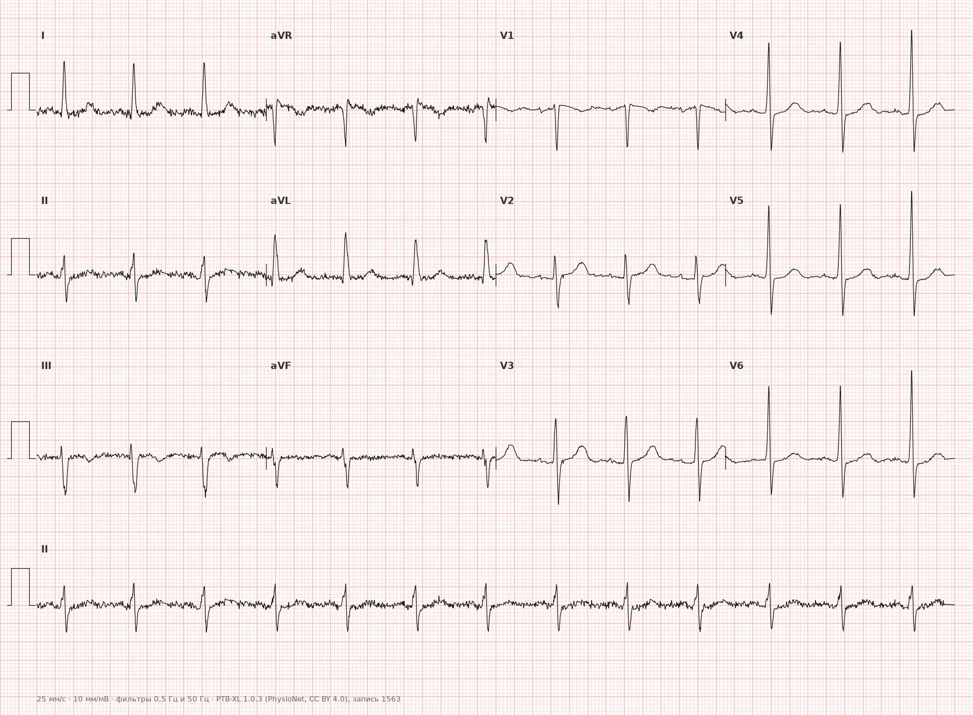
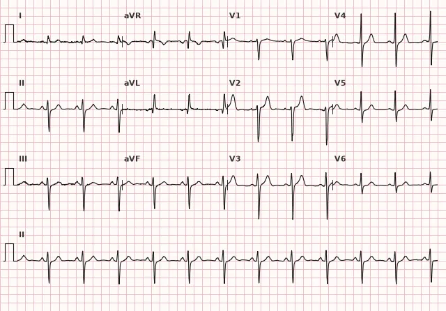
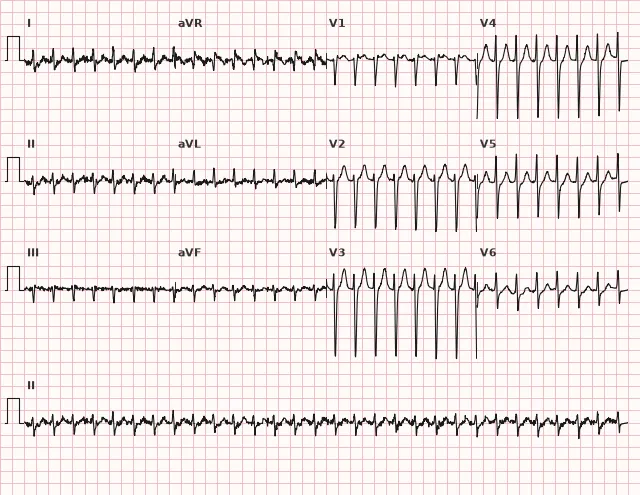
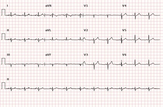
ECG from the open PTB-XL dataset (Wagner P. et al., Scientific Data, 2020; PhysioNet), CC BY 4.0 license. Rendering by WhiteCoat Pro.
Mechanism
The left bundle branch divides into anterior (anterosuperior) and posterior (posteroinferior) divisions. The anterior division is typically more vulnerable to ischemia and fibrosis due to its smaller diameter and less favorable, variable blood supply — branches of the left anterior descending and circumflex arteries are more frequently involved. The posterior division is thicker and more often has a dual blood supply from the right coronary and circumflex artery territories, making it less frequently damaged.
In anterior fascicular block, initial ventricular depolarization proceeds via the intact posterior fascicle — the vector is directed inferiorly and to the right (toward the inferior and posterior walls), producing initial q waves in I/aVL and r waves in II/III/aVF. Subsequently, the excitation wave spreads through the myocardium to the anterolateral wall, which is normally activated via the anterior fascicle — this terminal vector is directed leftward and superiorly, forming a tall R in I/aVL and a deep S in II/III/aVF. The net result is left axis deviation.
Causes
- Idiopathic conduction system fibrosis (Lenègre–Lev disease) — a frequent cause in elderly patients without structural heart disease
- Coronary artery disease, myocardial infarction (especially anterior/anteroseptal)
- Arterial hypertension with left ventricular hypertrophy
- Aortic valve calcification and fibrous skeleton calcification
- Cardiomyopathies
- Myocarditis
- Congenital heart defects, particularly atrioventricular septal defect (ostium primum), and status post cardiac surgery
- May occur as a normal variant in healthy individuals, especially in the elderly, in the absence of structural heart disease
Differential diagnosis
Inferior myocardial infarction (prior) — rS in inferior leads supports the diagnosis of LAFB but does not rule out a concomitant infarction: pathological Q/QS waves can coexist with fascicular block, and the resulting QRS morphology depends on the combination of the scar and the conduction defect. Diagnosis of infarction relies on Q-wave duration and amplitude, presence of QS waves, dynamic changes compared to previous ECGs, reciprocal ST-T changes, and clinical data (troponin in acute settings), rather than simply contrasting 'rS versus Q'.
Other causes of left axis deviation (left ventricular hypertrophy, nonspecific intraventricular conduction delay, ventricular pacing) — one must evaluate the concomitant QRS morphology, complex width, and clinical picture; isolated axis deviation without typical qR in aVL and rS in III does not justify an LAFB diagnosis.
Left posterior fascicular block — produces the opposite picture: right axis deviation, rS in I/aVL, qR in II/III/aVF.
Vertical or normal electrical axis with an S-type pattern — characterized by a lesser degree of deviation (does not reach −45°) and the absence of characteristic qR morphology in aVL.
Left ventricular hypertrophy — may coexist, but presents with its own voltage criteria (Sokolow–Lyon index, Cornell criteria), which are assessed separately.
Clinical significance and management
Isolated LAFB typically does not require treatment or pacing by itself, but necessitates clinical evaluation and a search for structural heart disease: assess history for CAD, hypertension, cardiomyopathies; in newly diagnosed block in a patient with chest pain, rule out acute coronary syndrome (ST-T analysis in conjunction with clinical presentation, troponin). The prognosis is determined by the underlying pathology and the presence of other conduction disorders; in observational studies, isolated LAFB is associated with an increased risk of subsequent atrial fibrillation, heart failure, and other cardiovascular events, so this finding should not be considered completely benign — it warrants follow-up rather than immediate interventions.
Clinical significance increases markedly when combined with right bundle branch block — this combination is considered a bifascicular block and indicates more extensive conduction system disease. Indications for pacing are determined by documented high-degree AV block or proven infra-Hissian conduction disturbance; in unexplained syncope with bifascicular block, systematic evaluation is indicated, often including an electrophysiological study, while empirical pacing without confirmed block is permissible only in specific guideline-defined scenarios.
LAFB can mask or mimic axis changes in inferior myocardial infarction and complicate the interpretation of LVH criteria — this must be taken into account during comprehensive tracing analysis.
Common interpretation errors
- Diagnosing based solely on borderline left axis deviation (from −30° to −44°) without typical qR/rS morphology and without excluding other causes
- Confusing the rS complex in lead III with a pathological Q wave in inferior infarction without assessing Q-wave duration, amplitude, and dynamics
- Treating S III > S II as a mandatory diagnostic requirement rather than a supportive sign
- Labeling PR interval prolongation in bifascicular block as 'trifascicular block' without proven involvement of the third fascicle
- Ignoring coexisting right bundle branch block, which alters the clinical significance of the finding
'Left-left-left': axis to the left, qR in I/aVL (vector to the left), rS in III (posterior fascicle activates first)
1. What is the primary ECG feature of left anterior fascicular block?
The most specific criterion is an axis near −45° or more to the left, but the diagnosis is made only in combination with typical qR/rS morphology and after excluding other causes of left axis deviation.
2. What QRS morphology is typical for lead III in LAFB?
In leads III, II, and aVF, an rS complex is formed; typically, the S wave in lead III is deeper than in lead II, but this is a supportive rather than a mandatory sign.
3. What is clinically significant when detecting LAFB combined with right bundle branch block?
The combination of LAFB and right bundle branch block constitutes a bifascicular block, which requires risk assessment for progression to complete AV block.
Frequently asked questions
Is Left Anterior Fascicular Block Dangerous?
Isolated LAFB in an individual without known structural heart disease typically does not require treatment or pacing per se. However, it is a finding that warrants investigating the underlying cause: it can be a marker of latent conduction system disease and has been associated in observational studies with a future risk of atrial fibrillation and heart failure.
How to Distinguish LAFB from Inferior Myocardial Infarction?
In LAFB, an rS complex is typically recorded in leads III and aVF, which supports the diagnosis but does not rule out a concurrent inferior myocardial infarction — a pathological Q/QS wave may coexist with the fascicular block. They are differentiated by Q-wave duration and amplitude, the presence of QS, dynamic changes compared to previous ECGs, reciprocal changes, and the clinical presentation.
Does the QRS Broaden in Left Anterior Fascicular Block?
No, the QRS duration in isolated LAFB generally remains within normal limits (< 120 ms) because the bulk of the ventricles depolarizes in a timely manner via intact conduction pathways. A wider complex indicates concurrent right bundle branch block or another conduction disturbance.
Is a Pacemaker Necessary for Left Anterior Fascicular Block?
LAFB itself is not an indication for cardiac pacing. Indications are determined in documented high-grade AV block or proven infra-hissian conduction disturbance; in unexplained syncope in the setting of bifascicular block, systematic evaluation and often electrophysiological study are indicated, whereas empiric pacing without confirmed block is reserved for specific situations outlined in guidelines.
What Electrical Axis Deviation is Characteristic of LAFB?
The classic and most specific criterion is an axis of approximately −45° or more to the left combined with typical QRS morphology. An axis from −30° to −44° is borderline and does not confirm the diagnosis on its own; some guidelines allow a threshold of −30° but designate it as less specific.
ECG Course
Interpretation from basics to myocardial infarction — learn to read any ECG using a systematic algorithm.
- Video lessons and theory for every topic
- Simulator case studies under every lesson
- Unlimited ECG simulator practice
- Study guide: "All ECGs in 25 minutes"
ECG Strip Simulator
ECG, radiology, histology, pathomorphology, pharmacology — thousands of flashcards with detailed explanations.
- Real ECG strips, not textbook drawings
- Instant answers and explanations
- Streaks and progress tracking in your dashboard
Related terms
More in the section "Bundle Branch and Fascicular Blocks"
Sources
- Goldberger's Clinical Electrocardiography, latest edition
- Surawicz B., Knilans T. Chou's Electrocardiography in Clinical Practice
- Orlov V.N. Guide to Electrocardiography
- AHA/ACCF/HRS Recommendations for the Standardization of ECG, 2009
Educational material for medical students and physicians. Does not replace clinical guidelines and physical examination; the ECG must always be interpreted in conjunction with the clinical picture.