ECG criteria
- QRS duration ≥ 120 ms for complete RBBB; 110–119 ms with appropriate morphology indicates incomplete RBBB
- In lead V1 (less commonly V2), a terminal positive notch: rSR', rsR', or a wide notched R
- In leads I and V6, a wide terminal notched S wave with a duration of > 40 ms or a duration greater than the R wave in that lead (terminal deviation to the right and anteriorly)
- Intrinsicoid deflection time in V1 > 50 ms
- Secondary ST-T changes: ST depression and/or T-wave inversion in V1–V3, directed oppositely to the terminal portion of the QRS
- Initial QRS activation, including septal activation, is usually preserved in isolated RBBB; conduction delay primarily affects the terminal part of the QRS — however, concomitant diseases and associated blocks may alter it, which should be evaluated separately rather than as an absolute rule
What it looks like on ECG
The key lead is V1 (sometimes V2 is more informative). The classic picture: a narrow initial r wave, a small S wave, followed by a wide notched R' wave — an rSR' or rsR' type complex. Variants include a wide notched monophasic R or an M-shaped complex. In leads I, aVL, and V6, there is a wide, often notched S wave lasting more than 40 ms (reflecting delayed right ventricular depolarization directed to the right and anteriorly at the end of the QRS).
| Lead | Typical pattern |
|---|---|
| V1–V2 | rSR', rsR', or wide notched R |
| I, aVL, V6 | wide notched S (>40 ms) |
| V1–V3 | sloping ST depression, inverted or biphasic T wave (discordant to the QRS direction) |
In isolated RBBB, initial interventricular septal depolarization is usually not significantly impaired; therefore, the Q wave in leads where it normally appears (II, III, aVF, V5–V6) retains its diagnostic value more often than in left bundle branch block. However, this is not an absolute rule: concomitant infarction, hypertrophy, scar, or associated conduction disorders can also alter the initial part of the QRS, so evaluation is always performed alongside clinical presentation and ECG dynamics, rather than based on a single Q wave. Isolated RBBB typically does not cause significant deviation of the electrical axis of the heart. Marked axis deviation in RBBB requires separate evaluation and may indicate concomitant left anterior or posterior hemiblock (bifascicular block), but is confirmed by separate fascicular block criteria, as axis deviation itself can be caused by ventricular hypertrophy, infarction, changes in cardiac position, and other factors.
Real ECGs: 7 examples
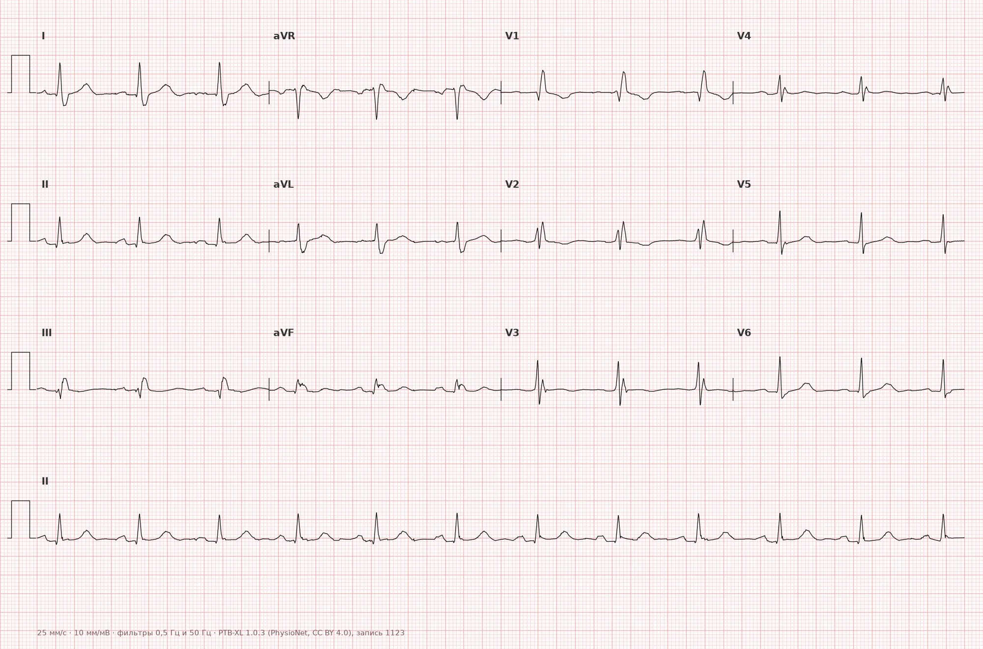
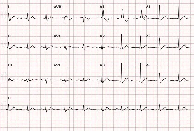
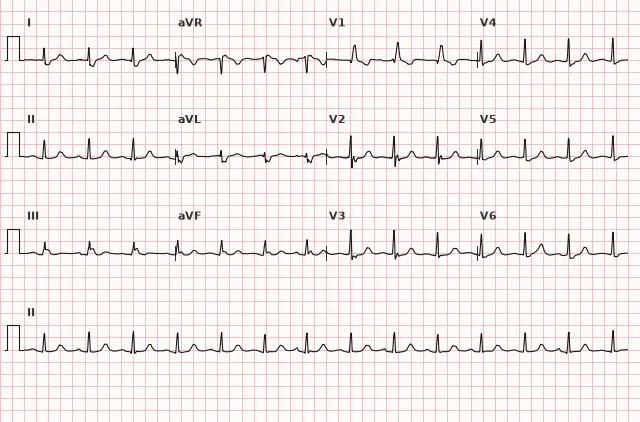
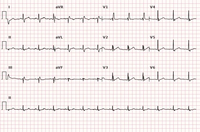
ECG from the open PTB-XL dataset (Wagner P. et al., Scientific Data, 2020; PhysioNet), CC BY 4.0 license. Rendering by WhiteCoat Pro.
Mechanism
The right bundle branch is thin and more susceptible to injury than the left; it is supplied predominantly by branches of the left anterior descending artery (LAD), though a variable contribution from branches of the right coronary artery is also possible. When it is blocked, the impulse travels via the left bundle branch to activate the interventricular septum and left ventricle normally and on time. The right ventricle depolarizes with a delay — not directly via the conduction system, but slowly, cell by cell, through the myocardium from the left ventricle. This prolongs the QRS and creates a late vector deviation to the right and anteriorly — resulting in the terminal R' in V1 and wide S in I, V6.
Causes
- Normal variant in healthy individuals (isolated incomplete or complete RBBB without structural heart disease)
- Congenital heart disease (atrial septal defect, Tetralogy of Fallot, post-surgical repair)
- Right ventricular overload and hypertrophy (COPD, cor pulmonale, pulmonary embolism)
- Coronary artery disease, myocardial infarction (especially anterior, involving the left anterior descending artery)
- Myocarditis, cardiomyopathies, cardiac sarcoidosis, arrhythmogenic right ventricular cardiomyopathy
- Age-related degenerative changes of the conduction system (Lenegre-Lev disease)
- Iatrogenic cause: right heart catheterization, cardiac surgery
- Hyperkalemia and certain intoxications (sodium channel blockers) can produce a similar morphology
Differential diagnosis
- Brugada type 1 pattern: coved ST-segment elevation ≥ 2 mm in V1–V2 (including high right precordial lead placement) transitioning into a T-wave inversion. Differentiation from RBBB is not based solely on the presence or absence of a wide S in I and V6 — the entire QRS-ST-T pattern, electrode placement, and clinical context must be evaluated; phenotypic overlap with RBBB is possible
- Left bundle branch block: deep wide S or QS in V1, wide monophasic R without a notch in I and V6; vector direction is opposite to RBBB
- Right ventricular hypertrophy: can produce a similar rSR' pattern in V1, but the QRS is usually not widened, and right axis deviation is present along with other signs of overload
- Dextrocardia and incorrect lead placement (V1–V2): can mimic a notched R, but verification of lead placement and correct leads alters the picture
- Myocardial infarction: in isolated RBBB, the initial part of the QRS is usually preserved, so pathological Q waves and ST changes in corresponding leads generally retain diagnostic value more often than in LBBB — however, secondary ST-T changes inherent to the block itself, especially in right precordial leads, can mask or mimic ischemia, so evaluation must be performed in conjunction with clinical findings, ECG dynamics, and troponins
Clinical significance and management
Isolated RBBB without structural heart disease is usually benign and does not require specific treatment. In newly diagnosed RBBB, the physician should search for the cause: assess for signs of right heart overload, coronary artery disease, pulmonary pathology, congenital defects — review medical history and perform echocardiography as indicated. RBBB combined with left anterior or posterior hemiblock constitutes a bifascicular block — this combination requires vigilance regarding the risk of progression to complete AV block, particularly in the presence of syncope. Isolated RBBB is not an independent criterion for STEMI; however, in the presence of ongoing ischemic symptoms, RBBB (new or previously known) is considered a high-risk feature: according to the 2003 ESC guidelines [Note: 2023 ESC guidelines context], this warrants urgent evaluation and, given the appropriate clinical picture, immediate coronary angiography/reperfusion strategy — on par with other occlusion equivalents. RBBB itself does not replace standard ST-segment and Q-wave evaluation. QRS duration and the presence of RBBB are taken into account when selecting patients for cardiac resynchronization therapy (they respond less favorably than patients with LBBB).
Common interpretation errors
- Mistaking a Brugada type 1 pattern for RBBB due to a similar appearance in V1
- Failing to measure the true QRS duration and confusing incomplete RBBB with complete RBBB
- Forgetting to check leads I and V6 — without a wide notched S, the diagnosis of RBBB is questionable
- Interpreting secondary ST depression and T-wave inversion in V1–V3 as ischemia without accounting for them as an expected consequence of the block itself
- Failing to notice concomitant axis deviation and omitting separate fascicular block criteria when bifascicular block is suspected
MaRRoW: Morphology rSR' — RBBB has an R' (rabbit ears) in V1 (right), LBBB has a wide S in V1
1. Which finding in lead V1 is characteristic of complete RBBB?
A notched terminal R' in V1 (rSR', rsR') is a classic sign of delayed right ventricular depolarization in RBBB.
2. What is the minimum QRS duration required to diagnose complete RBBB?
Complete RBBB is diagnosed when the QRS duration is ≥ 120 ms; a duration of 110–119 ms with appropriate morphology is termed incomplete block.
3. Why does the Q wave generally retain its diagnostic value for evaluating infarction in isolated RBBB, unlike in LBBB?
In isolated RBBB, primarily the terminal part of ventricular depolarization is blocked, while initial septal activation is usually preserved — however, the final interpretation should always be supplemented by clinical presentation and ECG dynamics.
Frequently asked questions
Is right bundle branch block dangerous?
Isolated RBBB in a person without heart disease is usually benign and does not affect prognosis. Danger arises when RBBB is combined with left bundle branch block (bifascicular block), occurs acutely in the setting of ACS, or is accompanied by structural heart disease — in such cases, the cause must be investigated and the risk of block progression assessed.
How to distinguish complete RBBB from incomplete?
The distinction is determined by the QRS duration in the presence of the corresponding RBBB morphology: a QRS duration ≥ 120 ms corresponds to complete block, 110–119 ms with a typical rSR'/rsR' in V1 and a wide terminal S in I and V6 corresponds to incomplete block. At the same time, an isolated rSr' in V1 without a wide S in I, V6 and without other criteria does not equal a diagnosis of incomplete RBBB and may be a non-specific normal variant.
Can myocardial infarction be diagnosed in the presence of RBBB?
RBBB generally distorts the initial part of QRS less than LBBB, therefore a pathological Q wave and ST-T changes in corresponding leads are evaluated, but not in isolation: secondary ST-T changes from the block itself can mask or mimic ischemia. The diagnosis is confirmed by clinical presentation, serial ECGs, and troponin levels, and if ACS is suspected, urgent invasive management is indicated.
How does RBBB differ from Brugada syndrome on ECG?
In Brugada syndrome type 1, a coved ST elevation ≥ 2 mm transitioning into a negative T wave is characteristic in V1–V2. The distinction from RBBB is not limited solely to the presence or absence of a wide notched S in I and V6 — the entire QRS-ST-T pattern, electrode placement, and clinical context must be evaluated, as phenotypic overlap is possible.
Does RBBB require treatment?
The block itself is not treated — the underlying condition is treated if identified. In asymptomatic isolated RBBB without structural heart disease, special monitoring is generally not required.
ECG Course
Interpretation from basics to myocardial infarction — learn to read any ECG using a systematic algorithm.
- Video lessons and theory for every topic
- Simulator case studies under every lesson
- Unlimited ECG simulator practice
- Study guide: "All ECGs in 25 minutes"
ECG Strip Simulator
ECG, radiology, histology, pathomorphology, pharmacology — thousands of flashcards with detailed explanations.
- Real ECG strips, not textbook drawings
- Instant answers and explanations
- Streaks and progress tracking in your dashboard
Related terms
More in the section "Bundle Branch and Fascicular Blocks"
Sources
- Goldberger's Clinical Electrocardiography, 9th ed.
- Surawicz B., Knilans T. Chou's Electrocardiography in Clinical Practice, 6th ed.
- AHA/ACCF/HRS Recommendations for the Standardization and Interpretation of the ECG, 2009
- Orlov V.N. Guide to Electrocardiography
Educational material for medical students and physicians. Does not replace clinical guidelines and physical examination; the ECG must always be interpreted in conjunction with the clinical picture.