ECG criteria
- QRS duration ≥120 ms with typical morphology (complete LBBB); 110–119 ms with the same morphology indicates incomplete LBBB. The rhythm can be sinus, atrial, atrial fibrillation, or another supraventricular rhythm — this is not a mandatory criterion
- In V1 (sometimes V2) — a QS or rS complex with a wide, deep S wave; the r wave is small or absent
- In I, aVL, V5–V6 — a wide, often notched or slurred R wave at the peak (M-shaped); septal q waves are typically absent in I and V5–V6, although isolated small q waves (especially in aVL) do not exclude LBBB
- The intrinsicoid deflection time in V5–V6 is prolonged, typically >60 ms
- Secondary discordant ST-T changes: the ST segment is directed oppositely to the dominant QRS wave (ST depression and inverted T wave with a dominant R wave, ST elevation and upright T wave with a dominant S wave); T wave polarity may vary, while marked concordance or excessive discordance is suspicious for acute ischemia
- The electrical axis is usually normal; marked leftward deviation may indicate concomitant left anterior hemiblock or structural myocardial disease
What it looks like on ECG
The key feature is QRS widening ≥120 ms for complete block (110–119 ms indicates incomplete LBBB). The morphology of the complex changes fundamentally:
| Lead | Typical morphology |
|---|---|
| V1–V2 | QS or rS, wide deep S wave, small or absent r wave |
| I, aVL, V5–V6 | wide R wave, often notched/slurred ("M"-shaped) |
| V3–V4 | transition zone shifted to the left, a small r/S wave may persist |
The q wave in I and V5–V6 is normally caused by septal activation from left to right; in LBBB, the septum is activated from right to left, which is why the septal q wave typically disappears in these leads — its appearance in a patient with LBBB is suspicious for a prior myocardial infarction. In aVL, morphology is more variable, and isolated small q waves do not rule out the diagnosis on their own.
The ST segment and T wave are normally discordant to the main QRS wave: where R dominates (I, aVL, V5–V6) — ST is usually depressed, T is inverted; where S dominates (V1–V3) — ST is elevated, T is upright. The shape and polarity of the T wave may vary; strict concordance in every lead is not required. These are secondary repolarization changes, not ischemia, but they are precisely what masks true STEMI in patients with suspected ACS and necessitate the use of Sgarbossa criteria.
Real ECGs: 8 examples
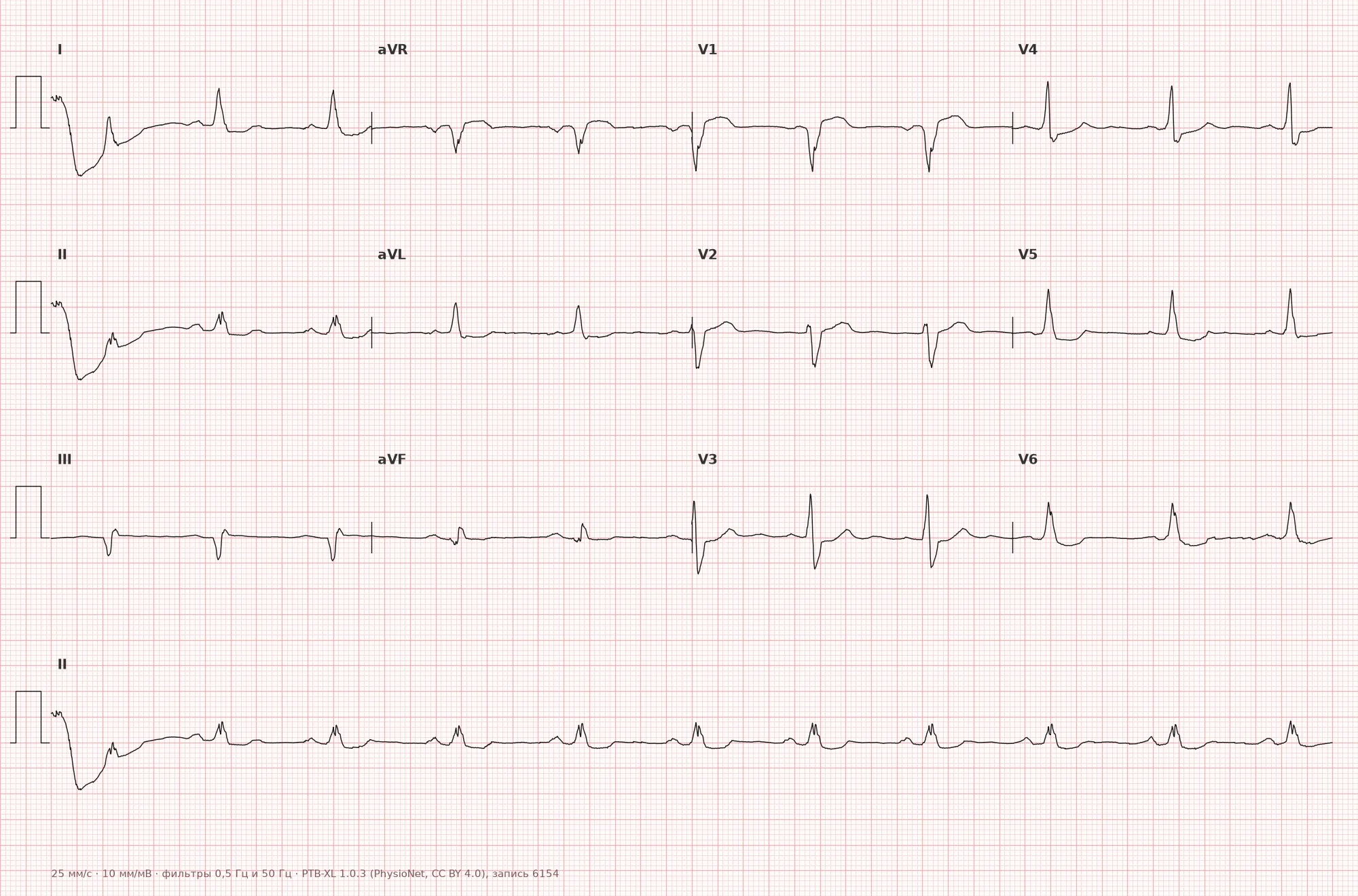
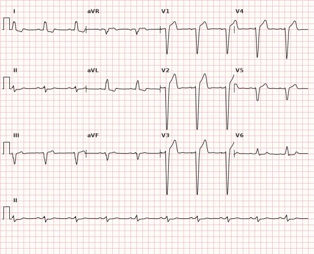
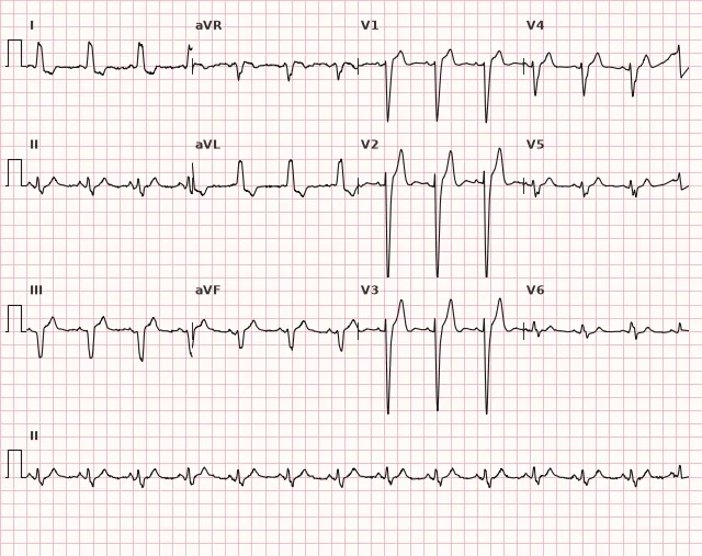
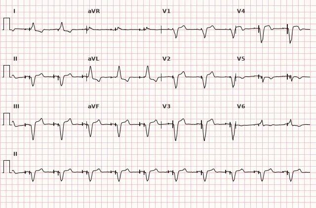
ECG from the open PTB-XL dataset (Wagner P. et al., Scientific Data, 2020; PhysioNet), CC BY 4.0 license. Rendering by WhiteCoat Pro.
Mechanism
The left bundle branch fails to conduct the impulse. Activation first spreads via the right bundle branch to the right ventricle and interventricular septum (from right to left — hence the disappearance of the septal q wave on the left), and then slowly crosses the septal myocardium, outside the specialized conduction system, to reach the left ventricle. This leads to two consequences: a marked prolongation of QRS (abnormal slow spread through the myocardium instead of rapid conduction via Purkinje fibers) and asynchronous, delayed contraction of the left ventricle. LBBB may be a marker and cause of LV mechanical dyssynchrony, but by itself is not an indication for cardiac resynchronization therapy (CRT). In a typical patient with symptomatic heart failure and reduced LVEF despite optimal medical therapy, the consideration for CRT is primarily driven by true LBBB morphology and QRS ≥150 ms (for QRS 120–149 ms, indications are less compelling); CRT is also separately discussed in situations associated with expected significant ventricular pacing or high-grade AV block, and in several other clinical scenarios according to current guidelines.
Causes
- Coronary artery disease, prior myocardial infarction
- Arterial hypertension with LV hypertrophy and fibrosis
- Dilated cardiomyopathy
- Valvular heart disease (aortic stenosis, mitral regurgitation)
- Degenerative conduction system fibrosis (Lenègre-Lev disease)
- Myocarditis
- Complication of cardiac surgery (aortic valve replacement, TAVI)
- Sometimes — without structural heart disease, especially in young individuals
Differential diagnosis
- Right bundle branch block: in V1 — rSR' or wide R, in V6 — wide S; the pattern is the opposite of LBBB.
- Nonspecific intraventricular conduction delay: QRS is wide, but the morphology matches neither typical LBBB nor RBBB.
- Ventricular pacing (artificial pacemaker): the wide QRS resembles LBBB, but pacemaker spikes are visible before the complexes on the tracing.
- Hyperkalemia with marked QRS widening: the characteristic LBBB morphology is absent, there is a tall, peaked T wave, and the P wave may disappear.
- Anterior myocardial infarction with a normal conduction system: QRS width, presence or absence of pathological Q waves do not rule out MI by themselves; signs are assessed based on the affected territory and the dynamics of ST-T and QRS in the corresponding leads. In LBBB, special criteria (Sgarbossa/Smith) and a clinical algorithm are used to detect concomitant ischemia rather than direct morphological comparison.
Clinical significance and management
LBBB itself is a finding that requires searching for the cause, rather than a standalone diagnosis. A new or unknown-onset LBBB in a patient presenting with acute coronary syndrome does not diagnose STEMI by itself and is not an automatic indication for immediate PCI. The decision depends on ongoing ischemic symptoms, hemodynamic stability, ECG dynamics, echocardiography, and troponin levels. In suspected ACS, modified Sgarbossa (Smith) criteria are used as an additional tool: concordant ST elevation ≥1mm in leads with a dominant R wave, concordant ST depression ≥1mm in V1–V3, or discordant ST elevation ≥1mm with an ST/S ratio ≤−0.25 (Smith criterion) in leads with a dominant S wave — these are evaluated alongside clinical findings, serial ECGs, and troponins rather than in isolation. A positive result increases the likelihood of acute coronary occlusion but does not prove it with absolute certainty; a negative result does not rule out MI due to limited sensitivity. In the presence of persistent ischemic symptoms, hemodynamic instability, or high probability of occlusion, an emergency invasive strategy is indicated regardless of formal criteria. An old LBBB without dynamic changes and without compelling ACS symptoms does not require emergency PCI by itself. In heart failure with reduced LVEF and LBBB, indications for cardiac resynchronization therapy are evaluated considering symptoms on optimal therapy, QRS width (primarily ≥150 ms), and true bundle branch block morphology; indications for CRT are also separately discussed when significant ventricular pacing or high-grade AV block is expected. Echocardiography is indicated to assess LV structure and function in newly diagnosed LBBB.
Common interpretation errors
- Attempting to diagnose LVH using voltage criteria in the presence of LBBB — standard indices (Sokolov–Lyon, Cornell) are unreliable in LBBB.
- Interpreting standard discordant ST-T changes as ischemia without applying Sgarbossa/Smith criteria, or conversely, treating a new LBBB in ACS as an automatic equivalent of STEMI without assessing clinical presentation and dynamic changes.
- Confusing incomplete LBBB (QRS 110–119 ms) with complete LBBB (QRS ≥120 ms) — management and clinical significance differ.
- Failing to notice pacemaker spikes and mistaking paced rhythm for spontaneous LBBB.
- Evaluating pathological Q waves in the inferior leads (II, III, aVF) as a sign of prior MI without considering that LBBB itself can distort initial QRS vectors.
"WiLLiaM": in V1 — W-shaped QS, in V6 — M-shaped wide R (mnemonic in English, helps remember based on the letter shape in the corresponding lead)
1. What is the minimum QRS duration threshold used to diagnose complete left bundle branch block?
According to the 2009 AHA/ACCF/HRS guidelines, complete LBBB is diagnosed when QRS ≥120 ms; 110–119 ms corresponds to incomplete block.
2. Which QRS morphology is typical for lead V1 in left bundle branch block?
In LBBB, the right ventricular lead V1 shows QS or rS with a wide, deep S wave because LV activation is directed away from this lead.
3. What is the modified Smith criterion for detecting acute coronary occlusion in the presence of LBBB?
The modified Smith criterion is ST elevation ≥1 mm with ST/S ≤−0.25; it increases the likelihood of acute coronary occlusion, but is used in conjunction with clinical presentation and other findings.
Frequently asked questions
How to distinguish LBBB from myocardial infarction on an ECG?
In LBBB, secondary discordant ST-T changes are expected: the ST segment is directed opposite to the dominant QRS wave. When ACS is suspected in the presence of LBBB, concomitant acute ischemia or occlusion is suggested by concordant ST elevation ≥1 mm in leads with a dominant R wave, concordant ST depression ≥1 mm in V1–V3, or discordant ST elevation ≥1 mm with ST/S ≤−0.25 (Smith criterion) in leads with a dominant S wave. These criteria are used alongside clinical presentation, serial ECGs, troponin, and imaging, as a negative result does not rule out MI.
Is left bundle branch block dangerous by itself?
Isolated LBBB without structural heart disease and without symptoms can be a benign finding. However, it frequently accompanies organic myocardial disease and requires echocardiography to rule out reduced contractility and investigate the cause.
Can left ventricular hypertrophy be diagnosed in the presence of LBBB?
Standard voltage criteria for LVH (Sokolow–Lyon, Cornell) are unreliable in the presence of LBBB due to the altered sequence of ventricular activation. The diagnosis of LVH in LBBB is established via echocardiography or cardiac MRI.
What to do if LBBB is newly detected in an asymptomatic patient?
Assessment of the clinical context is required (symptoms, history of CAD, hypertension, valvular disease), along with echocardiography to evaluate LV structure and function, application of Sgarbossa/Smith criteria combined with ECG dynamics and troponin if ACS is suspected, and a cardiology consultation.
What is the difference between complete and incomplete LBBB?
The criterion is QRS duration: in complete LBBB, QRS is ≥120 ms; in incomplete LBBB, 110–119 ms with the same characteristic morphology in V1 and V6. Incomplete block is less common and defined less strictly.
ECG Course
Interpretation from basics to myocardial infarction — learn to read any ECG using a systematic algorithm.
- Video lessons and theory for every topic
- Simulator case studies under every lesson
- Unlimited ECG simulator practice
- Study guide: "All ECGs in 25 minutes"
ECG Strip Simulator
ECG, radiology, histology, pathomorphology, pharmacology — thousands of flashcards with detailed explanations.
- Real ECG strips, not textbook drawings
- Instant answers and explanations
- Streaks and progress tracking in your dashboard
Related terms
More in the section "Bundle Branch and Fascicular Blocks"
Sources
- AHA/ACCF/HRS Recommendations for the Standardization and Interpretation of the Electrocardiogram, 2009
- Surawicz B., Knilans T. Chou's Electrocardiography in Clinical Practice
- Orlov V.N. Guide to Electrocardiography
- ESC Guidelines for the management of acute coronary syndromes, 2023
Educational material for medical students and physicians. Does not replace clinical guidelines and physical examination; the ECG must always be interpreted in conjunction with the clinical picture.