ECG criteria
- The QRS complex occurs earlier than the expected sinus beat (shortened RR interval preceding the premature beat)
- QRS is narrow, morphology matches sinus complexes (in the absence of aberrancy)
- A P' wave is present before the QRS — of altered shape, polarity, or with a different PQ interval compared to the sinus beat (in atrial premature beats)
- In junctional (AV) premature beats, the P' wave may be absent, negative in II, III, aVF before the QRS, buried within the QRS, or inverted after the QRS
- The pause following the premature beat is usually an incomplete compensatory pause (the sum of the pre- and post-extrasystolic intervals is less than two sinus cycles) due to discharge or phase resetting of the sinus node, although a full pause is also possible; the origin of a premature beat should not be determined by a single pause alone
- Blocked SVPB is possible — a P' wave is present, but the QRS is absent (falling into the refractory period of the AV node)
- With aberrant conduction, the QRS may be widened and resemble a ventricular premature beat — search for a P' wave preceding the complex
What it looks like on ECG
An SVPB is a premature complex that "wedges" into the sinus rhythm ahead of time. It is recognized by three elements: prematurity, the shape of the P' wave, and the width of the QRS.
P' wave. In an atrial premature beat, the shape of P' differs from the sinus P — it can be positive, biphasic, or negative depending on the location of the ectopic focus. The PR interval may differ from the sinus interval; with a very early premature beat, it is often prolonged due to decremental conduction through the AV node, but it can also be shorter or virtually identical to the sinus interval. The P' wave is best visualized in leads II and V1, and sometimes it superimposes on the preceding T wave (deforming it, creating a "bump" on T).
In a premature beat originating from the AV junction (junctional variant of SVPB), the atria depolarize retrogradely: P' is negative in II, III, aVF, and may precede the QRS with a short PQ (< 120 ms), coincide with the QRS (not visible), or follow it (negative wave in the ST segment).
QRS complex. Usually narrow (< 120 ms) and similar in shape to sinus complexes because ventricular activation proceeds via the normal His-Purkinje system. If the ventricles have not fully emerged from the refractory period following the previous contraction, aberrant conduction may occur — the QRS may widen resembling bundle branch block, most commonly right bundle branch block; morphology is assessed using standard criteria for intraventricular conduction block rather than a single feature in V1.
Pause. Following an SVPB, the pause is most often an incomplete compensatory pause: retrograde penetration of the ectopic impulse into the sinus node results in its discharge or phase resetting, and the next sinus cycle begins earlier than after a full compensatory pause. However, this does not always happen — the impulse may fail to reach the sinus node, finding it in the refractory phase, or it may not alter its cycle, in which case the pause is fully compensatory. Therefore, the origin of a premature beat cannot be determined solely by the pause.
| Sign | SVPB | VPB |
|---|---|---|
| QRS | narrow (unless aberrant) | usually widened (often ≥ 120 ms), but width is not an absolute criterion — morphology, P waves, timing of onset, and potential fusion and capture complexes must be evaluated |
| P' wave | often present (altered) or buried | usually absent before QRS |
| Pause | mostly incomplete compensatory | mostly complete compensatory |
Real ECGs: 8 examples
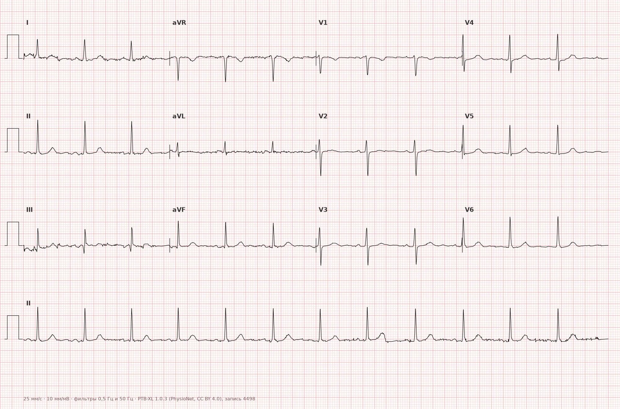
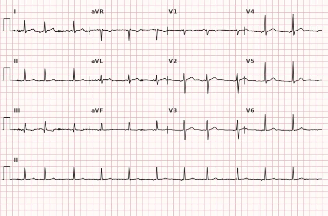
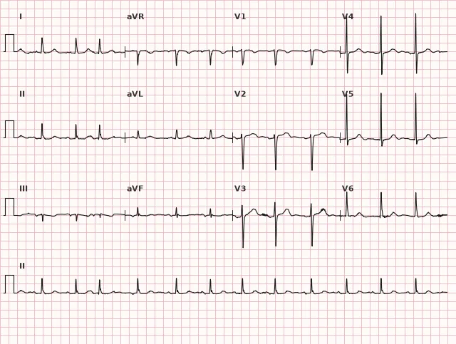
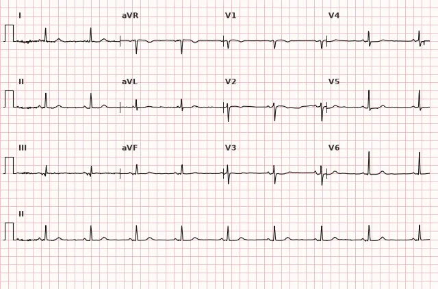
ECG from the open PTB-XL dataset (Wagner P. et al., Scientific Data, 2020; PhysioNet), CC BY 4.0 license. Rendering by WhiteCoat Pro.
Mechanism
The basis of SVPB is an ectopic automaticity focus or a re-entry mechanism at the level of the atria or AV junction, which generates an impulse earlier than the sinus node does. The impulse propagates to the atria (hence the altered shape of P') and further via the normal conduction system to the ventricles, so the QRS remains narrow. If the impulse catches the AV node or the His-Purkinje system in the relative refractory period, conduction to the ventricles is slowed or blocked asynchronously, resulting in an aberrant (wide) complex or a blocked premature beat without a QRS. An incomplete compensatory pause occurs when the ectopic impulse retrogradely penetrates the sinus node and causes its discharge or phase resetting, causing the next sinus cycle to trigger ahead of schedule. However, this does not always happen: in a junctional premature beat, retrograde conduction to the sinus node may be absent, resulting in a fully compensatory pause.
Causes
- Functional: stress, fatigue, sleep deprivation, excess caffeine, nicotine, alcohol
- Electrolyte disturbances (hypokalemia, hypomagnesemia)
- Structural heart diseases: atrial enlargement, valvular heart disease, cardiomyopathies
- Coronary artery disease, arterial hypertension with atrial hypertrophy and overload
- Chronic pulmonary pathology (COPD, cor pulmonale)
- Thyrotoxicosis
- Use of sympathomimetics, theophylline, certain psychostimulants
- Inflammatory changes in the atria (myocarditis, postoperative states)
- Can occur in healthy individuals without structural heart disease
Differential diagnosis
Ventricular premature beat. Typically features a wide QRS and the absence of a P' wave preceding the complex, with a usually complete compensatory pause, though none of these features are absolute. Narrow SVPB with aberrancy is distinguished primarily by the presence of a P' wave before the QRS and bundle branch block morphology assessed by standard criteria. Ashman phenomenon is another consideration — functional aberrancy (usually resembling right bundle branch block) occurring after a long cycle-short cycle sequence; it is most commonly observed in atrial fibrillation, but rhythm irregularity alone is insufficient for it.
Sinus arrhythmia. RR fluctuations are smooth, the P wave does not change shape, and there is no prematurity.
Blocked atrial premature beat. The ECG shows only a premature P' wave, often buried in the preceding T wave or sometimes in the ST segment, without a subsequent QRS — this can be mistaken for sinoatrial block or a sinus pause if T or ST wave deformation is not recognized.
Wandering atrial pacemaker. The shape of P changes gradually from cycle to cycle without prematurity, and the rate remains virtually unchanged.
Parasystole. Intervals between ectopic complexes are multiples of a common divisor, and there is no fixed coupling interval with the preceding sinus complex, unlike typical extrasystoles.
Clinical significance and management
Rare isolated SVPBs in individuals without structural heart disease have no clinical significance and require no treatment — this is a variant frequently encountered during 24-hour Holter monitoring in healthy subjects. It is important for the physician to:
- Evaluate the frequency and number of SVPBs per day (based on Holter monitoring data), and the presence of grouped forms (couplets, runs)
- Echocardiography — to assess structure, atrial size, cardiac function, and valves in frequent or symptomatic premature beats
- Screening for ischemia — based on symptoms and risk factors using appropriate methods (stress or imaging tests as indicated)
- Rule out electrolyte disturbances and thyrotoxicosis
- Assess the association with provoking factors (caffeine, alcohol, stress) and eliminate them if possible
- There is no single universally accepted threshold for "frequent" SVPB; an increased risk of future atrial fibrillation has been described with a high SVPB burden and/or runs, but results should be interpreted taking into account monitoring duration, the presence of structural heart disease, and AF risk factors
- Specific antiarrhythmic therapy is generally not indicated for asymptomatic cases. If symptoms persist after eliminating triggering factors, a beta-blocker is typically considered in the absence of contraindications; antiarrhythmic drugs and ablation are rare options for carefully selected patients following consultation with an arrhythmologist
Common interpretation errors
- Mistaking a blocked atrial premature beat for a sinus pause or SA block by failing to notice a deformed T or ST wave
- Confusing an aberrant SVPB with a ventricular premature beat due to a wide QRS without searching for a P' wave before the complex
- Failing to notice an altered P' wave superimposed on the preceding T wave
- Drawing conclusions about the origin of a premature beat solely based on the type of compensatory pause, without accounting for the P' wave and QRS morphology
- Ignoring frequent SVPBs as a "harmless finding" without assessing atrial structure and AF risk in a patient with risk factors
1. Which feature most reliably supports a supraventricular origin of a premature beat?
A premature P' wave before QRS is the most convincing sign of supraventricular origin, although its absence does not rule out SVES (junctional extrasystole or a concealed P' are possible).
2. Which pause most commonly occurs after a supraventricular extrasystole?
When the impulse retrogradely penetrates the sinus node, it discharges or phase-shifts it, and the pause is usually incomplete; however, the type of pause is not an absolute criterion for the origin of the extrasystole.
3. What happens during a blocked atrial extrasystole?
A premature atrial impulse catches the AV node in its refractory period and is not conducted to the ventricles—the ECG shows only a deformation of the T wave (less commonly ST) without a subsequent QRS.
Frequently asked questions
How to differentiate a supraventricular extrasystole from a ventricular one?
The most convincing sign is the presence of a premature altered P' wave before QRS, which supports a supraventricular origin, but its absence does not rule out SVES (P' may be hidden in the preceding T wave or within the QRS itself). QRS width is of secondary importance: SVES is typically narrow, but can be wide in aberrant conduction, so it is evaluated alongside morphology and context. The compensatory pause is the least reliable sign and has no independent diagnostic value.
Is supraventricular extrasystole dangerous?
Rare isolated SVES in a healthy individual are harmless and require no treatment. Frequent SVES can be a risk marker for the future development of atrial fibrillation and warrants searching for a structural cause (atrial enlargement, valvular pathology) and follow-up.
What is a blocked atrial extrasystole?
This is an SVES where the ectopic atrial impulse occurs so early that it catches the AV node in a state of refractoriness and is not conducted to the ventricles. The premature P' wave is often hidden within the T wave but may also be located in the ST segment; it is not followed by a conducted QRS.
Why is the pause in SVES often an incomplete compensatory pause?
An ectopic impulse from the atria frequently manages to reach the sinus node retrogradely and cause its discharge or phase shift prematurely. Therefore, the next sinus cycle is triggered earlier, and the sum of the intervals before and after the extrasystole is less than two normal RR cycles. However, this does not always happen: if the impulse does not reach the sinus node or does not alter its cycle, the pause becomes full—which is why the origin of an extrasystole is not determined by a single pause alone.
Can SVES be wide (with aberration)?
Yes. If the ventricles have not fully recovered from the previous contraction (relative refractoriness of the His-Purkinje system), conduction occurs asymmetrically, and the QRS widens, most commonly mimicking right bundle branch block. Differentiating it from a ventricular extrasystole is aided by the presence of a P' wave before the complex and evaluating morphology against standard bundle branch block criteria.
ECG Course
Interpretation from basics to myocardial infarction — learn to read any ECG using a systematic algorithm.
- Video lessons and theory for every topic
- Simulator case studies under every lesson
- Unlimited ECG simulator practice
- Study guide: "All ECGs in 25 minutes"
ECG Strip Simulator
ECG, radiology, histology, pathomorphology, pharmacology — thousands of flashcards with detailed explanations.
- Real ECG strips, not textbook drawings
- Instant answers and explanations
- Streaks and progress tracking in your dashboard
Related terms
More in the "Supraventricular Arrhythmias" section
Sources
- Goldberger A. Clinical Electrocardiography: A Simplified Approach
- Hampton J. The ECG Made Easy
- Surawicz B., Knilans T. Chou's Electrocardiography in Clinical Practice
- Orlov V.N. Guide to Electrocardiography
Educational material for medical students and physicians. Does not replace clinical guidelines and physical examination; the ECG must always be interpreted in conjunction with the clinical picture.