ECG criteria
- Ventricular rate is usually >100 bpm; assessed via RR intervals on a technically acceptable ECG
- QRS complex is narrow (<120 ms) unless aberrant conduction or preexisting bundle branch block is present
- Rhythm is regular (AVNRT, AVRT, atrial tachycardia) or irregular (atrial fibrillation, atrial flutter with variable conduction, multifocal atrial tachycardia)
- P wave may be visible before QRS, inside QRS, after QRS, or absent (depending on the mechanism)
- PR interval may be normal, shortened, or prolonged (depending on the mechanism and conduction)
- P:QRS ratio may be 1:1, >1:1 (atrial flutter), variable, or without distinct P waves (atrial fibrillation)
- In aberrant conduction, QRS >120 ms, but features of supraventricular origin are preserved (bundle branch block pattern, Ashman phenomenon)
What it looks like on ECG
SVT is an umbrella term encompassing several tachycardia mechanisms originating above the ventricles. A common feature is a ventricular rate typically >100 bpm and a narrow QRS (<120 ms), barring aberration.
Main variants of SVT:
| Mechanism | HR | Regularity | P waves | QRS |
|---|---|---|---|---|
| Sinus tachycardia | typically 100–180 (range depends on age and exertion) | regular | normal P waves (II, aVF +), preceding each QRS | narrow |
| AVNRT | 140–250 | regular | P waves 'pseudo-r'' in V1 or 'pseudo-S' in II, III, aVF (within/immediately after QRS) | narrow |
| AVRT (orthodromic) | 150–250 | regular | retrograde P wave is typically visible after QRS; RP is short, but the value is variable (can be longer with a slow accessory pathway) and is interpreted alongside other findings | narrow |
| Atrial tachycardia | 120–250 | regular | P wave before QRS, morphology differs from sinus | narrow |
| Atrial flutter | 240–340 (atria), ventricles depend on conduction | regular or irregular | sawtooth F waves (II, III, aVF) | narrow |
| Atrial fibrillation | atria 350–600, ventricles variable | irregular | f waves, without distinct P waves | narrow |
| Multifocal atrial tachycardia | 100–150 | irregular | ≥3 P-wave morphologies, irregular PP and PR intervals, isoelectric line between P waves | narrow |
Heart rate alone does not differentiate sinus tachycardia from SVT — rate ranges overlap, and the diagnosis is established based on a combination of features (onset/offset, P-wave morphology, response to maneuvers). With aberrant conduction (Ashman phenomenon, preexisting bundle branch block), QRS may be wide (>120 ms), typically with a right bundle branch block pattern. Differentiation from ventricular tachycardia requires additional criteria (Brugada, Vereckei).
Real ECGs: 8 examples
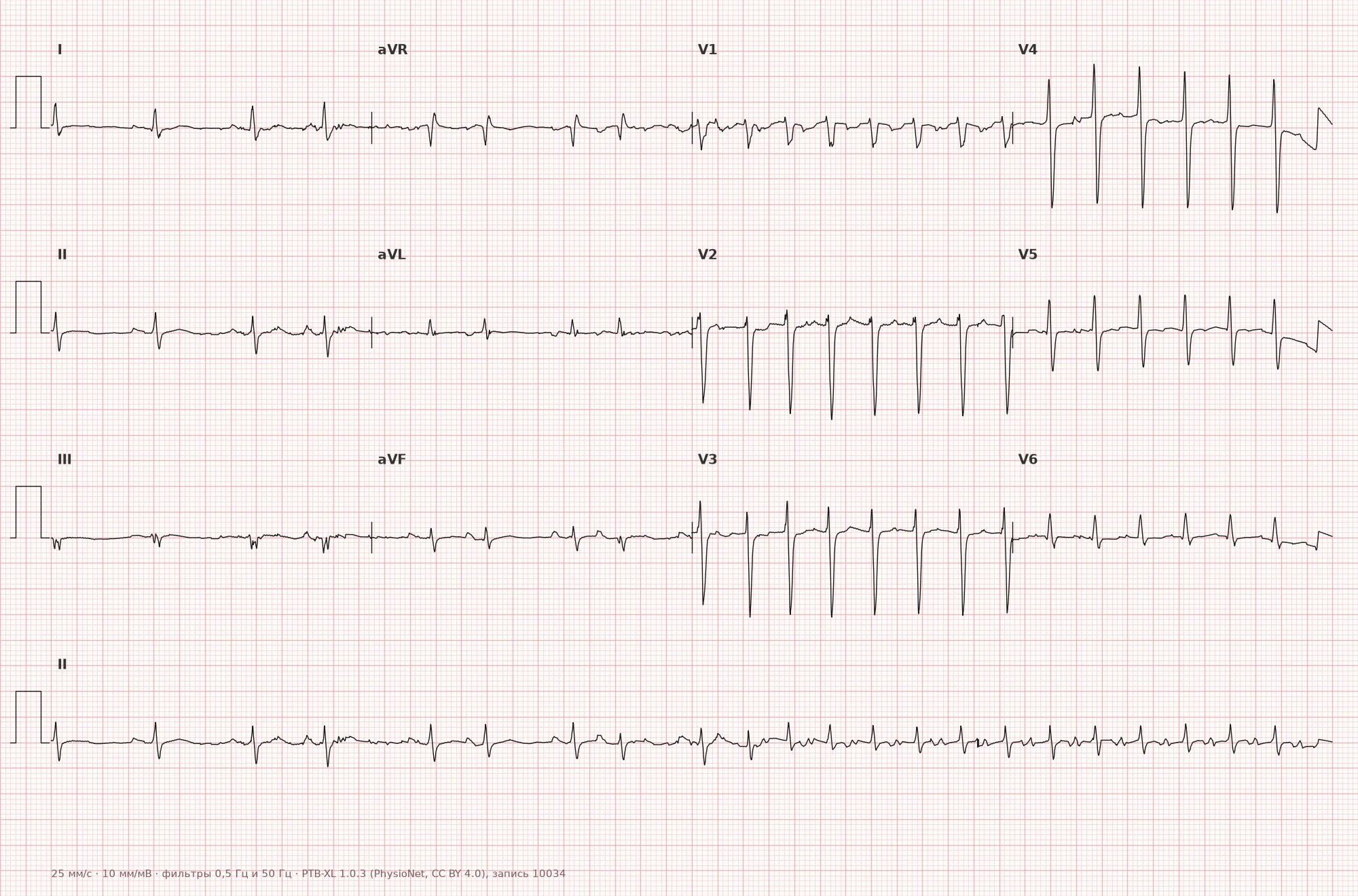
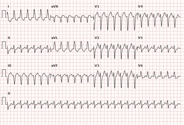
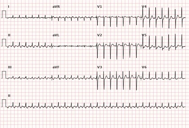
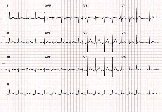
ECG from the open PTB-XL dataset (Wagner P. et al., Scientific Data, 2020; PhysioNet), CC BY 4.0 license. Rendering by WhiteCoat Pro.
Mechanism
SVT mechanisms are divided into three groups:
- Automatism: accelerated spontaneous impulse formation in the sinus node (sinus tachycardia), ectopic atrial foci (atrial tachycardia), or the AV node (junctional tachycardia). Catecholamines, hypoxia, and electrolyte disturbances increase automatism.
- Re-entry: circulation of an impulse along an anatomical or functional loop. AVNRT utilizes dual AV nodal pathways (slow and fast). AVRT uses a loop through the AV node, bundle of His, ventricles, and an accessory pathway back to the atria. Atrial flutter is macro-re-entry in the right atrium (typically around the tricuspid annulus).
- Triggered activity: early or delayed afterdepolarizations generate premature impulses. Occurs in digoxin toxicity, hypokalemia, hypomagnesemia.
Atrial fibrillation is associated with multiple micro-re-entrant circuits and ectopic foci (frequently in the pulmonary vein ostia).
Causes
Physiological and functional:
- physical exertion, stress, pain
- fever
- hypovolemia, anemia, hypoxia
- thyrotoxicosis
- stimulant use (caffeine, nicotine, cocaine, amphetamines)
- alcohol (especially in AF)
Cardiac:
- WPW syndrome (AVRT)
- CAD, myocardial infarction
- arterial hypertension, LV hypertrophy
- valvular heart disease (mitral stenosis — AF, flutter)
- cardiomyopathies
- myocarditis, pericarditis
- cor pulmonale (in PE, COPD)
Extracardiac:
- electrolyte disturbances (hypokalemia, hypomagnesemia)
- Pulmonary embolism
- sepsis
- digoxin toxicity, antiarrhythmic drugs
- idiopathic forms (especially in young individuals without structural heart disease)
Differential diagnosis
Ventricular tachycardia (VT): QRS is wide (>120 ms) with atypical morphology, AV dissociation, capture and fusion beats. SVT with aberration has a typical bundle branch block pattern (usually right), Ashman phenomenon (lengthening of the first QRS after a short cycle). Brugada and Vereckei criteria help differentiate.
Sinus tachycardia vs AVNRT/AVRT: sinus tachycardia starts and ends gradually, P waves are normal (positive in II, aVF). AVNRT/AVRT have sudden onset/offset, P waves are hidden within the QRS or immediately after. Heart rate ranges overlap, making rate a supportive rather than definitive feature.
AVNRT vs AVRT: in typical AVNRT, P waves are hidden within the QRS or immediately after (pseudo-r' in V1, pseudo-S in II, III, aVF). In orthodromic AVRT, the P wave is visible after QRS, RP is short, but its value is not absolute and is evaluated alongside other findings.
Atrial flutter with 2:1 conduction vs. sinus tachycardia: A heart rate of exactly 150 bpm (with a flutter rate of 300 bpm) is suspicious for flutter. Looking for F-waves (sawtooth pattern in II, III, aVF) and the adenosine test (temporary AV block unmasking F-waves) confirm the diagnosis.
Atrial fibrillation vs. multifocal atrial tachycardia (MAT): Both are irregular. AF is characterized by the absence of distinct P waves and the presence of f-waves. MAT shows P waves of varying morphology (≥3 types), a variable PR interval, and an isoelectric line preserved between P waves.
Clinical significance and management
Hemodynamic significance depends on heart rate, attack duration, rhythm (regular/irregular), left ventricular function, circulating blood volume, and comorbidities rather than heart rate alone. A high heart rate statistically more often reduces ventricular filling and coronary perfusion, potentially causing myocardial ischemia, pulmonary edema, and syncope. However, management is dictated by clinical signs of instability rather than a fixed heart rate threshold. Patients with coronary artery disease, heart failure, or valvular stenosis experience decompensation more rapidly at the same heart rate.
Management in unstable patients (hypotension, pulmonary edema, anginal pain, altered mental status): immediate synchronized electrical cardioversion. Initial energy depends on the device and protocol: typically 50–100 J for regular narrow-complex tachycardia, often 200 J biphasic for AF, while lower energies may be used for flutter.
Management in stable patients:
- Regular narrow-complex tachycardia (AVRT, AVNRT, atrial tachycardia): vagal maneuvers under ECG and BP monitoring. The modified Valsalva maneuver (with passive leg raise in the supine position after straining) is preferred as it is more effective than the classic one. Carotid sinus massage is permissible only in the absence of contraindications: carotid bruit, previous stroke/TIA, or known significant carotid atherosclerosis — in these cases, it is not performed. If vagal maneuvers are ineffective, give adenosine 6 mg IV push; if ineffective, 12 mg, followed by another 12 mg. Adenosine terminates AVNRT and AVRT (by blocking the AV node), slows AV conduction in flutter/AF (unmasking waves); it generally does not terminate automatic focal atrial tachycardia, but may temporarily slow the ventricular response, and occasionally terminates adenosine-sensitive atrial tachycardia. An alternative when adenosine is ineffective or contraindicated is verapamil, diltiazem, or IV beta-blockers, but only for stable regular narrow-QRS tachycardia without signs of pre-excitation and in the absence of hypotension, shock, and decompensated heart failure with reduced EF (verapamil and diltiazem are contraindicated in these settings). In irregular wide-complex tachycardia or suspected pre-excited AF, these drugs must not be used; when in doubt regarding the diagnosis or in unstable patients, specialist consultation and/or synchronized cardioversion are preferred.
- Irregular narrow-complex tachycardia (AF with rapid ventricular response): rate control (beta-blockers, non-dihydropyridine calcium channel blockers, digoxin) or cardioversion taking into account episode duration and stroke risk. If AF duration is >24 hours (or unknown), cardioversion requires either at least 3 weeks of therapeutic anticoagulation or a TOE-guided strategy to rule out left atrial appendage thrombus; the absence of a thrombus does not obviate the need for anticoagulation before and after the procedure. Following cardioversion, anticoagulation is typically continued for at least 4 weeks, and thereafter based on stroke risk stratification.
- Atrial flutter: the same anticoagulation principles as in AF apply — assessing episode duration (known/unknown onset) and stroke risk, a strategy of at least 3 weeks of anticoagulation or TOE prior to early cardioversion, and continuing anticoagulation post-procedure according to indications. Pharmacological rate control is feasible, but cardioversion in flutter is usually more effective than in AF.
WPW syndrome with AF (pre-excited AF): AV nodal blocking agents (adenosine, verapamil, diltiazem, digoxin, beta-blockers) and intravenous amiodarone are contraindicated, as all of them can enhance conduction via the accessory pathway and trigger ventricular fibrillation. In stable patients, procainamide or ibutilide is used (depending on availability and protocol); in unstable patients, immediate synchronized electrical cardioversion.
Recurrence prevention: lifestyle modification (avoiding triggers), beta-blockers, calcium channel blockers, antiarrhythmic drugs (flecainide, propafenone, sotalol, amiodarone). Catheter ablation is a radical treatment for AVNRT, AVRT, and typical atrial flutter (efficacy >90%).
Common interpretation errors
- Missing 2:1 atrial flutter: A regular heart rate of 150 bpm is mistaken for sinus tachycardia. Look for F-waves in II, III, aVF (especially buried within the T wave); perform a vagal maneuver or adenosine test to transiently slow AV conduction and unmask flutter waves.
- SVT with aberrancy mistaken for VT: A wide QRS (>120 ms) does not always indicate a ventricular origin. Look for bundle branch block patterns (especially right bundle branch block — rsR' in V1), Ashman phenomenon (widened QRS following a short RR interval), and sudden onset after an atrial extrasystole. For any regular wide-complex tachycardia, assume VT until proven otherwise (it is safer to treat as VT).
- Ignoring pseudo-r' in V1 and pseudo-S in inferior leads: These signs (retrograde P wave superimposed on the terminal portion of the QRS) are characteristic of AVNRT and help differentiate it from sinus tachycardia and atrial tachycardia.
- Overlooking WPW syndrome in AF: AF with a heart rate >200 bpm and variable QRS width (alternating narrow and wide complexes, sometimes with a delta wave) is suspicious for pre-excited atrial fibrillation. Until the mechanism is clarified, AV nodal blockers and IV amiodarone are withheld — the tachycardia is managed as potentially dangerous (risk of VF).
- Misinterpreting multifocal atrial tachycardia: An irregular rhythm with visible P waves of varying morphology is mistaken for AF. The distinction: in MAT, the isoelectric line between P waves is preserved, ≥3 P-wave morphologies are visible, PP intervals are irregular, and the PR interval is variable. In AF, the isoelectric line undulates (f-waves) and distinct P waves are absent.
1. A patient presents with a regular tachycardia, heart rate of 180 bpm, and narrow QRS complexes. Leads II, III, and aVF show a small negative deflection immediately following each QRS complex (pseudo-S). The most likely diagnosis is:
Pseudo-S waves in the inferior leads (II, III, aVF) represent a retrograde P wave superimposed on the terminal part of the QRS complex. This is a characteristic feature of typical (slow-fast) AVNRT, where the atria and ventricles are activated almost simultaneously.
2. During a tachycardia at 200 bpm, a typical left bundle branch block on the monitor transitions to a narrow QRS at the same heart rate while the atrioventricular association remains unchanged. How should this episode be interpreted?
A documented transition from typical bundle branch block to a narrow QRS complex with preserved, unchanged AV association indicates functional rate-dependent bundle branch block and a supraventricular origin of the rhythm. Isolated narrow complexes, evaluated without analyzing AV association and morphology, do not rule out VT by themselves; capture and fusion complexes, conversely, support the diagnosis of VT.
3. A patient with WPW syndrome develops an irregular tachycardia with a heart rate of 220 bpm. The ECG shows variable QRS width, and some complexes exhibit a delta wave. Which of the following is contraindicated?
This is pre-excited atrial fibrillation in WPW syndrome. Verapamil, along with other AV nodal blocking agents (adenosine, diltiazem, beta-blockers, digoxin) and intravenous amiodarone, is contraindicated: blocking the AV node diverts impulses down the accessory pathway, accelerates the ventricular response, and can precipitate ventricular fibrillation. Treatment consists of procainamide, ibutilide, or cardioversion.
Frequently asked questions
How to distinguish SVT from VT when QRS is wide?
Use the Brugada or Vereckei criteria. Signs of SVT with aberrancy include a typical bundle branch block pattern (most commonly right bundle branch block — rsR' in V1), Ashman phenomenon (first wide QRS after a short cycle length), and rate dependency (QRS narrows when heart rate slows). Signs of VT include AV dissociation, capture beats, fusion complexes, and atypical QRS morphology. When in doubt, assume VT and manage accordingly.
Can verapamil be used for any SVT?
No. Verapamil and other AV nodal blockers are contraindicated in tachycardia in a patient with WPW syndrome and atrial fibrillation, as well as in hypotension, shock, and decompensated heart failure. AV nodal blockade in pre-excited AF can direct impulses through the accessory pathway, accelerate the ventricular response, and cause ventricular fibrillation. In stable pre-excited AF, intravenous procainamide or ibutilide is used; in unstable patients, synchronized cardioversion. Verapamil can be used in stable regular narrow-complex tachycardia, including typical AVNRT and orthodromic AVRT, only after ruling out pre-excited AF/wide-complex tachycardia and the listed contraindications.
Why does adenosine not always terminate atrial tachycardia?
Adenosine blocks the AV node, interrupting the re-entry loop in AVNRT and AVRT, where the AV node is an obligatory part of the circuit. Automatic focal atrial tachycardia originates from an ectopic focus independent of the AV node, so it typically does not terminate, although adenosine transiently slows the ventricular response and unmasks frequent P waves. Less commonly, adenosine-sensitive triggered or reciprocal atrial tachycardia may terminate.
Is SVT with a heart rate of 180 dangerous?
The prognosis depends on the mechanism, duration, and symptoms, not just the heart rate. Tolerance is individual: even without underlying structural heart disease, prolonged tachycardia at 180 bpm can cause myocardial ischemia or, over time, tachycardia-induced cardiomyopathy (dilation and reduced ejection fraction). In coronary artery disease, such a heart rate rapidly provokes ischemia, and in chronic heart failure, pulmonary edema. Persistent tachycardia at 180 bpm requires medical evaluation, especially if accompanied by chest pain, dyspnea, syncope, or hypotension.
What to do if vagal maneuvers and adenosine fail?
If AVNRT/AVRT fail to terminate, in the presence of hemodynamic stability and absence of contraindications, intravenous beta-blockers (metoprolol, esmolol) or non-dihydropyridine calcium channel blockers (verapamil, diltiazem) are used. If medical therapy is ineffective or in case of hemodynamic instability, synchronized electrical cardioversion is performed. After termination, consider catheter ablation for recurrence prevention.
ECG Course
Interpretation from basics to myocardial infarction — learn to read any ECG using a systematic algorithm.
- Video lessons and theory for every topic
- Simulator case studies under every lesson
- Unlimited ECG simulator practice
- Study guide: "All ECGs in 25 minutes"
ECG Strip Simulator
ECG, radiology, histology, pathomorphology, pharmacology — thousands of flashcards with detailed explanations.
- Real ECG strips, not textbook drawings
- Instant answers and explanations
- Streaks and progress tracking in your dashboard
Related terms
More in the "Supraventricular Arrhythmias" section
Sources
- Page R.L. et al. 2015 ACC/AHA/HRS Guideline for the Management of Adult Patients With Supraventricular Tachycardia
- Orlov V.N. Guidelines for Electrocardiography, 2020
- Surawicz B., Knilans T.K. Chou's Electrocardiography in Clinical Practice, 6th ed., 2008
- ESC Guidelines for the diagnosis and management of atrial fibrillation, 2024
Educational material for medical students and physicians. Does not replace clinical guidelines and physical examination; the ECG must always be interpreted in conjunction with the clinical picture.