ECG criteria
- The rhythm is usually regular, with similar RR intervals; there is no strict universal RR variability threshold — regularity is assessed across consecutive intervals and in a clinical context
- Heart rate is typically 150–220 bpm — this is a general reference point rather than a diagnostic criterion: values both below and above this range can occur in adults
- The QRS complex is usually narrow (less than 120 ms) unless accompanied by bundle branch block, aberrant conduction, or pre-excitation
- P wave: either not visible (merged with the QRS in typical AVNRT) or located immediately after the QRS (pseudo-r' in V1, pseudo-S in II, III, aVF in AVNRT), or before the QRS with a short RP interval in orthodromic AVRT
- Sudden onset and sudden termination of the episode (on a long rhythm strip or by history) — a feature typical of re-entrant mechanisms, though not obligatory for all supraventricular tachycardias
- Absence of the preceding gradual "warm-up" phase characteristic of sinus tachycardia
- Response to vagal maneuvers or adenosine: termination of tachycardia (in re-entry involving the AV node) or transient slowing with unmasking of atrial activity (in atrial tachycardia); the response should be evaluated together with the ECG, not in isolation
What it looks like on ECG
The ictal ECG shows a narrow-QRS tachycardia with a rate most commonly between 150 and 220 bpm. The rhythm is usually strictly regular, with RR intervals remaining consistent from beat to beat; there is no strict variability threshold, and minor RR fluctuations do not rule out PSVT. Regularity of a narrow-QRS tachycardia aids in differential diagnosis but is not specific for PSVT: sinus tachycardia, atrial flutter with fixed AV conduction, and certain atrial tachycardias can present similarly. Therefore, additional assessment includes the onset/offset mode, visible atrial activity, and the response to vagal maneuvers or adenosine.
The P wave is a key yet often elusive element. In typical AV nodal re-entrant tachycardia (AVNRT), the atrium and ventricle are activated almost simultaneously, causing the P wave to merge with the QRS or appear as a small notching at its terminal portion: in lead V1, this appears as a pseudo-r' (a false small wave following R), and in the inferior leads II, III, aVF, as a pseudo-S (a shallow deflection following R). The RP interval in this case is short, less than 70 ms.
In orthodromic AV re-entrant tachycardia (AVRT, involving an accessory pathway), the retrograde P wave is usually visible after the QRS but may merge with its terminal portion; the RP interval is frequently > 70 ms and often shorter than the PR interval, though these ratios are approximate and overlap significantly with other mechanisms — a short RP can also occur in AVRT, and in atypical AVNRT the RP may be long. Intracardiac EPS is often required to definitively differentiate the mechanisms based on RP/PR.
In atrial tachycardia, the P wave precedes the QRS, its morphology differs from sinus rhythm, and the PR interval may be prolonged depending on the heart rate.
The transition zone and P-wave axis help suggest the site of origin, but on a standard 12-lead ECG, the P wave is frequently obscured by the tachycardia, and reliably differentiating the mechanism without intracardiac EPS is not always possible. The ST segment and T wave during tachycardia often exhibit secondary rate-dependent changes (ST depression, T-wave inversion), but ischemia cannot be ruled out based on the ECG alone: it may coexist with PSVT, especially in the presence of symptoms, risk factors, persistent changes after rhythm restoration, or elevated troponin — which warrants clinical evaluation.
Real ECGs: 8 examples
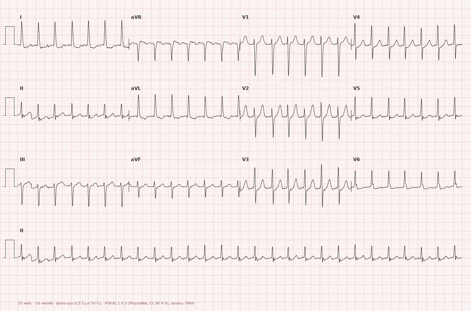
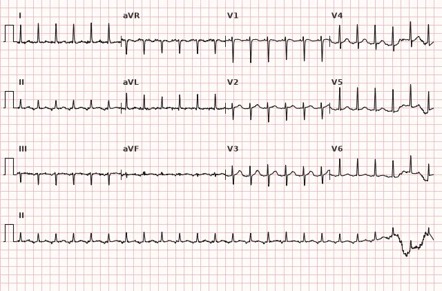
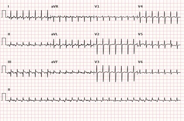
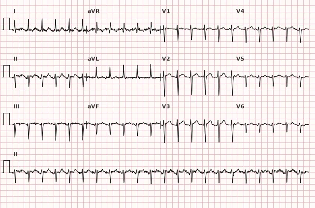
ECG from the open PTB-XL dataset (Wagner P. et al., Scientific Data, 2020; PhysioNet), CC BY 4.0 license. Rendering by WhiteCoat Pro.
Mechanism
The majority of paroxysmal SVTs are driven by a re-entry mechanism. Most commonly, this is AV nodal re-entrant tachycardia: the AV node functionally contains two conduction pathways with different speeds and refractory periods (fast and slow), forming a circuit for excitation. Less commonly, AV re-entrant tachycardia occurs via an anatomical accessory pathway between the atrium and ventricle (orthodromic variant — conduction down the AV node, up the accessory pathway). A third mechanism is atrial tachycardia, caused by focal ectopic activity or micro-re-entry within the atrium that does not directly involve the AV node. Ventricular activation typically proceeds via the normal His-Purkinje system, preserving a narrow QRS, but the QRS can be wide in the presence of aberrant conduction, pre-existing bundle branch block, electrolyte disturbances, or pre-excitation (antidromic AVRT typically presents with a wide QRS). Therefore, any wide-QRS tachycardia should initially be managed as ventricular tachycardia until proven otherwise.
Causes
- Functionally separate fast and slow AV nodal pathways (dual physiology) — the basis of AVNRT, often in the absence of structural heart disease
- Additional atrioventricular pathways (manifest in WPW syndrome or concealed) — the basis of AVRT
- Structurally normal heart — seen in the majority of patients, frequently young and middle-aged adults
- Triggering factors: emotional stress, physical exertion, caffeine, alcohol, sleep deprivation
- Organic heart disease (CAD, valvular defects, cardiomyopathies) — less common as a background
- Pregnancy (increased frequency of paroxysms due to hormonal and hemodynamic changes)
- Electrolyte imbalances, thyrotoxicosis — acting as triggering rather than causative factors
Differential diagnosis
- Sinus tachycardia — P waves of sinus morphology preceding each QRS, gradual onset and termination, HR variability with respiration and exertion; heart rate alone (even exceeding 150–160 bpm) does not allow for a diagnosis of sinus tachycardia or the exclusion of PSVT — P-wave morphology, regularity, clinical context, and onset/offset dynamics must be assessed
- Atrial flutter with 2:1 conduction — F waves at a rate of approximately 300 bpm; a sawtooth pattern in leads II, III, aVF is primarily characteristic of typical counterclockwise flutter, whereas in atypical flutter the F-wave morphology may differ, and with 2:1 conduction, the waves are frequently poorly discernible; slowing AV conduction (vagal maneuvers, adenosine) unmasks the flutter waves
- Atrial tachycardia with irregular conduction (multifocal atrial tachycardia) — irregular rhythm, varying P-wave morphologies
- Ventricular tachycardia with retrograde atrial activation — typically wide QRS; a narrow QRS makes VT rare but does not completely rule it out (fascicular and other narrow-QRS ventricular tachycardias are described) — the diagnosis is determined by combining features and the clinical context. In SVT with concomitant bundle branch block or antidromic AVRT, the QRS may be wide; in such cases, it is safer to initially treat the rhythm as VT until proven otherwise and apply wide-QRS tachycardia differentiation criteria (Brugada algorithm, Vereckei)
- Inappropriate sinus tachycardia — persistently elevated heart rate without a paroxysmal pattern, P waves retain sinus morphology
Clinical significance and management
Most paroxysmal SVT episodes are not directly life-threatening, but they cause significant symptoms: palpitations, anxiety, shortness of breath, presyncope, and in patients with structural heart disease or extremely high heart rates — arterial hypotension and myocardial ischemia.
The first step is to record a 12-lead ECG during the episode if possible, which is essential for subsequent verification of the mechanism. In stable hemodynamics, the preferred initial maneuver is the modified Valsalva maneuver. Carotid sinus massage should be performed only by trained medical personnel, unilaterally in alternation, after ruling out contraindications (a carotid bruit without exclusion of significant stenosis, prior stroke or TIA), and with continuous rhythm monitoring. Vagal maneuvers may terminate tachycardia involving the AV node in the re-entry circuit or transiently slow AV conduction and unmask P waves in atrial tachycardia or atrial flutter.
Adenosine is used for hemodynamically stable regular narrow-complex tachycardia after ineffective vagal maneuvers: it is administered as a rapid intravenous bolus with continuous ECG recording, blood pressure monitoring, and readiness for defibrillation and resuscitation. It is not administered in irregular wide-complex tachycardia and suspected atrial fibrillation with pre-excitation—AV nodal blockade can enhance conduction over the accessory pathway and precipitate ventricular fibrillation. Use with caution or according to specific protocols in severe asthma and marked bronchoembolism/bronchospasm, sinus node dysfunction or high-degree AV block without a pacemaker, after heart transplantation (increased hypersensitivity), and during dipyridamole therapy (potentiated effect). When conditions are met, adenosine blocks AV nodal conduction and usually terminates AVNRT and orthodromic AVRT; in atrial tachycardia, it usually only slows AV conduction and unmasks atrial activity without terminating the arrhythmia, although certain focal atrial tachycardias (especially adenosine-sensitive ones) may be terminated by it—therefore, termination of tachycardia per se does not automatically prove AVNRT or AVRT, and the response must be interpreted alongside the ECG.
In unstable hemodynamics (hypotension, pulmonary edema, ischemia, altered mental status), emergency synchronized electrical cardioversion is indicated.
For recurrence prevention and definitive treatment, catheter ablation should be considered, which leads to complete elimination of the arrhythmia in the majority of patients with AVNRT and AVRT.
Common interpretation errors
- Omitting a concealed P wave within the QRS complex and interpreting the rhythm simply as "tachycardia" without determining the mechanism
- Diagnosing sinus tachycardia or ruling out PSVT based solely on heart rate without considering P wave morphology, regularity, and the onset/offset dynamics of the episode
- Failing to account for the possibility of a wide QRS in antidromic AVRT or concomitant bundle branch block—leading to misinterpretation as ventricular tachycardia or vice versa; when in doubt, it is safer to treat a wide tachycardia as ventricular in origin
- Interpreting secondary ST depression during tachycardia as an absolute sign of ischemia without clinical context and evaluation after rhythm termination
- Administering adenosine without readiness to document the response (ECG recording at the time of administration) and without accounting for contraindications (irregular wide-complex tachycardia/suspected AF with pre-excitation, severe asthma, sinus node dysfunction)
1. Which feature is most characteristic of typical paroxysmal supraventricular tachycardia on an ECG?
Typical regular narrow-complex PSVT is characterized by sudden onset and termination of the episode, unlike sinus tachycardia with gradual heart rate dynamics; a wide QRS does not rule out a supraventricular mechanism in the presence of aberrancy or pre-excitation.
2. Why is the P wave often not visible on the ECG in typical AV nodal reentrant tachycardia?
In typical AVNRT, the re-entry circuit within the AV node leads to near-simultaneous activation of the atria and ventricles, so the P wave is masked by the QRS complex.
3. Which effect of adenosine is most informative for differentiating the mechanism of a narrow-complex tachycardia?
Blockade of AV conduction by adenosine typically terminates tachycardias that utilize the AV node in the re-entry circuit, whereas in atrial tachycardia or flutter, it usually only temporarily slows the ventricular response, helping to reveal true atrial activity.
Frequently asked questions
How to distinguish PSVT from sinus tachycardia?
PSVT typically begins and ends abruptly, and the P wave is often concealed or has abnormal morphology. Sinus tachycardia accelerates and decelerates gradually, the P wave retains normal sinus morphology before each QRS, and the heart rate varies with respiration and exertion. Heart rate alone is not a reliable criterion: sinus tachycardia can exceed 150 bpm, and some PSVTs run slower.
Is paroxysmal supraventricular tachycardia dangerous?
In most patients with a structurally normal heart, PSVT is not directly life-threatening, but it can cause significant symptoms and a drop in blood pressure. In organic heart disease or very high heart rates, ischemia and decompensation are possible, requiring emergency termination.
Why is the P wave not visible in PSVT?
In typical AV nodal reentrant tachycardia, the atria and ventricles are activated almost simultaneously via a re-entry mechanism; therefore, the P wave is superimposed on the QRS complex and becomes invisible or is seen only as a small notch at its terminal portion.
Can AVNRT be distinguished from AVRT on a standard ECG?
Roughly yes—based on the position and distance of the P wave from the QRS (RP interval), but these relationships show significant overlap between mechanisms, and an intracardiac electrophysiological study is often required to reliably differentiate them.
What does adenosine do in PSVT?
Adenosine transiently blocks conduction through the AV node. If the tachycardia utilizes the AV node as part of the re-entry circuit (AVNRT, orthodromic AVRT), it is usually terminated. If the AV node is not part of the circuit (atrial tachycardia, atrial flutter), adenosine typically only slows the ventricular response, unmasking atrial activity, although certain focal atrial tachycardias may also be terminated.
ECG Course
Interpretation from basics to myocardial infarction — learn to read any ECG using a systematic algorithm.
- Video lessons and theory for every topic
- Simulator case studies under every lesson
- Unlimited ECG simulator practice
- Study guide: "All ECGs in 25 minutes"
ECG Strip Simulator
ECG, radiology, histology, pathomorphology, pharmacology — thousands of flashcards with detailed explanations.
- Real ECG strips, not textbook drawings
- Instant answers and explanations
- Streaks and progress tracking in your dashboard
Related terms
More in the "Supraventricular Arrhythmias" section
Sources
- Goldberger's Clinical Electrocardiography, latest edition
- Hampton. The ECG in Practice
- Surawicz B., Knilans T. Chou's Electrocardiography in Clinical Practice
- Orlov V.N. Guide to Electrocardiography
Educational material for medical students and physicians. Does not replace clinical guidelines and physical examination; the ECG must always be interpreted in conjunction with the clinical picture.