ECG criteria
- Clear organized P waves are absent; instead, fine or coarse fibrillatory activity may be recorded, best seen in V1 and inferior leads. f waves may be indistinguishable in certain leads
- f waves: irregular in morphology, amplitude, and rate; the approximate atrial rate is about 350–600 bpm, but it often cannot be reliably measured on a surface ECG
- Ventricular rhythm is usually absolutely irregular, with no reproducible RR pattern; the coincidence of individual intervals does not rule out AF
- The QRS complex is usually narrow (<120 ms) unless accompanied by bundle branch block, aberrant conduction, or pre-excitation
- Ventricular rate is variable: without rate control, it often exceeds 100–110 bpm; with treatment or impaired AV conduction, it may be less than 100 bpm
- With a very rapid ventricular response, f waves may be masked within the QRS and ST-T segments, but RR irregularity persists
- The diagnosis is confirmed on an ECG recording of at least 30 seconds or on a standard 12-lead ECG with characteristic features; a single short fragment may be insufficient
- Classification by f wave amplitude into coarse and fine forms is not standardized and is not mandatory for diagnosis; if a reference point is needed, AF is conventionally considered coarse with an f wave amplitude of approximately >0.5 mm (>0.05 mV, mostly in V1) and fine with a smaller amplitude, taking lead dependence into account
What it looks like on ECG
Clear organized P waves are absent — instead, continuously varying fibrillatory activity (f waves) is recorded, chaotic in shape, amplitude, and intervals. The approximate atrial rate is about 350–600 bpm, but it often cannot be reliably measured on a surface ECG due to wave fusion and low amplitude. Fibrillatory activity is often better seen in V1 and inferior leads (II, III, aVF), but its prominence depends on the lead and the specific patient — f waves may be indistinguishable in individual leads.
The diagnosis is confirmed by a combination of features — the absence of organized P waves and RR irregularity — on an ECG recording of at least 30 seconds or on a standard 12-lead ECG; a single short rhythm strip fragment may be insufficient, especially if the irregularity is ambiguous.
The main diagnostic feature is the ventricular rhythm, which is usually absolutely irregular, with no reproducible RR pattern (irregularly irregular). The coincidence of individual intervals on a short fragment does not rule out AF; this distinguishes AF from most other rhythm disorders where RR intervals are at least partially predictable.
The QRS complex is generally narrow unless there is pre-existing bundle branch block, rate-dependent block, aberrant conduction (Ashman phenomenon), or pre-excitation syndrome (WPW). The ST segment and T wave are assessed as usual, but their analysis is hindered by tachysystole and superimposed f waves.
| Sign | Characteristic |
|---|---|
| P wave | Absent (no organized P waves) |
| Atrial activity | f waves, approximately 350–600 bpm, irregular, rate often unmeasurable |
| Ventricular rhythm | Usually absolutely irregular |
| QRS | Usually narrow |
| HR | Any — from bradysystole to marked tachysystole |
Based on the ventricular response rate, AF is classified into normosystolic, bradysystolic, and tachysystolic forms. Division by f wave amplitude into coarse and fine forms is not standardized and is not required for diagnosis, but is traditionally mentioned as a conventional reference.
Real ECGs: 8 examples
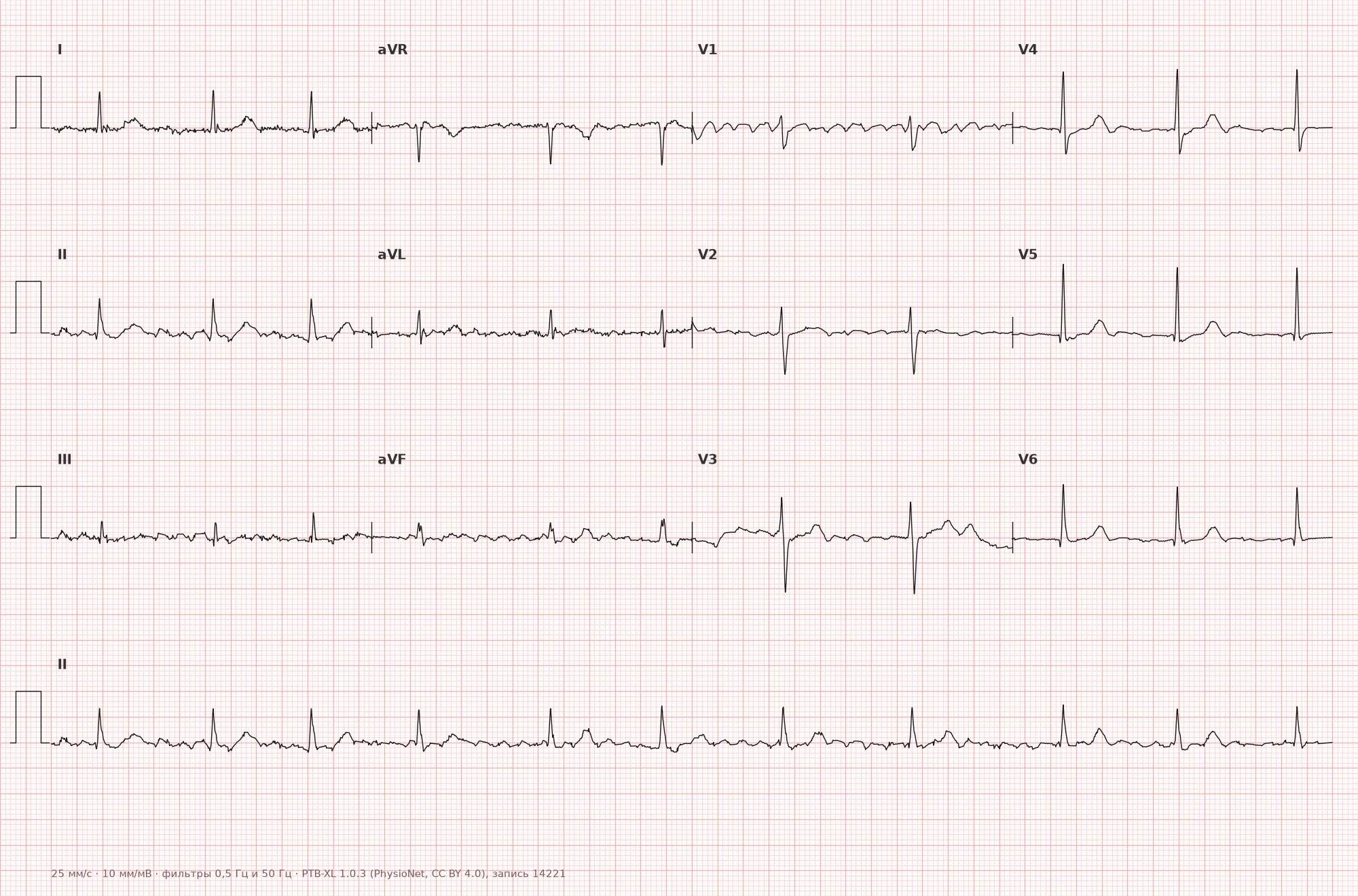
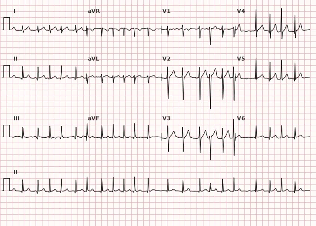
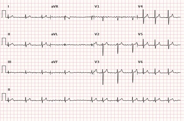
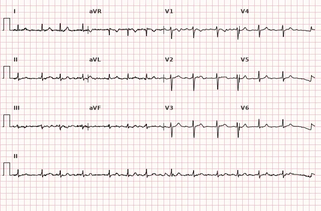
ECG from the open PTB-XL dataset (Wagner P. et al., Scientific Data, 2020; PhysioNet), CC BY 4.0 license. Rendering by WhiteCoat Pro.
Mechanism
The basis of AF is multiple chaotic re-entry wavelets circulating in the atria simultaneously, often initiated by ectopic foci located predominantly in the ostia of the pulmonary veins. The atria do not contract in a coordinated manner, their mechanical function is lost, which promotes blood stasis and thrombus formation, especially in the left atrial appendage.
The AV node receives numerous irregular impulses and transmits them to the ventricles selectively and unpredictably — hence the absolute irregularity of the ventricular rhythm. The rate of conduction to the ventricles depends on AV node refractoriness, autonomic nervous system tone, and the action of drugs affecting AV conduction.
Causes
- Arterial hypertension
- Mitral valve disease, especially mitral stenosis
- Coronary artery disease, myocardial infarction
- Heart failure of any etiology
- Left atrial enlargement
- Hyperthyroidism
- Sick sinus syndrome (tachy-brady syndrome)
- Pre-excitation/WPW syndrome associated with AF is not a universal cause, but the combination drastically alters risk and management when AF occurs
- Significant alcohol consumption ("holiday heart"); individual patients may have specific triggers, including energy drinks or caffeine, but regular caffeine consumption is not considered a proven general cause of AF
- Chronic obstructive pulmonary disease, pulmonary embolism, chronic cor pulmonale
- Postoperative period, especially after cardiac surgery
- Obesity, obstructive sleep apnea syndrome
- Idiopathic (lone) form in individuals without structural heart disease
Differential diagnosis
- Atrial flutter — classical flutter typically produces regular sawtooth F waves of identical morphology and amplitude (rate usually around 300 bpm), but in atypical flutter their morphology may differ from the classic sawtooth; the ventricular rhythm in flutter is often regular (e.g., 2:1), but with variable AV conduction, RR intervals become irregular.
- Multifocal atrial tachycardia (MAT) — atrial rate >100 bpm, at least three P wave morphologies, isoelectric intervals between them, irregular PP, PR, and RR intervals — in contrast to the complete absence of organized P waves in AF.
- Sinus arrhythmia — P waves are preserved and of normal morphology, only the PP interval changes, usually related to respiration.
- Frequent atrial extrasystoles with aberrancy — against the background of a generally regular sinus rhythm, occasional premature irregular complexes appear rather than continuous total irregularity.
- Tremor or motion artifact — can mimic the absence of P waves and baseline wander, but careful analysis of other leads usually reveals true sinus rhythm with P waves.
Clinical significance and management
AF increases the risk of cardioembolic stroke and systemic thromboembolism due to blood stasis in the atria, especially in the left atrial appendage, and can also lead to tachycardiomyopathy during prolonged uncontrolled tachysystole.
During ECG interpretation, the physician must: confirm the diagnosis using the described criteria on a recording of at least 30 seconds or on a standard 12-lead ECG, assess the ventricular response rate and QRS width (rule out concurrent bundle branch block, pre-excitation, or aberrancy), and document the episode duration if known (first-diagnosed, paroxysmal, persistent, long-standing persistent, permanent — based on clinical data, not a single ECG).
In cases of hypotension, shock, myocardial ischemia, pulmonary edema, or other hemodynamic instability, immediate synchronized electrical cardioversion is indicated without waiting for anticoagulation.
In a stable condition, the strategy for cardioversion depends on the duration of AF and thromboembolic risk. If AF has lasted for more than 24 hours (or duration is unknown), elective cardioversion is usually deferred until at least 3 weeks of therapeutic anticoagulation or performed earlier after ruling out a thrombus in the left atrium and its appendage using transesophageal echocardiography. After cardioversion, anticoagulation is continued for at least 4 weeks, and thereafter based on the calculated stroke risk. If AF duration is ≤24 hours, the decision on pre-cardioversion anticoagulation is also determined by the patient's thromboembolic risk rather than solely by the time from arrhythmia onset.
The risk of thromboembolism is assessed using the CHA2DS2-VA score (excluding sex, per ESC 2024 guidelines) or CHA2DS2-VASc in other current guidelines; when long-term anticoagulation is indicated, direct oral anticoagulants are generally preferred, except in mechanical heart valves and moderate-to-severe mitral stenosis, where vitamin K antagonists are used. Next, a rhythm control or rate control strategy is chosen, and reversible causes (thyrotoxicosis, electrolyte imbalances, alcohol) are investigated and corrected.
In pre-excited AF (WPW syndrome), AV nodal blockers are contraindicated: beta-blockers, verapamil, diltiazem, digoxin, and adenosine; intravenous amiodarone should also be avoided. These drugs can accelerate conduction via the accessory pathway and lead to ventricular fibrillation — urgent specialist consultation and appropriate protocol medications/electrical cardioversion are required.
Common interpretation errors
- Mistaking fine-wave AF for a total absence of atrial activity or confusing it with atrial asystole.
- Failing to distinguish AF from atrial flutter with irregular AV conduction — one must look for at least segments of regular sawtooth F waves.
- Forgetting to check QRS duration and missing concomitant WPW syndrome or bundle branch block, which changes management.
- Mistaking an artifact (tremor, electrical interference, motion artifact) for AF — one should examine multiple leads simultaneously.
- Evaluating rhythm rate and making a diagnosis based on too short a recording segment (less than 30 seconds), without considering RR variability across the entire available tracing.
"No P — irregular, irregular — no P" (irregularly irregular without organized P waves)
1. Which ECG feature is most specific for atrial fibrillation?
The absence of coordinated atrial activity (no organized P waves) and usually absolute ventricular rhythm irregularity are the key and most specific signs of AF.
2. Which drug is contraindicated in atrial fibrillation in the setting of WPW syndrome?
Verapamil and other AV nodal blockers in pre-excited AF can enhance conduction through the accessory pathway and provoke ventricular fibrillation.
3. How does AF differ from multifocal atrial tachycardia?
In multifocal atrial tachycardia, P waves persist but vary in shape (at least 3 morphologies) with irregular PP, PR, and RR intervals, whereas in AF, organized atrial activity and P waves are absent.
Frequently asked questions
How to distinguish atrial fibrillation from atrial flutter?
In AF, atrial activity is represented by chaotic f waves that vary in shape and amplitude, and the ventricular rhythm is typically completely irregular. In typical atrial flutter, F waves are regular, identical, and sawtooth-shaped, and the ventricular rhythm is often regular (e.g., with 2:1 conduction), although in atypical flutter and variable AV conduction, the picture may be less typical.
Is atrial fibrillation dangerous?
Yes, primarily due to the risk of thromboembolism—most notably cardioembolic stroke—as well as the potential development of tachycardia-induced cardiomyopathy during prolonged uncontrolled tachyarrhythmia. The level of risk is assessed using clinical scales rather than the ECG itself.
Can the P wave be seen in atrial fibrillation?
In true AF, clear organized P waves are absent; fibrillatory waves may be visible only in certain leads (most often V1 and inferior leads) or may be virtually indistinguishable, especially in fine AF.
Why is the ventricular rhythm irregular in AF?
The AV node receives numerous chaotic impulses from the atria and conducts them to the ventricles unpredictably, depending on the current refractoriness of the node, which is why RR intervals typically do not repeat.
How long must the ECG recording be to confirm a diagnosis of AF?
The diagnosis is confirmed by a recording of at least 30 seconds duration (single-lead recording) or on a standard 12-lead ECG with characteristic features present. Too short a tracing with similar irregularity may be insufficient for a diagnosis.
ECG Course
Interpretation from basics to myocardial infarction — learn to read any ECG using a systematic algorithm.
- Video lessons and theory for every topic
- Simulator case studies under every lesson
- Unlimited ECG simulator practice
- Study guide: "All ECGs in 25 minutes"
ECG Strip Simulator
ECG, radiology, histology, pathomorphology, pharmacology — thousands of flashcards with detailed explanations.
- Real ECG strips, not textbook drawings
- Instant answers and explanations
- Streaks and progress tracking in your dashboard
Related terms
More in the "Supraventricular Arrhythmias" section
Sources
- Hampton J. The ECG Made Easy
- Surawicz B., Knilans T. Chou's Electrocardiography in Clinical Practice
- Orlov V.N. Guide to Electrocardiography
- 2024 ESC Guidelines for the diagnosis and management of atrial fibrillation
Educational material for medical students and physicians. Does not replace clinical guidelines and physical examination; the ECG must always be interpreted in conjunction with the clinical picture.