ECG criteria
- The QRS complex is premature, occurring earlier than the expected sinus complex
- QRS duration is usually ≥ 0.12 s, and its morphology differs from the underlying (sinus) rhythm; with a fascicular or His-bundle origin, the QRS may be relatively narrow
- There is usually no sinus P wave preceding the PVC; an accidentally superimposed atrial P wave or a P wave hidden in the preceding T wave is possible. A retrograde P wave (due to ventriculoatrial conduction) appears after the PVC's QRS and may deform the ST segment / T wave
- The T wave is typically discordant (directed opposite to the main QRS vector) in pure ventricular origins, though this is a supportive rather than a mandatory sign
- Following a PVC, there is frequently a full compensatory pause: the interval from the preceding to the following sinus P wave equals twice the baseline PP interval; with retrograde atrial conduction, the pause may be non-compensatory
- With a typical full compensatory pause, the sinus node is not reset by the ventricular impulse; a single PVC episode does not constitute AV dissociation as such
- Fusion complexes may occur when sinus and ectopic activation coincide
- Morphology in V1 gives only an approximate localization: a RBBB-like pattern often suggests a left ventricular origin, while a LBBB-like pattern points to a right ventricular or septal origin; exact localization requires analyzing all 12 leads and remains presumptive
What it looks like on ECG
A PVC is a premature QRS complex that occurs earlier than the next expected sinus impulse. The QRS is typically wide and bizarre (≥ 0.12 s) because activation propagates through the ventricular myocardium rather than the specialized conduction system; if the focus is located high up — near the bundle of His or fascicles — the QRS may be relatively narrow. There is usually no sinus P wave preceding the complex, but an accidentally superimposed atrial P wave or a P wave hidden in the preceding T wave is possible. A retrograde P wave, resulting from ventriculoatrial conduction, appears after the PVC's QRS and can deform the ST segment or T wave. The T wave is generally discordant to the main QRS vector: if the QRS points upward, the T wave points downward, and vice versa, although this is not mandatory and depends on the site of origin and baseline repolarization.
A full compensatory pause is frequently observed after a PVC: if the ventricular impulse does not reach the sinus node retrogradely and reset it, the sinus node continues firing at its intrinsic rate, while the ventricles "skip" the regular beat as they are in the refractory period. In this case, the interval between the sinus P waves (or sinus R waves, if reliably identifiable) surrounding the PVC equals two baseline PP/RR intervals. Retrograde conduction to the atria or sinus node is possible and sometimes alters the timing of the next sinus impulse, making the pause non-compensatory.
Based on morphology in the chest leads, PVCs are conventionally divided into:
| Morphology in V1 | Presumed site of origin |
|---|---|
| RBBB-like (wide R in V1) | Left ventricle (more common) |
| LBBB-like (wide S in V1) | Right ventricle or septum (more common) |
This is a rough guideline: the exit point, scar, propagation pathways, and electrode placement affect the morphology, so reliable localization requires analyzing all 12 leads.
If all PVCs in a patient have the same morphology in a given lead, the ectopy is monomorphic; if they vary in shape, it is polymorphic. Multifocality requires convincing evidence of different foci (e.g., varying coupling intervals combined with different morphologies) rather than merely different QRS shapes, since morphology is also influenced by the exit site, conduction pathways, and fusion complexes. Regarding their timing relative to sinus beats, one distinguishes bigeminy (a PVC after every sinus beat), trigeminy (after every two sinus beats), and couplets (two consecutive PVCs). Three or more consecutive ventricular complexes at a rate usually ≥ 100 bpm constitute a run/episode of ventricular tachycardia; at a lower rate, this sequence may correspond to an accelerated idioventricular rhythm.
Real ECGs: 8 examples
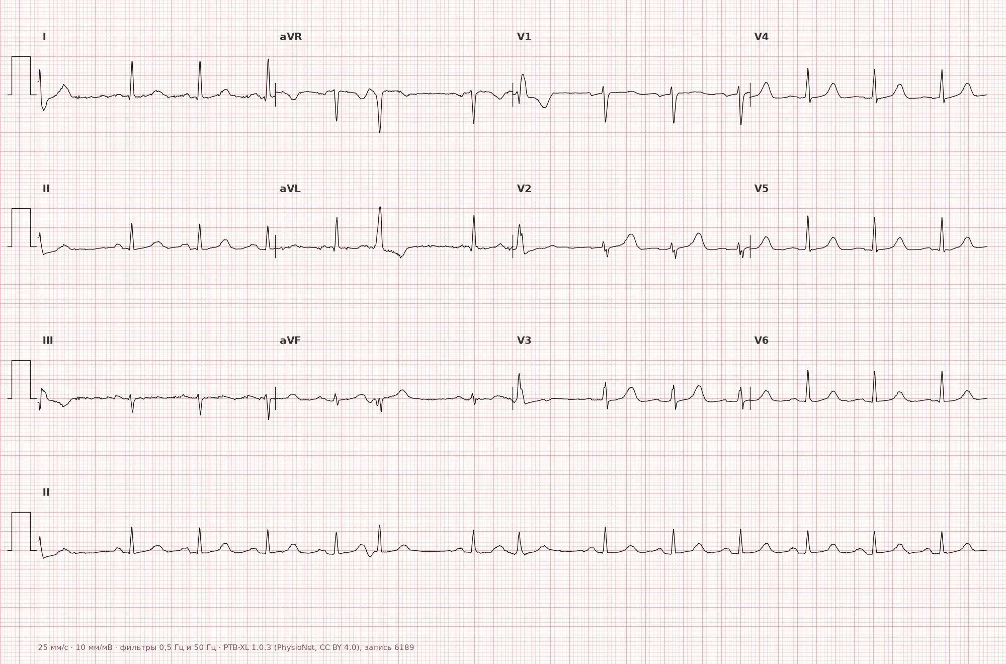
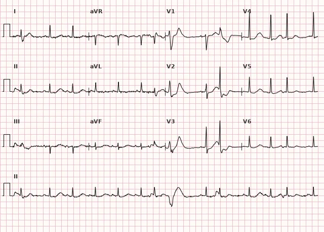
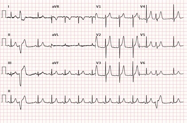
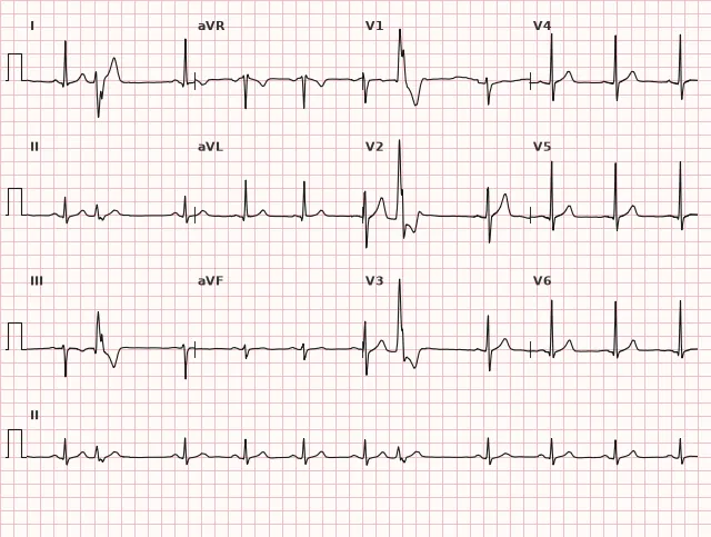
ECG from the open PTB-XL dataset (Wagner P. et al., Scientific Data, 2020; PhysioNet), CC BY 4.0 license. Rendering by WhiteCoat Pro.
Mechanism
PVCs originate from an ectopic focus in the ventricular myocardium or Purkinje fibers that depolarizes earlier than the arrival of the next sinus impulse. Mechanisms vary: enhanced normal automatism, abnormal automatism in damaged tissue, triggered activity (early or delayed afterdepolarizations), or re-entry at the border of a scar, fibrosis zone, or ischemia.
Activation spreads slowly through the ventricles rather than via the specialized conduction system, directly through the myocardium — hence the typically wide and bizarre QRS; with a focus close to the His-Purkinje system, propagation is faster and the QRS may be narrower. Retrograde conduction to the atria and sinus node is possible but often blocked or delayed in the AV node, so the sinus rhythm is typically not reset — resulting in a full compensatory pause; with successful retrograde conduction, the pause may be non-compensatory.
Causes
- In healthy individuals: functional PVCs associated with stress, sleep deprivation, excess caffeine, alcohol, smoking
- Coronary artery disease, including prior myocardial infarction (scar as a re-entry substrate)
- Structural myocardial diseases: cardiomyopathies (dilated, hypertrophic, arrhythmogenic right ventricular cardiomyopathy), myocarditis, valvular heart disease
- Electrolyte disturbances: hypokalemia, hypomagnesemia
- Heart failure, mitral valve prolapse
- Digoxin toxicity, proarrhythmic effects of certain drugs
- Idiopathic PVCs from the right ventricular outflow tract in individuals without structural heart disease
- Increased ventricular load in pulmonary hypertension, hypertrophy
Differential diagnosis
- Premature atrial complex with aberrant conduction — wide QRS, but a P wave is often visible before the complex (may deform the preceding T wave); QRS exhibits aberrant conduction morphology (RBBB or LBBB pattern, with Ashman phenomenon more frequently showing an RBBB pattern); non-compensatory pause
- Fusion complex (fusion of sinus and ventricular activation) — intermediate in QRS morphology and duration between a normal and a purely ectopic beat
- Ventricular escape beat — also a wide QRS without a P wave, but occurs later than scheduled (after a pause) rather than prematurely
- Ashman phenomenon — aberrant conduction following a "long cycle – short cycle" sequence, frequently seen in atrial fibrillation but not exclusively; the wide complex results from functional refractoriness of the conduction system rather than an ectopic ventricular focus; differentiation relies on assessing the entire RR sequence and clinical context rather than merely the presence or absence of a post-complex pause
- Motion artifact or muscle tremor — can mimic a wide, bizarre complex; identified by simultaneous "noise" in multiple leads, baseline instability, mismatch with the monitor pulse, and disappearance on repeat recording
Clinical significance and management
The prognostic significance of a PVC is determined not by its mere presence, but by the structural state of the heart. In patients without structural heart disease, rare monomorphic PVCs are generally benign. In the presence of structural heart disease (CAD, cardiomyopathy, reduced ejection fraction), frequent and/or polymorphic PVCs, couplets, and short-coupled PVCs can trigger ventricular tachycardia and ventricular fibrillation, especially in an acute ischemic setting; the isolated "R-on-T" phenomenon is no longer considered an independent marker of sudden cardiac death risk and does not mandate urgent antiarrhythmic therapy — risk is assessed based on the clinical context, ejection fraction, presence of a scar, and documented non-sustained or sustained ventricular tachycardia.
During ECG interpretation, the physician needs to: evaluate the morphology (mono- or polymorphic), frequency, and pattern (single, paired, bigeminy), as well as the presence of periods of ventricular tachycardia. Next, determine the structural background: an echocardiogram to assess the ejection fraction and myocardial structure, and if necessary, Holter monitoring to evaluate the 24-hour PVC burden. A burden of approximately ≥ 10% of all daily complexes increases the likelihood of PVC-induced cardiomyopathy, especially with a reduced ejection fraction, though the percentage alone does not automatically establish a diagnosis. For symptomatic PVCs or left ventricular dysfunction, pharmacotherapy and/or catheter ablation of the arrhythmogenic focus are discussed—the choice depends on the extrasystole morphology, suspected substrate, and patient preferences; an asymptomatic high burden without a drop in EF requires individual assessment and clinical follow-up rather than mandatory treatment. Electrolytes (potassium, magnesium) and potential proarrhythmic drug effects should be checked first.
Common interpretation errors
- Mistaking an aberrantly conducted supraventricular extrasystole for a ventricular one (one must look for the P wave and assess the completeness of the compensatory pause)
- Missing a hidden P wave that distorts the preceding T wave, or a retrograde P wave following the QRS complex
- Failing to distinguish a single PVC from the onset of ventricular tachycardia (three or more consecutive ventricular complexes at a rate of generally ≥ 100/min is already VT, not extrasystoles)
- Automatically considering any PVC a "benign finding" without assessing cardiac structure and the daily arrhythmia burden
- Confusing a fusion beat with a pathological QRS morphology
1. Which feature is most characteristic of a ventricular extrasystole on an ECG?
A PVC originates from an ectopic focus in the ventricle, and the impulse propagates primarily outside the conduction system, typically resulting in a wide, bizarre QRS complex without a relation to the atrial P wave.
2. How many consecutive ventricular complexes (at a rate generally ≥ 100/min) are sufficient to diagnose ventricular tachycardia rather than an extrasystole?
One or two consecutive PVCs (a couplet) remain extrasystoles, whereas three or more consecutive ventricular complexes with a rate generally ≥ 100/min are classified as ventricular tachycardia.
3. What is characteristic of a full compensatory pause following a PVC?
If the ectopic ventricular impulse does not retrogradely reset the sinus node, the sinus rhythm continues at its regular rate, and the ventricles skip one cycle—making the total interval equal to two normal PP/RR intervals.
Frequently asked questions
How to distinguish a premature ventricular contraction from a supraventricular one with aberrant conduction?
Look for a P wave before the complex: in a supraventricular extrasystole, it is usually present (may be hidden in the preceding T wave); in a ventricular one, it is typically absent, and if a retrograde P wave is present, it appears after the QRS. The compensatory pause is also evaluated: it is usually full in PVCs and incomplete in supraventricular ones. Since no single sign is absolute, they are evaluated in combination.
Are premature ventricular contractions dangerous?
Occasional monomorphic PVCs in a person with a structurally normal heart are generally harmless. The risk increases in the presence of structural heart disease, a high daily arrhythmia burden, and polymorphic or paired PVCs—in which case an in-depth workup is warranted.
What is a compensatory pause in PVCs?
This is the pause following an extrasystole where the distance from the previous sinus complex to the next equals twice the normal PP/RR interval. It occurs because the sinus node is typically not reset by the retrograde impulse from the ventricle, and the ventricle simply "skips" the regular beat due to refractoriness. With retrograde conduction to the atria, the pause may be incomplete.
How do PVCs differ from ventricular tachycardia?
One or two consecutive PVCs constitute extrasystoles (single or paired/couplet). Three or more consecutive ventricular complexes at a rate generally ≥ 100 beats per minute are classified as ventricular tachycardia: non-sustained if the episode lasts less than 30 seconds, and sustained if duration is ≥ 30 seconds or if it requires termination earlier due to hemodynamic instability.
Should premature ventricular contractions be treated if there are no symptoms?
Management depends on cardiac structure, the 24-hour PVC burden on Holter monitoring, and the presence of high-risk morphological patterns. Asymptomatic, infrequent PVCs in a structurally normal heart typically require no treatment but should be monitored dynamically.
ECG Course
Interpretation from basics to myocardial infarction — learn to read any ECG using a systematic algorithm.
- Video lessons and theory for every topic
- Simulator case studies under every lesson
- Unlimited ECG simulator practice
- Study guide: "All ECGs in 25 minutes"
ECG Strip Simulator
ECG, radiology, histology, pathomorphology, pharmacology — thousands of flashcards with detailed explanations.
- Real ECG strips, not textbook drawings
- Instant answers and explanations
- Streaks and progress tracking in your dashboard
Related terms
More in the section "Ventricular Arrhythmias"
Sources
- Hampton J. The ECG in Practice
- Goldberger A. Clinical Electrocardiography: A Simplified Approach
- Surawicz B., Knilans T. Chou's Electrocardiography in Clinical Practice
- Orlov V.N. Guide to Electrocardiography
Educational material for medical students and physicians. Does not replace clinical guidelines and physical examination; the ECG must always be interpreted in conjunction with the clinical picture.