ECG criteria
- Repeating pattern: 2 consecutive complexes of the underlying rhythm, followed by 1 extrasystole, and so on cyclically; the underlying rhythm is usually sinus, but can be another rhythm (e.g., atrial fibrillation)
- The extrasystole is premature: shortened coupling interval preceding it compared to the basic RR cycle
- In ventricular trigeminy, the QRS of the extrasystole is usually ≥ 120 ms and deformed; it is typically not preceded by a related sinus P wave, though hidden or retrograde P waves and relatively narrow VEAs from the conduction system are possible—QRS width alone does not confirm the origin
- The morphology of the extrasystole may be identical in all cycles (monomorphic) or variable (polymorphic); different morphology suggests different foci (polytopicity), but morphology alone does not always establish this reliably
- A ventricular extrasystole is often followed by a full compensatory pause, though an incomplete pause and interpolated VEA without a pause are possible; the presence or absence of a pause is not a mandatory criterion for trigeminy
- For a reliable description, it is advisable to see several consecutive repetitions of the cycle; there is no fixed minimum number of cycles
- Differentiate from bigeminy (1:1 alternation) and paired extrasystoles (couplets)
What it looks like on ECG
The tracing shows a regularly repeating three-component cycle: the first and second complexes belong to the underlying rhythm (during sinus rhythm, with a normal P wave and narrow QRS, barring baseline bundle branch block), and the third is extrasystolic. In ventricular trigeminy, the extrasystole typically has a wide, deformed QRS (≥ 120 ms) and a discordant T wave; it is usually not preceded by a related sinus P wave, though a retrograde P wave may follow the QRS, hidden within the complex or the ST-T segment. The extrasystole is frequently followed by a compensatory pause, after which the cycle resumes with two complexes of the underlying rhythm; both an incomplete pause and an interpolated VEA without a pause are also possible.
In supraventricular trigeminy (atrial or junctional extrasystoles with the same repetition rhythm), the QRS of the extrasystole is usually narrow and morphologically similar to the underlying rhythm complex. In atrial extrasystole, the P′ wave is premature and altered in shape, sometimes hidden in the preceding T wave or merged with the QRS; in junctional extrasystole, the P wave may be absent, hidden in the QRS, or retrograde following the QRS. A narrow QRS supports a supraventricular origin, but is not mandatory: with aberrant conduction, it may be wide, so the P wave, coupling interval, and morphology across all 12 leads should be evaluated, rather than the complex width alone.
| Sign | Ventricular trigeminy | Supraventricular trigeminy |
|---|---|---|
| QRS of the extrasystole | usually ≥ 120 ms, deformed | usually narrow, but may be wide with aberration |
| P wave preceding the extrasystole | usually absent; retrograde P after QRS is possible | atrial: premature altered P′, sometimes hidden in T or QRS; junctional: may be absent, hidden, or retrograde following QRS |
| Pause after the extrasystole | often full compensatory, incomplete pause or interpolation possible | more frequently non-compensatory |
During Holter monitoring, trigeminy is often registered in runs, and may alternate with bigeminy or isolated extrasystoles over 24 hours.
Real ECGs: 8 examples
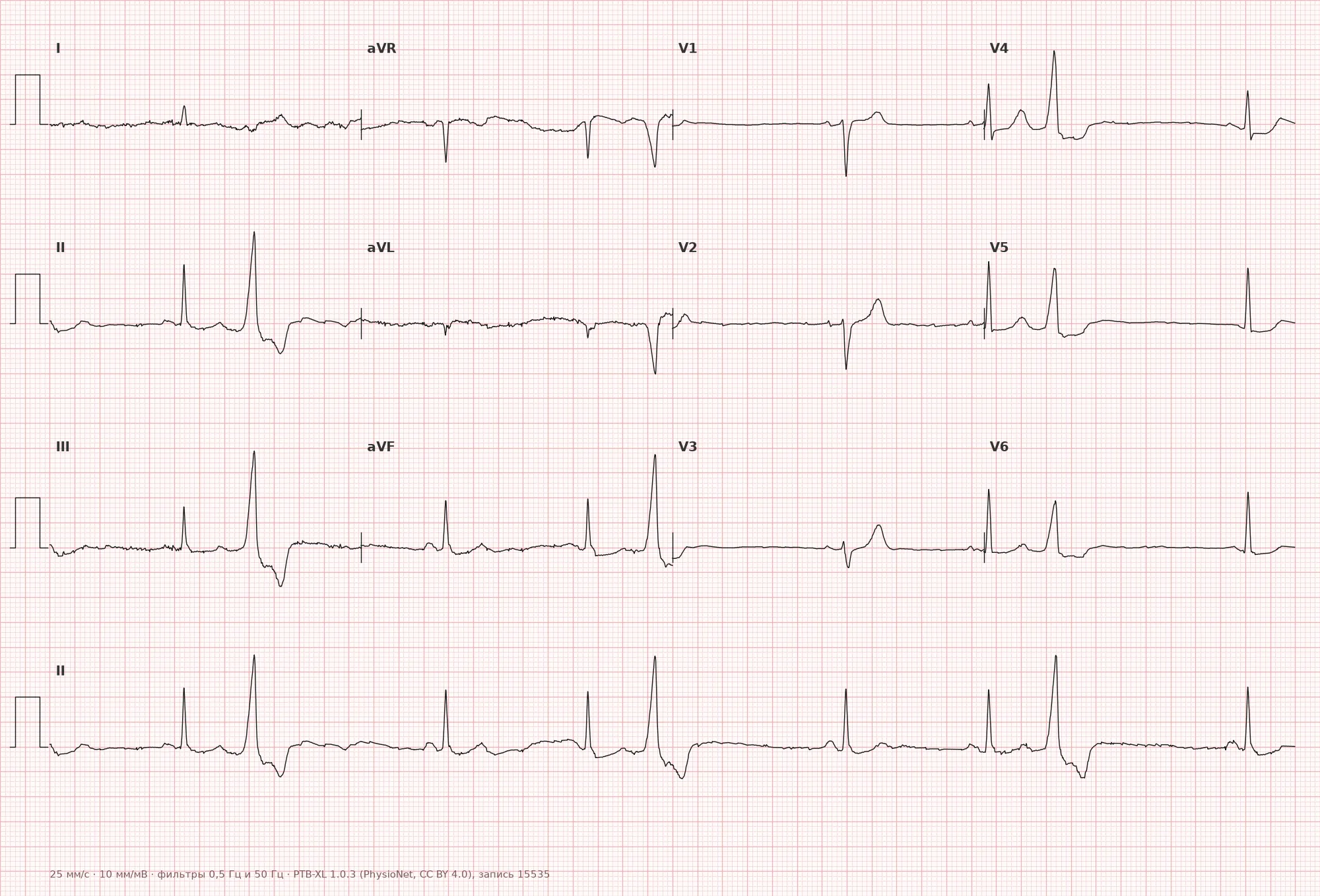
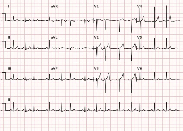
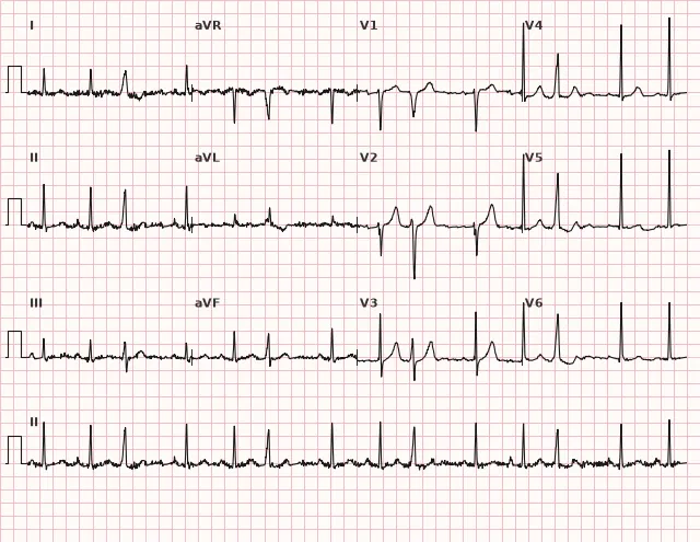
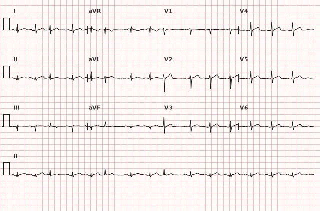
ECG from the open PTB-XL dataset (Wagner P. et al., Scientific Data, 2020; PhysioNet), CC BY 4.0 license. Rendering by WhiteCoat Pro.
Mechanism
The underlying mechanism involves an ectopic focus (in the ventricle, atrium, or AV junction) that generates an impulse coupled to the basic contraction rhythm—occurring after every second complex. The mechanism of the extrasystole itself may be related to re-entry, triggered activity, or abnormal automaticity; the periodicity of trigeminy reflects the reproducibility of extrasystole generation within this cycle and does not by itself establish a specific mechanism. The compensatory pause following a ventricular extrasystole (when present) occurs because the retrograde impulse does not conduct to the sinus node, and the next sinus impulse arrives at the expected time despite the preceding cycle being shorter; with interpolated VEAs, no pause occurs.
Causes
- Organic heart disease: coronary artery disease, prior myocardial infarction, cardiomyopathies, myocarditis
- Electrolyte disturbances: hypokalemia, hypomagnesemia
- Potential triggering factors in individual patients: stress, caffeine, alcohol, sleep deprivation—the association with symptoms and extrasystoles is assessed individually, with no proven universal causal link for all patients
- Use of certain medications (sympathomimetics, cardiac glycosides in toxic doses)
- Idiopathic extrasystole in individuals without structural heart disease, including young people and athletes
Differential diagnosis
- Bigeminy — an extrasystole following each complex of the underlying rhythm (1:1 ratio) rather than after two
- Quadrigeminy — an extrasystole following every three complexes of the underlying rhythm (3:1 ratio)
- Paired ventricular extrasystoles (couplet) — two consecutive ventricular ectopic complexes; may occur isolated or as part of a repeating pattern
- Aberrant conduction of a supraventricular extrasystole — wide QRS, but the complex is usually preceded by an atrial P wave distinct from the sinus P wave
- Ashman phenomenon — functional aberration of intraventricular conduction, typically a wide-complex beat following a short RR interval preceded by a long RR interval, most commonly seen in atrial fibrillation; lacks a fixed three-component periodicity
Clinical significance and management
Trigeminy itself does not determine prognosis. Its significance depends on the presence and severity of structural heart disease, daily extrasystole burden, left ventricular function, morphology (monomorphic or polymorphic), and clinical presentation (palpitations, reduced exercise tolerance).
The physician should: clarify the origin of the extrasystole (ventricular or supraventricular) based on QRS and P wave morphology across all 12 leads; perform echocardiography to assess cardiac structure and function; select ischemia testing based on clinical probability and symptoms, usually starting with non-invasive testing, and reserve invasive coronary angiography for appropriate indications; perform Holter monitoring to determine the 24-hour burden of extrasystoles and their relation to activity or sleep; check electrolytes (potassium, magnesium); evaluate reversible triggers (caffeine, alcohol, medications) individually. A daily VEA burden roughly ≥ 10% of all complexes increases the likelihood of VEA-induced cardiomyopathy—in such cases, ejection fraction and its dynamics are assessed. For symptomatic idiopathic VEA or VEA-induced cardiomyopathy, pharmacotherapy (e.g., beta-blocker) per indications and catheter ablation by an arrhythmologist are considered; treatment is not prescribed based on the mere fact of trigeminy alone.
Common interpretation errors
- Confusing trigeminy with bigeminy due to careless cycle counting on a short tracing
- Failing to distinguish between ventricular and supraventricular extrasystoles, relying solely on QRS width without assessing the P wave
- Mistaking random coincidence of rare single extrasystoles for a stable trigeminal pattern due to insufficient length of the analyzed segment
- Failing to evaluate extrasystole morphology across all leads, thus missing polymorphism
- Omitting the quantitative assessment (Holter) from the conclusion in persistent trigeminy, which is important for management
1. Which pattern corresponds to trigeminy?
Trigeminy is the regular repetition of a cycle consisting of two complexes of the underlying rhythm and one extrasystole.
2. Which sign favoring ventricular origin of an extrasystole in trigeminy is the most reliable?
A ventricular extrasystole typically presents with a wide, deformed QRS without a preceding related sinus P wave, although no single sign is absolute on its own.
3. What primarily determines the clinical significance of trigeminy?
Prognosis is determined by the underlying heart disease and the characteristics of the extrasystoles, rather than the repetition pattern itself.
Frequently asked questions
How does trigeminy differ from bigeminy?
In bigeminy, an extrasystole follows every complex of the underlying rhythm (1:1 ratio); in trigeminy, it follows every two such complexes (a three-beat cycle, 2:1 ratio).
Is trigeminy dangerous?
The repetition pattern itself does not determine prognosis. Risk depends on the underlying heart disease, daily extrasystole burden, left ventricular function, and their morphology — these are assessed using echocardiography and Holter monitoring.
Does trigeminy need to be treated if there are no symptoms and the heart is healthy?
In the absence of structural heart disease and symptoms, specific treatment is usually not required; observation and evaluation of potential triggers (caffeine, alcohol, electrolytes) are sufficient.
Can trigeminy be supraventricular?
Yes, a 2:1 repetition pattern is also possible with atrial or junctional extrasystoles. In this case, the extrasystolic QRS is usually narrow, but may be wide due to aberrant conduction, which makes evaluation of the P wave and coupling interval essential.
How to confirm the diagnosis of trigeminy?
A sufficiently long ECG rhythm strip or 24-hour Holter monitoring is required to observe a consistent three-beat cycle repetition rather than a chance coincidence of rare extrasystoles.
ECG Course
Interpretation from basics to myocardial infarction — learn to read any ECG using a systematic algorithm.
- Video lessons and theory for every topic
- Simulator case studies under every lesson
- Unlimited ECG simulator practice
- Study guide: "All ECGs in 25 minutes"
ECG Strip Simulator
ECG, radiology, histology, pathomorphology, pharmacology — thousands of flashcards with detailed explanations.
- Real ECG strips, not textbook drawings
- Instant answers and explanations
- Streaks and progress tracking in your dashboard
Related terms
More in the section "Ventricular Arrhythmias"
Sources
- Goldberger's Clinical Electrocardiography, current edition
- Hampton «The ECG in Practice»
- Surawicz, Knilans «Chou's Electrocardiography in Clinical Practice»
- Orlov, V.N. «Electrocardiography Guide»
Educational material for medical students and physicians. Does not replace clinical guidelines and physical examination; the ECG must always be interpreted in conjunction with the clinical picture.