ECG criteria
- Strict alternation: basic complex — ventricular extrasystole — basic complex — extrasystole, and so on; when the underlying rhythm is sinus, it is correct to specify "ventricular bigeminy in the setting of sinus rhythm"; in atrial fibrillation or another supraventricular rhythm, the conducted supraventricular complexes serve as the basic ones
- The extrasystolic QRS is usually wide (typically ≥ 120 ms) and deformed; the preceding P wave may be absent, buried in the preceding T wave, or dissociated; a narrow PVC originating from the His-Purkinje system is not ruled out, and the origin of the complex is assessed by a combination of features rather than QRS width alone
- The extrasystole occurs earlier than the expected next complex (shortened coupling interval)
- The ventricular extrasystole is frequently followed by a full compensatory pause, though an incomplete pause and interpolated PVC without a pause are also possible
- The T wave following a PVC is frequently discordant with the main QRS deflection; this supports a ventricular origin, though the absence of discordance does not rule it out
- To reliably characterize the rhythm as bigeminy, several consecutive cycles are desirable; a short fragment can be described as an episode of a bigeminal pattern
- The morphology of the extrasystole is identical across all cycles in monomorphic bigeminy; this indicates a common source or common exit pathway, but does not establish the exact anatomical localization from the ECG
What it looks like on ECG
The strip shows a repeating pair of complexes: the first is a narrow basic (usually sinus) QRS with a preceding P wave, and the second is a typically wide, deformed ventricular complex. The P wave preceding the extrasystole may be absent, hidden in the preceding T wave, fused with the QRS, or appear as a dissociated sinus P wave unrelated to this QRS; occasionally, a retrograde P wave appears after the PVC. The pair repeats consecutively, forming a "normal–extrasystole–normal–extrasystole" pattern.
The coupling interval (from the preceding basic QRS to the onset of the extrasystole) is usually constant in true extrasystole and may vary in parasystole. The extrasystole is frequently followed by a full compensatory pause: the interval from the basic complex preceding the PVC to the next basic complex is approximately equal to two basic PP or RR intervals. An incomplete compensatory pause and an interpolated PVC inserted between two basic complexes without a noticeable pause are possible.
| Element | Basic (sinus) complex | Extrasystolic complex |
|---|---|---|
| P wave | present, normal morphology | usually absent, hidden, or dissociated |
| QRS | narrow, typically < 120 ms | frequently wide, ≥ 120 ms, deformed; narrow PVC is possible |
| T wave | concordant with QRS | frequently discordant with the main deflection of its QRS, but may also be concordant |
| Pause after | none | frequently compensatory, but an incomplete pause or its absence (interpolation) is possible |
The morphology of the extrasystole roughly indicates the focus localization: a right bundle branch block pattern (wide R in V1) suggests the focus is more often in the left ventricle, while a left bundle branch block pattern (wide S in V1) suggests the right ventricle or interventricular septum. This is a tentative feature, not an absolute one.
Real ECGs: 8 examples
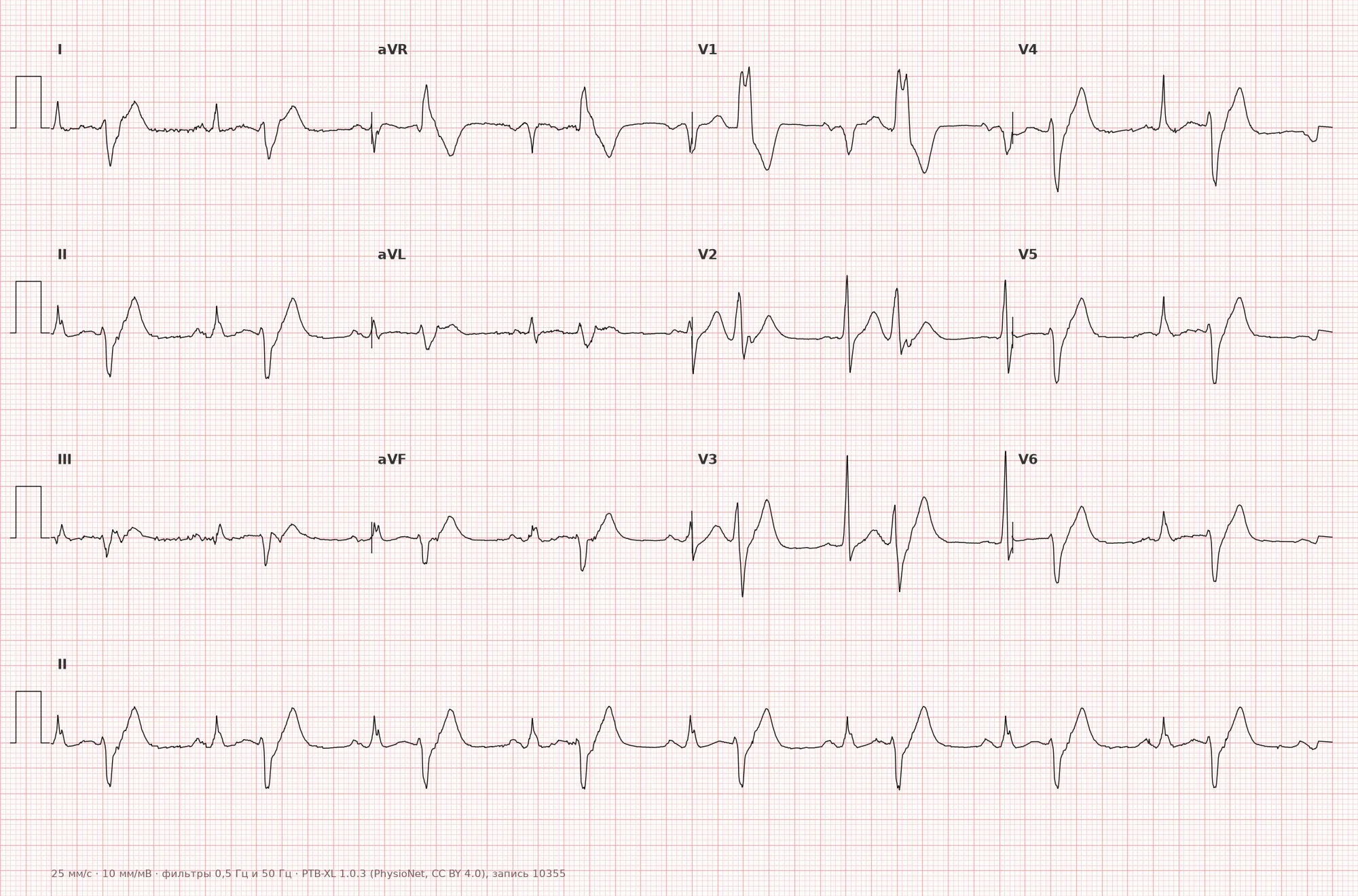
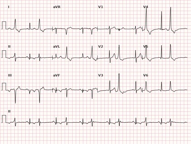
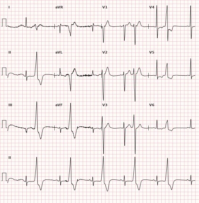
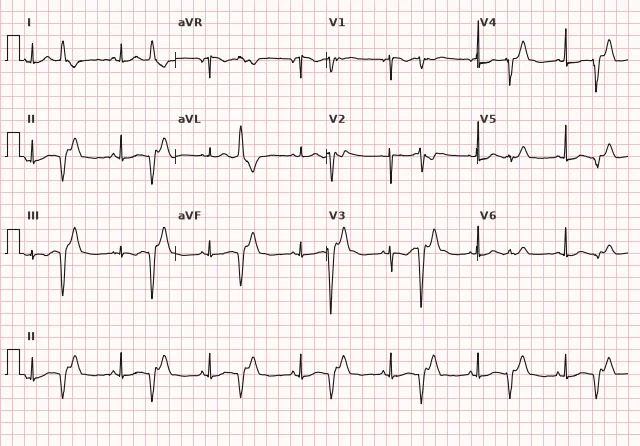
ECG from the open PTB-XL dataset (Wagner P. et al., Scientific Data, 2020; PhysioNet), CC BY 4.0 license. Rendering by WhiteCoat Pro.
Mechanism
The mechanism involves an ectopic focus in the ventricle (ventricular myocardium or Purkinje fibers) that generates an impulse with sufficient regularity relative to the underlying rhythm for every second complex to be an extrasystole. This may result from enhanced automaticity of the ectopic focus, triggered activity (early or delayed afterdepolarizations), or a re-entry mechanism within the ventricular myocardium. The strict 1:1 periodicity distinguishes bigeminy from a simple increase in the number of isolated extrasystoles and often indicates either a stable ectopic focus with a fixed excitability recovery time or a parasystolic focus not suppressed by the underlying rhythm (when the coupling interval is variable).
Causes
- Organic heart disease: coronary artery disease, post-infarction cardiosclerosis, cardiomyopathies
- Structurally normal heart (idiopathic ventricular extrasystole)
- Electrolyte disturbances: hypokalemia, hypomagnesemia
- Digoxin toxicity (classic cause of sustained bigeminy)
- Hypoxia, acidosis
- Increased sympathetic tone: stress, caffeine, stimulants
- Myocarditis, valvular heart disease
- Reperfusion period in acute myocardial infarction
Differential diagnosis
- Trigeminy — there are two basic complexes for every one extrasystole (2:1 ratio) rather than 1:1
- Coupled ventricular extrasystole (couplet) — two consecutive extrasystoles without an intervening basic complex
- Aberrant conduction of atrial extrasystoles — a wide complex is preceded by an ectopic P wave with altered morphology; differentiation sometimes requires multichannel recording and dynamic analysis
- Electrical alternans — alternation of amplitude or shape in consecutive complexes, typically without the appearance of premature wide ventricular complexes; QRS width alone does not define this phenomenon
- Parasystole with a fixed ratio — distinguished by a variable coupling interval and the greatest common divisor rule for inter-ectopic intervals
Clinical significance and management
The danger is determined not by the bigeminy pattern itself, but by the underlying heart disease, left ventricular function, and the total daily ventricular extrasystole burden. In a structurally normal heart, the prognosis is generally favorable. In unexplained left ventricular ejection fraction reduction and a PVC burden of approximately 10% or more, PVC-induced cardiomyopathy should be considered, with the risk typically higher at a burden exceeding 20%; assessment is performed taking into account arrhythmia duration, PVC morphology, symptoms, and LV function. This threshold alone is not an independent indication for ablation — the decision is made comprehensively.
The physician should: assess the width and morphology of the extrasystole, document the pattern on a standard ECG, refer the patient for Holter monitoring to assess the daily count and burden of extrasystole, evaluate electrolytes (potassium, magnesium) and medications (primarily digoxin), and perform echocardiography to evaluate ventricular structure and function. In symptomatic bigeminy, reduced ejection fraction, or high daily extrasystole burden, referral to an arrhythmologist is indicated to determine the strategy for pharmacotherapy or catheter ablation.
Common interpretation errors
- Mistaking a motion artifact for a wide complex and erroneously diagnosing bigeminy
- Confusing an aberrantly conducted atrial extrasystole with a ventricular one due to inattentive search for the ectopic P wave
- Failing to distinguish true bigeminy (fixed coupling interval) from parasystolic bigeminy with a variable interval
- Categorically dismissing the diagnosis of bigeminy upon encountering a short alternation fragment, without considering the possibility of an intermittent pattern
- Failing to evaluate the compensatory pause (or its absence in interpolated PVCs) and confusing it with other types of grouped extrasystoles
1. Which feature is mandatory for the diagnosis of ventricular bigeminy?
Bigeminy is defined by a strict 1:1 alternation of the basic complex and ventricular extrasystole.
2. Which classic drug can cause sustained ventricular bigeminy during toxicity?
Digoxin toxicity is a classic cause of persistent ventricular bigeminy and other ventricular arrhythmias.
3. How does ventricular bigeminy differ from paired ventricular extrasystoles (couplets)?
In a couplet, two extrasystoles occur consecutively, whereas in bigeminy, the extrasystole and the basic complex strictly alternate.
Frequently asked questions
Is ventricular bigeminy dangerous?
Bigeminy itself is an ECG pattern, not a diagnosis of immediate danger. The prognosis depends on the presence of underlying structural heart disease, left ventricular function, and the 24-hour burden of extrasystoles on Holter monitoring. In a structurally normal heart with a low extrasystole burden, the prognosis is generally favorable.
How to distinguish bigeminy from trigeminy?
In bigeminy, the ratio of basic complexes to extrasystoles is 1:1 — every second complex is an extrasystole. In trigeminy, there are two basic complexes for every extrasystole (a 2:1 ratio).
Can bigeminy be a normal variant?
Isolated ventricular extrasystoles, including bigeminy, can occur in individuals with a structurally normal heart. Evaluating the 24-hour ectopic burden and ruling out structural heart disease and electrolyte imbalances is essential.
How does bigeminy differ from aberrant conduction of supraventricular extrasystoles?
In a ventricular extrasystole, the P wave is usually absent, hidden, or dissociated before the wide QRS complex, whereas in an aberrantly conducted supraventricular extrasystole, the wide complex is preceded by an ectopic P wave, albeit with altered morphology. In complex cases, differentiation requires multi-channel recording and dynamic observation.
Is Holter monitoring necessary when bigeminy is detected on a resting ECG?
This is usually advisable, especially in the presence of symptoms, frequent or continuous bigeminy, suspected high ventricular ectopic burden, or a reduced ejection fraction. The specific type and duration of monitoring are chosen based on clinical needs rather than being routinely prescribed in every single case.
ECG Course
Interpretation from basics to myocardial infarction — learn to read any ECG using a systematic algorithm.
- Video lessons and theory for every topic
- Simulator case studies under every lesson
- Unlimited ECG simulator practice
- Study guide: "All ECGs in 25 minutes"
ECG Strip Simulator
ECG, radiology, histology, pathomorphology, pharmacology — thousands of flashcards with detailed explanations.
- Real ECG strips, not textbook drawings
- Instant answers and explanations
- Streaks and progress tracking in your dashboard
Related terms
More in the section "Ventricular Arrhythmias"
Sources
- Hampton J.R. The ECG Made Easy
- Surawicz B., Knilans T.K. Chou's Electrocardiography in Clinical Practice
- Orlov V.N. Guide to Electrocardiography
- AHA/ACCF/HRS Recommendations for the Standardization of ECG, 2009
Educational material for medical students and physicians. Does not replace clinical guidelines and physical examination; the ECG must always be interpreted in conjunction with the clinical picture.