ECG criteria
- P wave amplitude > 2.5 mm (0.25 mV) at a paper speed of 25 mm/s in the inferior leads II, III, and aVF is the primary and most standardized criterion; some guidelines lower the threshold to ≥ 2.0 mm. Lead II is usually the most convenient for measurement, but a tall P wave in III and aVF should not be considered secondary — the criterion is met if present in any of these leads.
- In an isolated typical P pulmonale pattern, P wave duration is usually not increased and remains within normal limits (< 120 ms); this is a typical but not mandatory sign — P wave widening does not rule out right atrial enlargement and may indicate combined biatrial enlargement.
- Pointed (peaked) P wave morphology in II, III, and aVF — hence the term "P pulmonale".
- In V1, an additional criterion is sometimes used: the amplitude of the initial positive phase of a biphasic P wave ≥ 1.5 mm; this sign is not equivalent to the primary amplitude criterion in the inferior leads. Changes in V2 may be present, but there is no separate standard threshold for V2.
- The P wave axis may deviate to the right (approximately > +75°) — an ancillary, optional sign that is not among the mandatory diagnostic criteria.
- Often associated with other signs of right-sided overload: right axis deviation, signs of right ventricular hypertrophy/overload.
- P wave amplitude is assessed in all inferior leads (II, III, aVF); the criterion is met if the threshold is exceeded in at least one of them, not just lead II.
What it looks like on ECG
Normally, the P wave is formed by the sequential depolarization of the right and then the left atrium; their vector sum produces a smooth, dome-shaped P wave with an amplitude of up to 2.5 mm, a duration usually of 80–110 ms, and no more than 120 ms in lead II.
In P pulmonale, the initial (right atrial) component of the P vector increases and becomes dominant. The result is a tall, peaked P wave with typically preserved duration:
| Lead | Sign |
|---|---|
| II, III, aVF | P wave is tall (> 2.5 mm, or ≥ 2.0 mm according to some authors), peaked; the criterion is met if present in at least one of these leads. |
| V1 | additional criterion: the initial positive phase of a biphasic P wave is increased (≥ 1.5 mm). |
| V2 | sometimes the initial positive phase is also increased, but there is no independent threshold. |
In isolated typical P pulmonale, P wave duration remains normal — this distinguishes the pattern from left atrial enlargement, where the P wave is widened and notched (P mitrale). However, ECG right atrial enlargement does not strictly require a normal P duration: in combined biatrial involvement or intraatrial conduction delays, the P wave may be widened, which does not exclude right atrial pathology. The inferior lead morphology is sometimes described as a tall, peaked P wave ("gothic spire") — this is not a standardized term, but merely a descriptive analogy.
Real ECGs: 8 examples
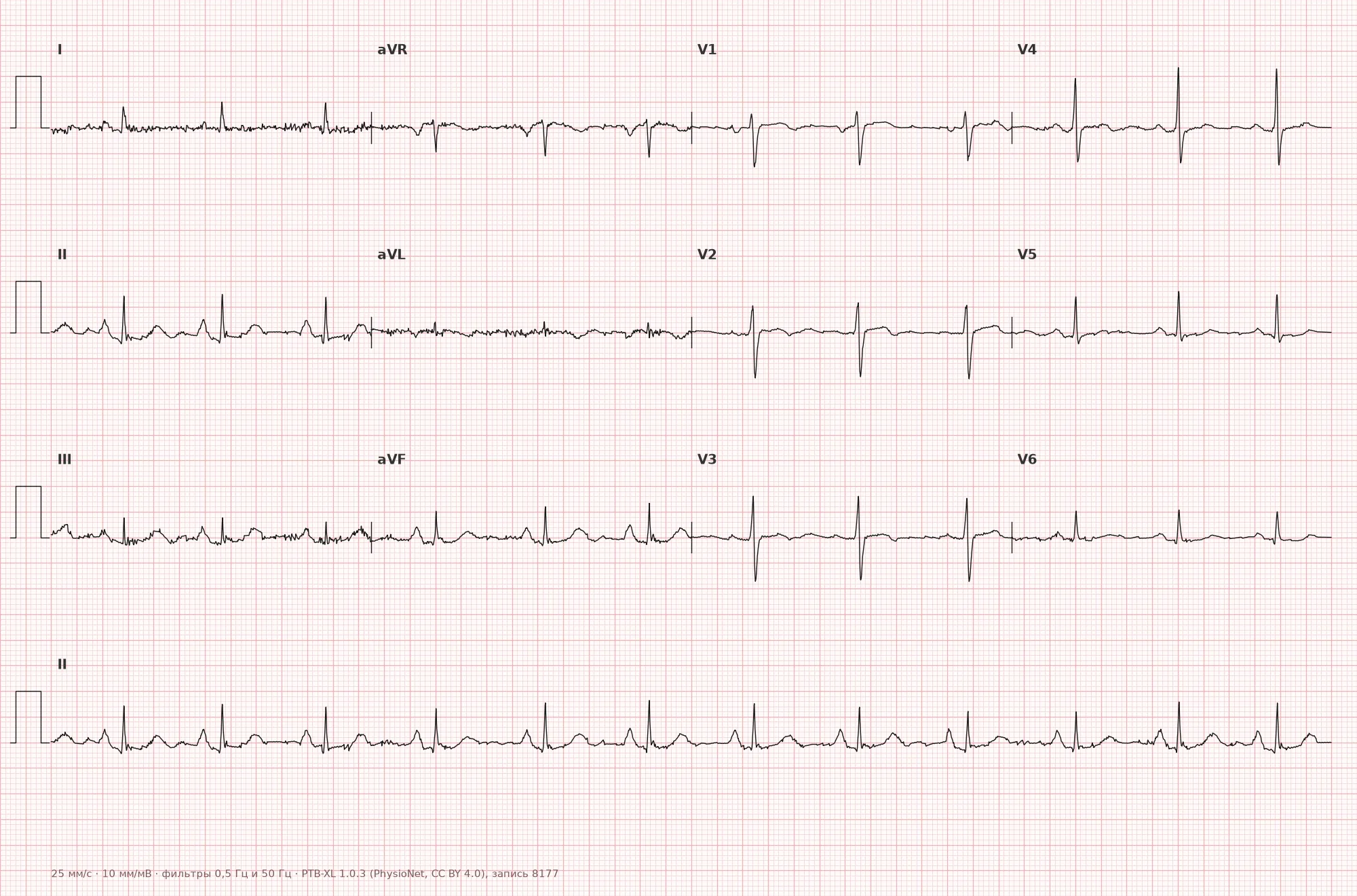
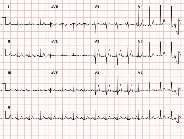
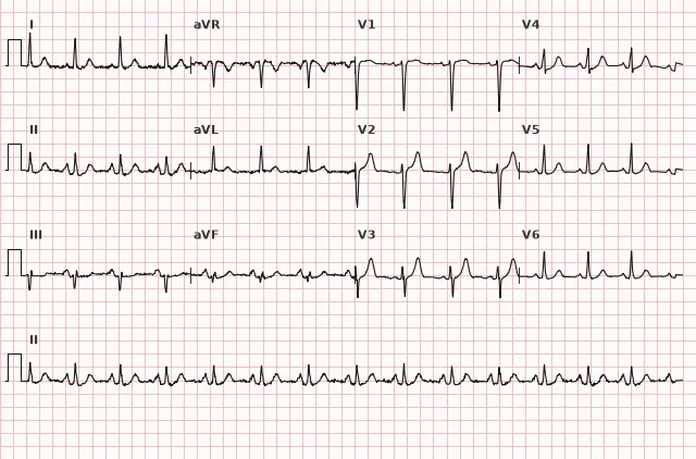
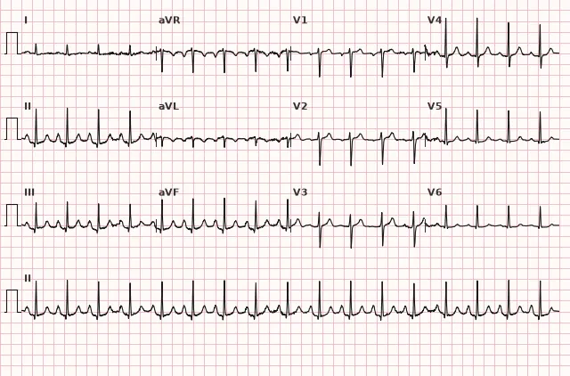
ECG from the open PTB-XL dataset (Wagner P. et al., Scientific Data, 2020; PhysioNet), CC BY 4.0 license. Rendering by WhiteCoat Pro.
Mechanism
Atrial depolarization on the ECG is the sum of the right and left atrial vectors, slightly offset in time: the right atrium activates first, forming the initial part of the P wave, while the left activates slightly later, forming the terminal part.
In pressure or volume overload of the right atrium, the amplitude of its electrical vector may increase, whereas depolarization time changes very little because the initial right atrial myocardial mass is small. Theoretically, this explains the tall, but not necessarily widened, P wave. However, P wave height and morphology correlate poorly with anatomical right atrial size and condition: in confirmed dilatation or hypertrophy, P pulmonale may be absent, and the sensitivity and specificity of the sign are limited. Therefore, P pulmonale is more correctly considered an electrocardiographic marker of possible right atrial overload or anomaly rather than proof of its enlargement — anatomical assessment is provided by echocardiography.
Causes
- Chronic obstructive pulmonary disease, emphysema, chronic cor pulmonale (hence the term "P pulmonale").
- Pulmonary hypertension of any etiology (primary, due to chronic thromboembolic disease, pulmonary vascular pathology).
- Tricuspid stenosis or regurgitation.
- Pulmonary stenosis, other congenital heart defects with right-sided overload (e.g., atrial septal defect).
- Tetralogy of Fallot and other congenital defects with right ventricular and atrial overload.
- Acute right heart overload in massive pulmonary embolism (PE) — high P pulmonale is inconsistent in this setting and is used neither to diagnose nor to rule out PE; ECG in acute PE is often normal or shows non-specific changes, and management is determined by clinical probability, D-dimer, and imaging rather than evaluation of a single P wave.
- Sometimes — a normal variant in healthy individuals without structural pathology.
Differential diagnosis
- Left atrial enlargement (P mitrale) — P wave is widened (≥ 120 ms), notched in lead II; in V1, the terminal negative force of the P wave is assessed (Morris criterion) — the product of the depth and duration of the terminal negative phase, with an absolute value of approximately ≥ 0.04 mm·s (roughly depth ≥ 1 mm and duration ≥ 40 ms); this sign indicates left atrial enlargement and is also not strictly specific. In isolated typical P pulmonale, P wave width remains normal, but P widening per se does not exclude concomitant right atrial pathology.
- Biatrial enlargement — a combination of a tall initial (right atrial) component and a widened terminal (left atrial) P wave component.
- Normal variant of a tall P wave — no clinical data indicating pulmonary or valvular disease, and no concomitant signs of right ventricular overload.
- Wandering atrial pacemaker or ectopic atrial rhythm — P wave morphology changes from beat to beat or differs in morphology and axis from sinus P waves, rather than simply having increased amplitude.
Clinical significance and management
P pulmonale is not life-threatening on its own and does not confirm anatomical right atrial enlargement — it is a non-specific electrocardiographic marker of possible overload or anomaly, serving as an indication to search for an underlying cause: chronic pulmonary disease, pulmonary hypertension, or right-sided valvular heart disease.
When P pulmonale is detected, the physician should evaluate the overall ECG for signs of right ventricular hypertrophy and overload (right axis deviation, tall R in V1, T wave inversion in the right precordial leads), correlate with the clinical picture (dyspnea, chronic cough, edema, history of COPD), and refer the patient for echocardiography to assess atrial dimensions, pulmonary artery pressure, and valve status. The sensitivity and specificity of ECG criteria for right atrial enlargement are limited, so the diagnosis is not established by ECG alone — echocardiography remains the primary confirmation method.
Common interpretation errors
- Confusing high P pulmonale with a normal variant of a tall P wave in tachycardia or sympathicotonia without assessing morphology and clinical context.
- Failing to measure P wave amplitude accurately using calibration (1 mm = 0.1 mV), overestimating or underestimating wave height by eye.
- Evaluating the criterion using lead II only and missing a tall P wave in lead III or aVF when amplitude in lead II is normal.
- Interpreting a tall P pulmonale as a reliable sign of acute PE or as proof of anatomical atrial dilatation, whereas it is merely a non-specific electrical marker.
- Considering P wave widening as definitive exclusion of right atrial pathology, failing to account for the possibility of combined biatrial enlargement.
"P pulmonale — tall, but not wide" — the P wave in inferior leads.
1. Which ECG sign is most characteristic of right atrial enlargement?
P pulmonale is an amplitude criterion in the inferior leads (II, III, aVF) with typically preserved P wave duration, in contrast to left atrial enlargement.
2. What pathology is most commonly associated with P pulmonale?
The name P pulmonale is historically associated specifically with chronic pulmonary pathology and right heart overload.
3. In which leads should the amplitude of the P wave be evaluated when right atrial enlargement is suspected?
The criterion for a tall P wave is evaluated in all inferior leads — II, III, and aVF; an increased amplitude in at least one of them is taken into account, not just in lead II.
Frequently asked questions
How to distinguish P pulmonale from P mitrale?
P pulmonale is a tall (> 2.5 mm in II, III, or aVF) P wave, usually of normal duration. P mitrale is a notched, wide (≥ 120 ms) P wave with a deep and wide negative phase in V1. The main differentiator is width, rather than height, although P wave widening does not in itself rule out concomitant right atrial pathology.
Is right atrial enlargement on an ECG dangerous?
The phenomenon itself is not directly life-threatening, but it indicates possible right heart overload and requires searching for the cause — chronic pulmonary pathology, pulmonary hypertension, or tricuspid/pulmonary valve disease.
Can P pulmonale be a normal variant?
A tall P wave is sometimes found as an individual variant, including in individuals with a more vertical heart position, without structural pathology. A diagnosis is not made based on a single ECG: technical recording errors must be ruled out, clinical presentation evaluated, and echocardiography performed if indicated.
What P wave amplitude threshold is considered diagnostic?
Most sources indicate greater than 2.5 mm in the inferior leads (II, III, aVF) at a paper speed of 25 mm/s, while some sources lower the threshold to ≥ 2.0 mm. Lead II is usually the most convenient for measurement, but the criterion is also met with an isolated increase in lead III or aVF.
Is echocardiography necessary when P pulmonale is suspected?
Yes, ECG criteria for right atrial enlargement have limited sensitivity and specificity and do not confirm anatomical atrial enlargement, which is why echocardiography remains the main method for confirming atrial dimensions and estimating pulmonary artery pressure.
ECG Course
Interpretation from basics to myocardial infarction — learn to read any ECG using a systematic algorithm.
- Video lessons and theory for every topic
- Simulator case studies under every lesson
- Unlimited ECG simulator practice
- Study guide: "All ECGs in 25 minutes"
ECG Strip Simulator
ECG, radiology, histology, pathomorphology, pharmacology — thousands of flashcards with detailed explanations.
- Real ECG strips, not textbook drawings
- Instant answers and explanations
- Streaks and progress tracking in your dashboard
Related terms
More in the section «Hypertrophy and Chamber Enlargement»
Sources
- Goldberger's Clinical Electrocardiography, textbook
- Surawicz, Knilans. Chou's Electrocardiography in Clinical Practice
- Orlov V.N. Guide to Electrocardiography
- AHA/ACCF/HRS Recommendations for the Standardization and Interpretation of the ECG, 2009
Educational material for medical students and physicians. Does not replace clinical guidelines and physical examination; the ECG must always be interpreted in conjunction with the clinical picture.