معايير تخطيط القلب
- A recognizable atrial impulse is conducted before every QRS; sinus rhythm is typical but not obligatory
- PR is usually prolonged beyond 200 ms and conduction is 1:1; slight physiological beat-to-beat PR variability is acceptable
- Every P wave is conducted to the ventricles (P:QRS ratio = 1:1), no dropped QRS complexes
- QRS complex of normal width (< 120 ms) unless there is concomitant bundle branch block
كيف يظهر في التخطيط
The PQ interval is measured from the onset of the P wave to the onset of the QRS complex. Normally it is 120–200 ms (3–5 mm). In first degree AV block, PQ is prolonged beyond 200 ms, but every recognizable P wave is conducted to the ventricles (1:1 conduction).
| المؤشر | طبيعي | إحصار أذيني بطيني من الدرجة الأولى |
|---|---|---|
| الفاصل PQ | 120–200 مللي ثانية | > 200 ملي ثانية |
| P wave (in sinus rhythm) | Positive in II, III, aVF | Unchanged |
| P:QRS ratio | 1:1 | 1:1 (every P conducted) |
| مركب QRS | < 120 ملي ثانية | Usually unchanged |
Measure PR in the lead with the most clearly defined onset of P and onset of QRS; this is often II or V1. Important: the PQ interval is in most cases stable from complex to complex (unlike the progressive prolongation in Wenckebach periodicity in second degree AV block, Mobitz type I), although small PR fluctuations are possible with changes in autonomic tone or heart rate. With marked PQ prolongation, the P wave may overlap the preceding T wave, creating a pseudo-notched T wave.
مخططات حقيقية: 8 أمثلة
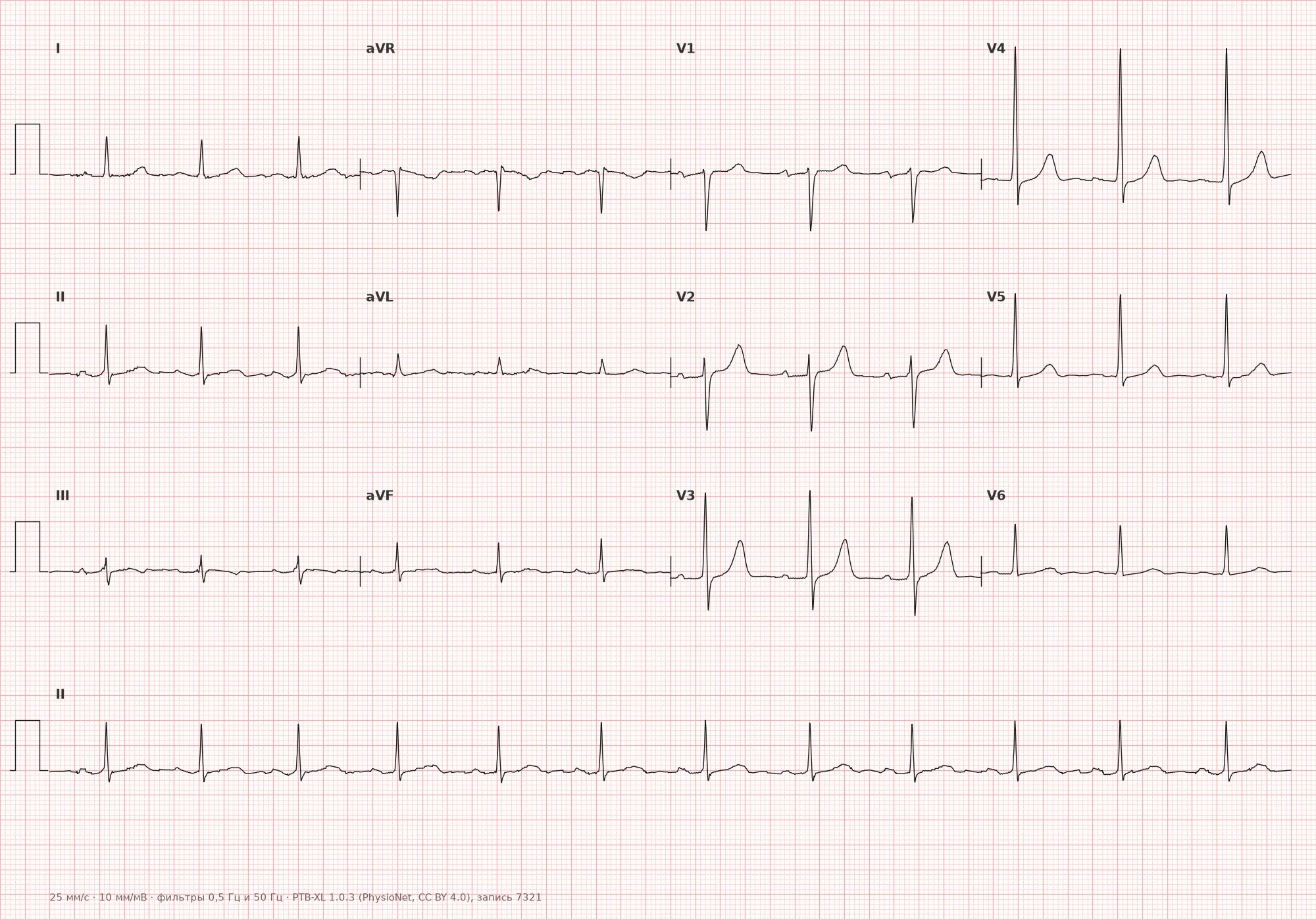
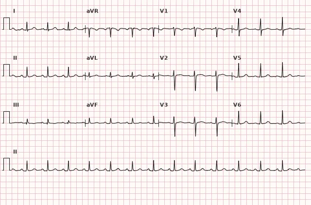
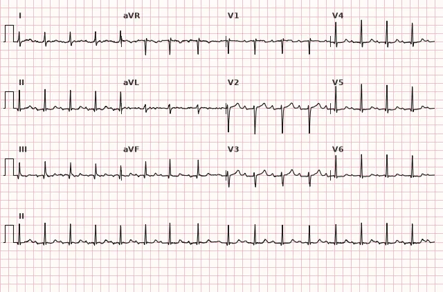
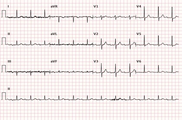
مخطط قلب من قاعدة البيانات المفتوحة PTB-XL (Wagner P. et al., Scientific Data, 2020; PhysioNet)، ترخيص CC BY 4.0. الرسم - منصة WhiteCoat Pro.
الآلية
The impulse from the sinus node spreads through the atrial myocardium to the AV node, where the main physiological delay occurs (normally 50–120 ms, which constitutes a significant part of the PQ interval), then it is conducted through the His–Purkinje system to the ventricular myocardium. The PR interval itself reflects the total time of intra-atrial, AV nodal, and His–Purkinje conduction. On a standard ECG the level of block is usually not determined; more often the delay is located in the AV node, but an infrahisian level or combined slowing is possible. Electrophysiological studies show that in most patients the delay is located in the compact portion of the AV node (AH interval prolonged), and such block responds well to atropine. Less commonly, prolongation occurs in the His–Purkinje system (HV interval), which indicates a more distal level of involvement and may be a harbinger of higher degrees of block.
الأسباب
الفسيولوجية
- High vagal tone (athletes, healthy young people, sleep)
- Use of parasympathomimetics, beta-blockers, non-dihydropyridine calcium channel blockers (verapamil, diltiazem), class III antiarrhythmics (amiodarone, sotalol), digitalis
Pathological
- Degenerative changes of the conduction system — Lev–Lenègre disease (sclerodegenerative involvement of the conduction system), age-related fibrosis
- Coronary artery disease, especially inferior wall infarction (occlusion of the right coronary artery supplying the AV node)
- Myocarditis and inflammatory heart diseases; in rheumatic fever, carditis with AV conduction disturbances is possible
- Infiltrative processes (sarcoidosis, amyloidosis, hemochromatosis)
- Hyperkalemia (in combination with QRS widening)
- After cardiac surgery (aortic valve replacement, correction of congenital defects) or in certain congenital defects
التشخيص التفريقي
Second degree AV block, Mobitz type I (Wenckebach): the PQ interval is not constant but gradually lengthens from cycle to cycle, followed by a dropped QRS (the P wave is not conducted). In first degree AV block, PQ is stably prolonged, but every P is conducted.
P wave partially overlapping T: if the P wave is inconspicuous and hidden at the end of the preceding T, PQ may be erroneously measured from the "hump" on T to QRS. Always look for the onset of P — it may be on the descending limb of T.
Bundle branch block: a QRS width > 120 ms results from intraventricular delay, but PQ may be normal. A prolonged PQ with bundle branch block means a combination of two conduction disturbances.
Normal upper limit of PQ in the young: in children and adolescents the upper limit of normal may be 180 ms. In the Russian Ministry of Health clinical guidelines on bradyarrhythmias (2020) and AHA/ACCF/HRS (2009), the 200 ms threshold applies to adults; in children, age-based nomograms are used.
الأهمية السريرية والتكتيكات
First degree AV block is usually benign and does not cause symptoms. Prevalence depends on age, population, and selection criteria and varies considerably between studies; it is more common in older people and in structural heart disease than in young healthy individuals. With normal or moderately prolonged PR, hemodynamic consequences are usually absent; with marked PR > 300 ms, AV dyssynchrony and symptoms are possible. Patients do not require treatment if there are no symptoms and no risk of progression.
When to be vigilant:
- The combination of PR > 200 ms with bifascicular block requires assessment of the clinical context and the risk of distal conduction disturbance; the term "trifascicular block" should not be used solely on the basis of PR prolongation.
- Marked PQ prolongation (> 300 ms) may be accompanied by complaints of palpitations or reduced exercise tolerance due to shortened diastolic filling and AV dyssynchrony (pseudopacemaker syndrome).
- Acute myocardial infarction: first degree AV block in inferior MI is most often reversible; in anterior MI it may indicate extensive involvement of the interventricular septum and a risk of complete AV block.
- Progressive PQ prolongation or progression to second–third degree AV block.
Management:
- Assess use of drugs that slow AV conduction (beta-blockers, digitalis, verapamil, amiodarone); consider dose adjustment or discontinuation.
- Workup is determined by the clinical context. Assess medications and electrolytes; evaluate for ischemia, myocarditis, or Lyme disease if there are symptoms, acute onset, relevant history, signs of inflammation/ischemia, or unexplained progressive block.
- If intermittent higher-degree block is suspected or if symptoms are present — ECG monitoring, the type and duration of which are chosen according to the clinical picture and the expected event rate.
- With PR usually more than 300 ms and symptoms clearly related to AV dyssynchrony, pacemaker implantation may be considered individually. When combined with bundle branch block, management is determined by symptoms, documented high-grade or infrahisian block, and/or electrophysiological study results; PR prolongation alone is not an indication for pacing.
Most patients with isolated first degree AV block are observed without treatment.
الأخطاء الشائعة أثناء قراءة تخطيط القلب
- Measuring PQ not from the onset of P: the onset of P may be gradual, especially in low-amplitude leads. Measure in the lead with a clear onset of the P wave (usually II or V1).
- Missing a P wave overlapping T: in tachycardia or with a long PQ, the P wave may "hide" in the preceding T wave. If suspected, analyze a long recording, the P morphology in other leads, and the P–QRS relationship; alternating T shape is not a reliable sign of P overlap and should not be used as a diagnostic criterion.
- Diagnosing "first degree AV block" in the absence of atrial activity: if the rhythm originates from the AV node (junctional rhythm), P may be absent or retrograde, and the term "first degree AV block" does not apply.
- Forgetting to check ECG calibration and paper speed: at 50 mm/s, the same 10 mm corresponds to 200 ms, not 400 ms. Always verify the recording speed.
- Overlooking functional block: PQ prolongation during beta-blocker use or in an athlete is not a disease but an adaptation or drug effect. After discontinuation of the drug, PQ may normalize.
"First degree — everyone gets through, but slowly": every P is conducted, but PQ > 200 ms (5 small squares).
1. On the ECG the PQ interval = 240 ms, there is a P wave before every QRS, the rhythm is regular, heart rate 68 per minute. What is the diagnosis?
فترة PQ > 200 ms (240 ms) مع مرور كل موجة P إلى البطينات (1:1) — هذا تعريف إحصار AV من الدرجة الأولى. النظم منتظم، ولا توجد دورية Wenckebach، ولا فقدان لـ QRS.
2. لدى المريض في ECG فترة PQ 160 ms في مسارات الأطراف و220 ms في المسارات الصدرية. أين يُقاس PQ بشكل صحيح؟
تُقاس فترة PQ في المسار الذي يظهر فيه بوضوح بداية موجة P وبداية مركّب QRS، عادةً II أو aVF أو V1. غالبًا ما يرتبط تباين القيم بعدم وضوح بداية P في مسارات مختلفة، وليس باختلاف حقيقي في زمن التوصيل.
3. في احتشاء عضلة القلب السفلي ظهر إحصار AV من الدرجة الأولى (PQ 240 ms). ما هو مستوى الإحصار الأرجح؟
في احتشاء عضلة القلب السفلي (انسداد الشريان التاجي الأيمن) غالبًا ما تتأثر العقدة AV، التي يغذيها هذا الشريان. الإحصار على مستوى العقدة AV عادةً قابل للعكس، ويستجيب جيدًا للأتروبين، وله إنذار جيد.
الأسئلة الشائعة
هل إحصار AV من الدرجة الأولى خطير؟
الإحصار AV المعزول من الدرجة الأولى عادةً غير خطير ولا يتطلب علاجًا. قد يكون نمطًا طبيعيًا لدى الرياضيين والشباب الأصحاء. يلزم الحذر عند اقترانه بإحصار الحزم، أو الإطالة الواضحة لـ PQ > 300 ms مع أعراض، أو احتشاء حاد، أو علامات تطور إلى درجات إحصار أعلى.
كيف نميّز إحصار AV من الدرجة الأولى عن إحصار AV من الدرجة الثانية Mobitz I؟
في إحصار AV من الدرجة الأولى تكون فترة PQ مطولة بثبات (> 200 ms)، وتُوصَّل كل موجة P إلى البطينين (1:1). في Mobitz I (Wenckebach) تطول فترة PQ تدريجيًا من دورة إلى أخرى، ثم يسقط مركّب QRS واحد (دورية Samoilov–Wenckebach)، وبعدها تقصر PQ وتتكرر الدورة.
هل يمكن أن يتحول إحصار AV من الدرجة الأولى إلى إحصار كامل؟
نادرًا ما يتطور إحصار AV المعزول من الدرجة الأولى إلى إحصار AV كامل إذا لم توجد عوامل إضافية. يزداد الخطر عند الاقتران بإحصارات الحزم (إحصار ثنائي الحزمة)، أو احتشاء عضلة القلب الحاد، أو التهاب عضلة القلب، أو الأمراض التنكسية لجهاز التوصيل. يُختار نوع المراقبة وضرورتها حسب الأعراض والتكرار المتوقع للأحداث؛ وفي الإغماء المصحوب بإحصار ثنائي الحزمة يلزم تقييم متخصص، غالبًا مع مراقبة أطول أو دراسة الفيزيولوجيا الكهربائية.
هل يجب علاج إحصار AV من الدرجة الأولى عند تناول حاصرات بيتا؟
إذا حدث إحصار AV من الدرجة الأولى على خلفية حاصرات بيتا ولم توجد أعراض، فلا حاجة إلى علاج نوعي. قيّم التأثير السريري ودواعي استخدام الدواء. عند الإطالة الواضحة لـ PQ (> 300 ms) أو ظهور درجات إحصار أعلى، فكّر في تقليل الجرعة أو استبدال الدواء.
ما هو إحصار AV من الدرجة الأولى «marked»؟
يُستخدم مصطلح «marked» (بالإنجليزية: marked) للإطالة الواضحة في PQ، عادةً > 300 ms. قد تؤدي هذه الإطالة إلى تقصير الانبساط، وتراكب انقباض الأذينين على انقباض البطينين، وأعراض تشبه متلازمة الناظمة القلبية. في حالات نادرة، عند PQ > 300 ms وأعراض مرتبطة بوضوح باختلال التزامن AV، يمكن مناقشة زرع ناظمة قلبية بشكل فردي.
دورة تخطيط القلب الكهربائي
التفسير من الأساسيات وحتى احتساء الاحتشاءات — لقراءة أي مخطط وفق خوارزمية دقيقة.
- دروس فيديو ونظرية لكل موضوع
- حالات تدريبية تحت كل درس
- جهاز محاكاة تخطيط القلب بلا حدود يومية
- دليل «جميع تخطيطات القلب في 25 دقيقة»
جهاز محاكاة التخطيطات
تخطيط القلب، الأشعة، علم الأنسجة، علم الأمراض، الصيدلة — آلاف البطاقات مع الشرح.
- مخططات حقيقية وليست مجرد صور من الكتب
- الإجابة والشرح الفوري
- السلاسل والتقدم في الحساب الشخصي
المصطلحات ذات الصلة
المزيد في قسم «إحصارات AV»
المصادر
- AHA/ACCF/HRS Recommendations for the Standardization and Interpretation of the Electrocardiogram, 2009
- ESC Guidelines on cardiac pacing and cardiac resynchronization therapy, 2021
- Surawicz B., Knilans T. Chou's Electrocardiography in Clinical Practice, 6th ed., 2008
- التوصيات السريرية «اضطرابات النظم البطيئة»، وزارة الصحة الروسية، 2020
مادة تعليمية لطلاب كليات الطب والأطباء. لا تحل محل التوصيات السريرية والفحص الطبي؛ يُفسر تخطيط القلب دائماً جنباً إلى جنبกับการحالة السريرية.