ECG criteria
- A recognizable atrial impulse precedes every QRS complex; sinus rhythm is typical but not mandatory
- The PR interval is usually prolonged (> 200 ms) with 1:1 conduction; minor physiological variability of the PR interval from cycle to cycle is acceptable
- Every P wave is conducted to the ventricles (P:QRS ratio = 1:1), with no dropped QRS complexes
- Normal QRS complex duration (< 120 ms) in the absence of concomitant bundle branch block
What it looks like on ECG
The PQ interval is measured from the onset of the P wave to the onset of the QRS complex. Normal values range from 120 to 200 ms (3–5 mm). In first-degree AV block, the PQ interval is prolonged beyond 200 ms, but every recognizable P wave is conducted to the ventricles (1:1 conduction).
| Parameter | Normal | First-degree AV block |
|---|---|---|
| PQ interval | 120–200 ms | > 200 ms |
| P wave (during sinus rhythm) | Positive in leads II, III, aVF | Unchanged |
| P:QRS ratio | 1:1 | 1:1 (every P wave is conducted) |
| QRS complex | < 120 ms | Usually unchanged |
Measure the PR interval in the lead where the onset of the P wave and the onset of the QRS complex are most clearly defined; this is often lead II or V1. Note: In most cases, the PQ interval remains stable from beat to beat (unlike the progressive PR prolongation seen in Mobitz type I Wenckebach periodicity in second-degree AV block), although minor PR fluctuations may occur with changes in autonomic tone or heart rate. When the PQ interval is markedly prolonged, the P wave may superimpose on the preceding T wave, creating a pseudobifid T wave.
Real ECGs: 8 examples
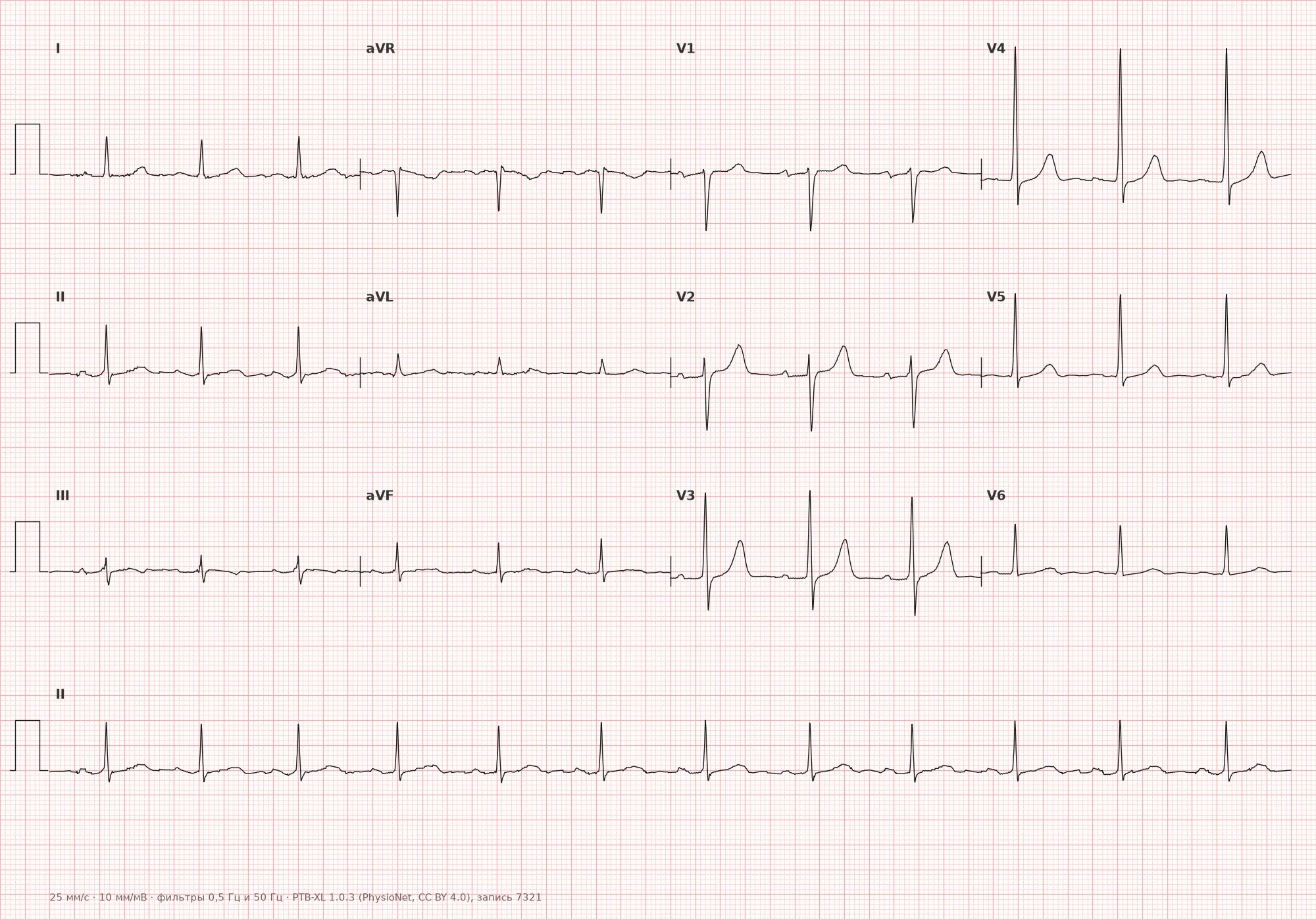
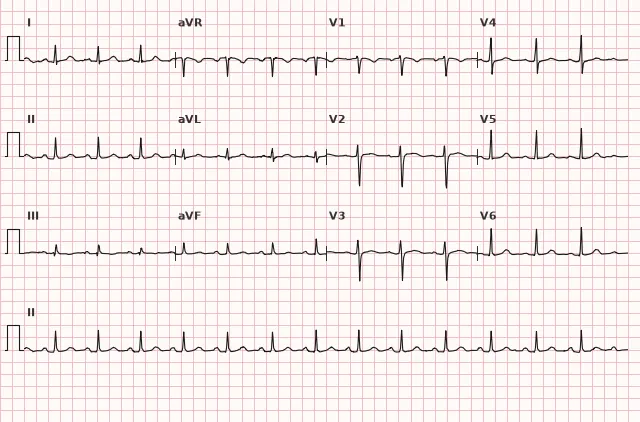
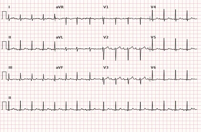
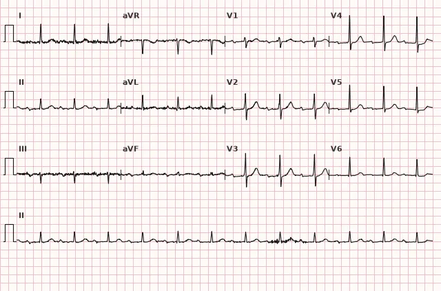
ECG from the open PTB-XL dataset (Wagner P. et al., Scientific Data, 2020; PhysioNet), CC BY 4.0 license. Rendering by WhiteCoat Pro.
Mechanism
The impulse originates in the sinus node, propagates through the atrial myocardium to the AV node, where the primary physiological delay occurs (normally 50–120 ms, accounting for a significant portion of the PQ interval), and is then conducted via the His-Purkinje system to the ventricular myocardium. The PR interval reflects the total time of intra-atrial, AV nodal, and His-Purkinje conduction. On a standard ECG, the exact level of the block is generally not determined; the delay is most frequently localized within the AV node, though an infra-hisian level or combined slowing is also possible. Electrophysiological studies demonstrate that in most patients the delay is localized in the compact AV node (prolonged A–H interval), and such block responds well to atropine. Less frequently, prolongation occurs within the His-Purkinje system (prolonged H–V interval), indicating a more distal level of disease and potentially heralding higher-grade block.
Causes
Physiological
- High vagal tone (athletes, healthy young individuals, sleep)
- Administration of parasympathomimetics, beta-blockers, nondihydropyridine calcium channel blockers (verapamil, diltiazem), class III antiarrhythmics (amiodarone, sotalol), digitalis
Pathological causes
- Degenerative changes of the conduction system — Lenègre-Lev disease (sclerodegenerative disease of the conduction system), age-related fibrosis
- Coronary artery disease, especially inferior wall myocardial infarction (occlusion of the right coronary artery supplying the AV node)
- Myocarditis and inflammatory heart diseases; rheumatic fever may cause carditis associated with AV conduction abnormalities
- Infiltrative processes (sarcoidosis, amyloidosis, hemochromatosis)
- Hyperkalemia (in combination with QRS widening)
- Post-cardiac surgery (aortic valve replacement, congenital heart defect repair) or associated with specific congenital heart diseases
Differential diagnosis
Second-degree AV block, Mobitz type I (Wenckebach): the PQ interval is not constant, but progressively lengthens from cycle to cycle until a QRS complex is dropped (the P wave is not conducted). In first-degree AV block, the PQ interval is consistently prolonged, but every P wave is conducted.
P wave partially superimposed on the T wave: if the P wave is subtle and obscured at the end of the preceding T wave, one might mistakenly measure the PQ interval from a notch on the T wave to the QRS complex. Always search for the onset of the P wave — it may be located on the downstroke of the T wave.
Bundle branch block: a QRS duration > 120 ms occurs due to intraventricular conduction delay, but the PQ interval may be normal. A prolonged PQ interval in the presence of bundle branch block indicates the coexistence of two conduction abnormalities.
Normal upper limit of PQ in the young: in children and adolescents, the upper limit of normal may be 180 ms. The Russian Ministry of Health clinical guidelines for bradyarrhythmias (2020) and AHA/ACCF/HRS guidelines (2009) apply the 200 ms threshold to adults; age-specific nomograms are used for children.
Clinical significance and management
First-degree AV block is generally benign and asymptomatic. Prevalence varies depending on age, population, and selection criteria, differing significantly across studies; it is more frequent in older adults and individuals with structural heart disease than in young, healthy subjects. With a normal or moderately prolonged PR interval, hemodynamic consequences are typically absent; however, with marked PR prolongation (> 300 ms), AV dyssynchrony and symptoms may occur. Patients do not require treatment in the absence of symptoms and risk factors for progression.
When caution is warranted:
- The combination of a PR interval > 200 ms with bifascicular block requires assessment of the clinical context and the risk of distal conduction disease; the term "trifascicular block" should not be used solely on the basis of PR prolongation.
- Marked PQ prolongation (> 300 ms) may be accompanied by complaints of palpitations or reduced exercise tolerance due to shortened diastolic filling and AV dyssynchrony (pseudo-pacemaker syndrome).
- Acute myocardial infarction: first-degree AV block in inferior MI is most often reversible, whereas in anterior MI it may indicate extensive interventricular septum involvement and the risk of complete AV block.
- Progressive PQ prolongation or progression to second- or third-degree AV block.
Management:
- Review medications that slow AV conduction (beta-blockers, digitalis, verapamil, amiodarone); consider dose adjustment or discontinuation.
- The diagnostic evaluation is determined by the clinical context. Assess medications and electrolytes; screen for ischemia, myocarditis, or Lyme disease when symptoms, acute onset, relevant history, signs of inflammation/ischemia, or unexplained progressive block are present.
- If intermittent higher-degree block is suspected or in the presence of symptoms, perform ECG monitoring, selecting the type and duration based on the clinical presentation and estimated event frequency.
- When the PR interval exceeds 300 ms and symptoms are clearly related to AV dyssynchrony, pacemaker implantation may be considered on an individual basis. When combined with bundle branch block, management is guided by symptoms, documented high-grade or infra-hisian block, and/or electrophysiological study results; PR prolongation alone is not an indication for pacing.
The majority of patients with isolated first-degree AV block are monitored without medical intervention.
Common interpretation errors
- Measuring PQ from a point other than the P wave onset: the onset of the P wave can be subtle, especially in low-amplitude leads. Measure in a lead where the onset of the P wave is distinct (usually lead II or V1).
- Missing a P wave superimposed on the T wave: during tachycardia or a prolonged PQ interval, a P wave may be "hidden" within the preceding T wave. When suspected, analyze a long rhythm strip, P-wave morphology in other leads, and P–QRS relationships; T-wave contour alterations are not a reliable sign of P-wave superimposition and should not be used as a diagnostic criterion.
- Diagnosing "first-degree AV block" in the absence of atrial activity: if the rhythm originates from the AV node (junctional rhythm), the P wave may be absent or retrograde, and the term "first-degree AV block" does not apply.
- Failing to check ECG calibration and speed: at a paper speed of 50 mm/s, the same 10 mm corresponds to 200 ms, not 400 ms. Always verify the recording speed.
- Failing to account for functional block: PQ prolongation occurring against the background of beta-blocker therapy or in an athlete is an adaptive response or drug effect, not a disease. The PQ interval may normalize upon drug withdrawal.
"First degree — everyone gets through, but slowly": every P wave is conducted, but PQ > 200 ms (5 small squares).
1. The ECG shows a PQ interval of 240 ms, a P wave precedes every QRS complex, the rhythm is regular, and the heart rate is 68 bpm. What is the diagnosis?
PQ interval > 200 ms (240 ms) with conduction of every P wave to the ventricles (1:1) is the definition of first-degree AV block. The rhythm is regular, Wenckebach periodicity is absent, and no QRS dropouts are present.
2. A patient's ECG shows a PQ interval of 160 ms in limb leads and 220 ms in chest leads. Where is it correct to measure the PQ interval?
The PQ interval is measured in the lead where the onset of the P wave and the onset of the QRS complex are clearly visible, usually II, aVF, or V1. The variation in values is often due to an unclear P wave onset in different leads rather than a true difference in conduction time.
3. First-degree AV block (PQ 240 ms) developed in the setting of an inferior myocardial infarction. What level of block is most likely?
In inferior myocardial infarction (right coronary artery occlusion), the AV node is most commonly affected as it receives its blood supply from this vessel. AV nodal block is usually reversible, responds well to atropine, and has a favorable prognosis.
Frequently asked questions
Is first-degree AV block dangerous?
Isolated first-degree AV block is generally benign and requires no treatment. It can be a normal variant in athletes and healthy young individuals. Vigilance is required when it is combined with bundle branch block, marked PQ prolongation > 300 ms with symptoms, acute myocardial infarction, or signs of progression to higher-degree block.
How to distinguish first-degree AV block from second-degree AV block Mobitz I?
In first-degree AV block, the PQ interval is stably prolonged (> 200 ms), and every P wave is conducted to the ventricles (1:1). In Mobitz I (Wenckebach), the PQ interval progressively lengthens from cycle to cycle until a QRS complex is dropped (Samoilov–Wenckebach periodicity), after which the PQ interval shortens and the cycle repeats.
Can first-degree AV block progress to complete block?
Isolated first-degree AV block rarely progresses to complete AV block in the absence of additional factors. The risk is higher when combined with bundle branch blocks (bifascicular block), acute myocardial infarction, myocarditis, or degenerative conduction system disease. The management and monitoring strategy are chosen based on symptoms and the estimated event frequency; syncope combined with bifascicular block requires specialized evaluation, often with prolonged monitoring or electrophysiological study.
Does first-degree AV block need to be treated when taking beta-blockers?
If first-degree AV block occurs during beta-blocker therapy and the patient is asymptomatic, specific treatment is not required. Assess the clinical efficacy and indications for the drug. In cases of marked PQ prolongation (> 300 ms) or the appearance of higher-degree block, consider reducing the dose or switching the medication.
What is marked first-degree AV block?
The term «marked» is used for pronounced PQ prolongation, typically > 300 ms. Such prolongation can lead to shortening of diastole, atrial systole overlapping with ventricular systole, and symptoms resembling pacemaker syndrome. In rare cases with a PQ > 300 ms and symptoms clearly related to AV dyssynchrony, pacemaker implantation may be considered individually.
ECG Course
Interpretation from basics to myocardial infarction — learn to read any ECG using a systematic algorithm.
- Video lessons and theory for every topic
- Simulator case studies under every lesson
- Unlimited ECG simulator practice
- Study guide: "All ECGs in 25 minutes"
ECG Strip Simulator
ECG, radiology, histology, pathomorphology, pharmacology — thousands of flashcards with detailed explanations.
- Real ECG strips, not textbook drawings
- Instant answers and explanations
- Streaks and progress tracking in your dashboard
Related terms
More in the «AV Blocks» section
Sources
- AHA/ACCF/HRS Recommendations for the Standardization and Interpretation of the Electrocardiogram, 2009
- ESC Guidelines on cardiac pacing and cardiac resynchronization therapy, 2021
- Surawicz B., Knilans T. Chou's Electrocardiography in Clinical Practice, 6th ed., 2008
- Clinical Practice Guidelines «Bradyarrhythmias», Russian Ministry of Health, 2020
Educational material for medical students and physicians. Does not replace clinical guidelines and physical examination; the ECG must always be interpreted in conjunction with the clinical picture.