ECG criteria
- Complete AV dissociation: the atrial rhythm (P or f/F waves) and the ventricular rhythm (QRS) are not related to each other
- The atrial rhythm is usually regular (P–P intervals are constant during sinus rhythm), and the atrial rate is generally faster than the ventricular rate; atrial fibrillation, atrial flutter, or an ectopic atrial rhythm may occur instead of sinus rhythm
- The ventricular escape rhythm is usually regular or nearly regular (R–R intervals have no fixed relationship to P–P intervals); moderate variability may occur due to autonomic tone or changes in the pacemaker site
- There is no constant relationship between P waves and QRS complexes; visible P–R intervals vary from complex to complex and may coincide by chance
- Replacement ventricular rhythm: narrower and faster (40–60 bpm, QRS <0.12 s) usually suggests a juxta- or intranodal level of escape, while wider and slower (20–40 bpm, QRS ≥0.12 s) indicates an infra-Hissian level; precise localization requires clinical context or EPS
- P waves superimposed on the QRS complex or T wave (distorting their contour) are sometimes visible
- Ventricular capture beats and fusion complexes, when their mechanism is confirmed, prove intermittent AV conduction; their absence alone does not rule out other forms of AV dissociation and is not a mandatory criterion for complete block
What it looks like on ECG
The main sign is complete independence of the atrial and ventricular rhythms.
Atrial rhythm: during sinus rhythm, P waves are regular, upright in lead II, inverted in aVR, and P–P intervals are equal. However, complete AV block can also coexist with other atrial rhythms — atrial fibrillation (f waves), atrial flutter, or an ectopic atrial rhythm. In these cases, the diagnosis is established by the presence of an independent, regular ventricular escape rhythm rather than the nature of the atrial activity.
Ventricular rhythm: QRS complexes are usually regular or nearly regular. The rate depends on the level of the escape pacemaker:
| Level of block | Escape rhythm | HR | QRS width |
|---|---|---|---|
| AV node (proximal to the bundle of His) | Junctional (from the AV junction) | 40–60 bpm | Narrow <0.12 s (unless there is concomitant bundle branch block) |
| Distal to the bundle of His (bundle branches, Purkinje fibers) | Idioventricular | 20–40 bpm | Wide ≥ 0.12 s |
P–R interval: varies from complex to complex (shorter then longer) because P waves and QRS complexes arise independently. P–P and R–R intervals are generated by two autonomous pacemakers and have no fixed phase relationship. At a specific ratio of rates (e.g., atrial rate 80 bpm, ventricular rate 40 bpm), R–R intervals may appear to be multiples of P–P intervals purely by coincidence — such a mathematical relation does not rule out complete block on its own, and the diagnosis is confirmed by the shift of P waves relative to QRS complexes on a long rhythm strip. P waves may fall on the ST segment, T wave, or remain "hidden" inside the QRS complex (making them invisible or distorting the complex). A coincidental P wave preceding a QRS complex does not imply conduction.
During sinus atrial rhythm, the atrial rate is typically faster than the ventricular rate, resulting in more P waves than QRS complexes.
Real ECGs: 7 examples
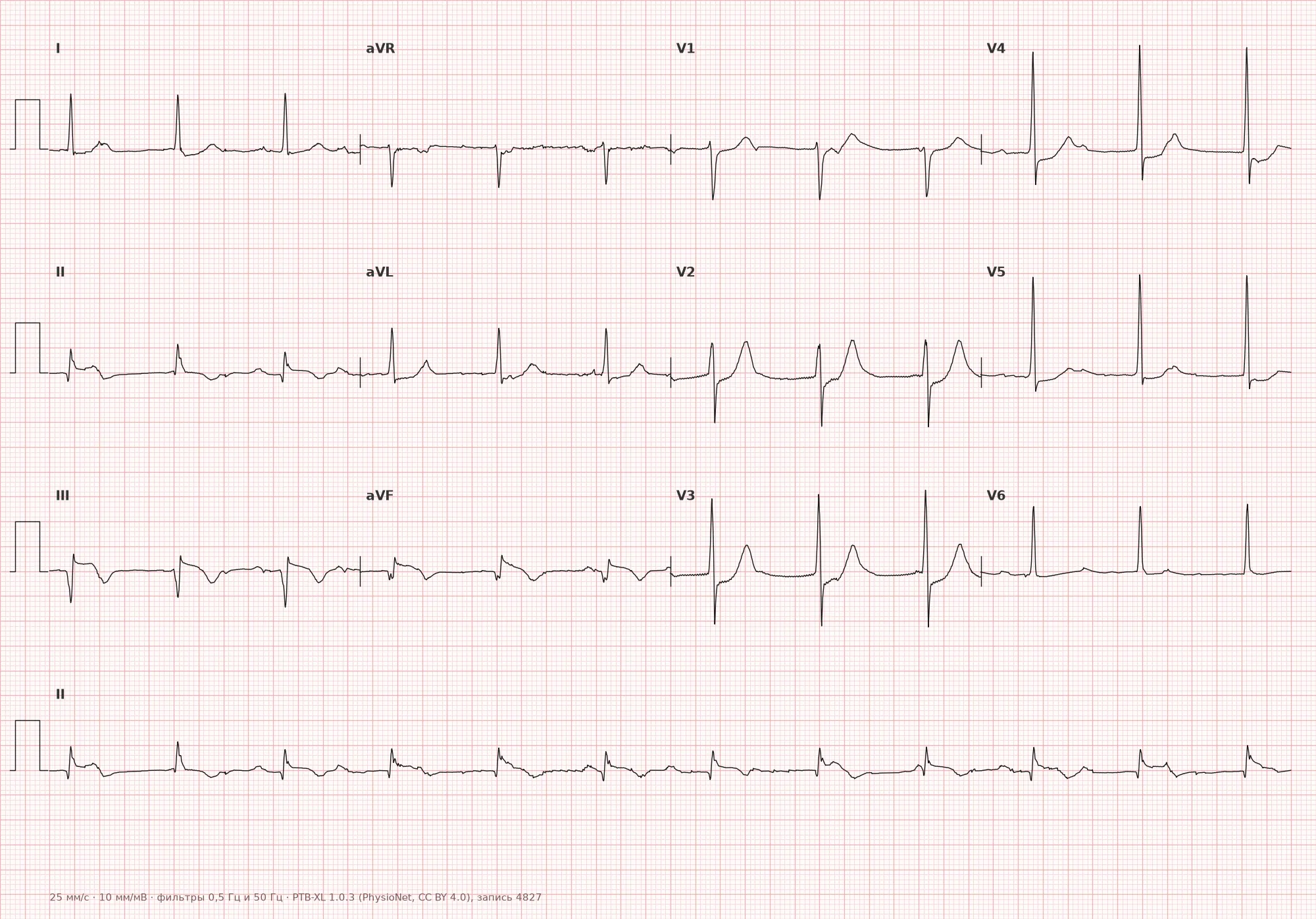
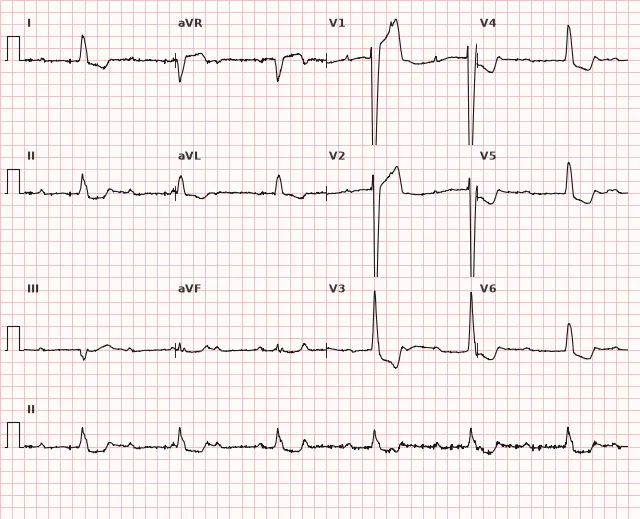
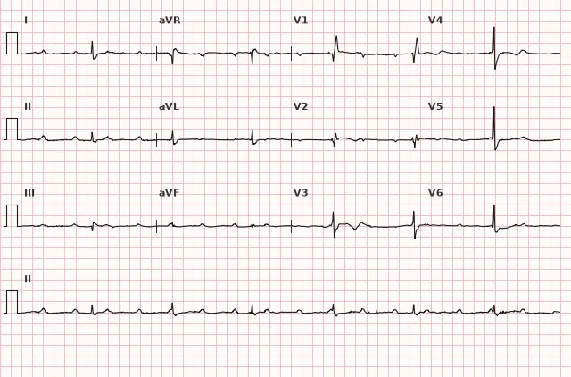
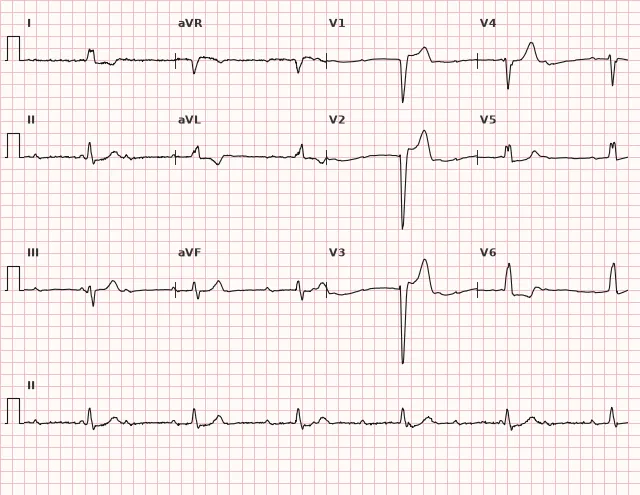
ECG from the open PTB-XL dataset (Wagner P. et al., Scientific Data, 2020; PhysioNet), CC BY 4.0 license. Rendering by WhiteCoat Pro.
Mechanism
Normally, the impulse from the sinus node travels through the AV node, the bundle of His, and its branches to the ventricular myocardium. In complete third-degree AV block, conduction is completely interrupted at one of the levels:
- AV node — most commonly due to functional causes (drug overdose, high vagal tone, inferior wall myocardial infarction).
- Bundle of His or both bundle branches — most commonly due to organic damage (anterior MI, degenerative changes of the conduction system, fibrosis).
The atrial rhythm continues independently of the ventricular rhythm: during sinus rhythm, regular P waves are seen on the ECG; during atrial fibrillation, f waves; and during atrial flutter, F waves. However, these impulses do not reach the ventricles. To prevent asystole, a subordinate pacemaker is activated (escape rhythm): junctional or idioventricular. The atria and ventricles operate at their own rhythms — resulting in AV dissociation.
The lower the level of the block, the slower the escape rhythm and the worse the hemodynamics.
Causes
Cardiac:
- Acute myocardial infarction (inferior — AV node, anterior — His–Purkinje system)
- Chronic coronary artery disease, cardiosclerosis
- Degenerative conduction system diseases (Lenègre's disease, Lev's disease)
- Myocarditis (diphtheritic, viral, Lyme disease, Chagas disease)
- Cardiac surgery (aortic valve replacement, VSD repair)
- Infiltrative diseases (sarcoidosis, amyloidosis, hemochromatosis)
- Isolated congenital complete AV block — most commonly associated with transplacental exposure to maternal anti-Ro/SSA and anti-La/SSB antibodies (can also occur with clinically asymptomatic maternal SLE); also found in complex congenital heart disease and following surgical repair
- Mitral or aortic annular calcification
Drug-induced and metabolic:
- Antiarrhythmics (beta-blockers, nondihydropyridine calcium channel blockers, digoxin, amiodarone)
- Hyperkalemia
- Overdose of AV nodal blocking drugs
Functional (reversible):
- Increased vagal tone (in young athletes — rare)
- Obstructive sleep apnea — may cause predominantly nocturnal episodes of bradycardia and AV block; in persistent daytime complete AV block, sleep apnea is not considered the sole cause, and a structural, ischemic, drug-induced, or metabolic etiology must be investigated
Differential diagnosis
High-degree AV block (second-degree): periodically, a QRS complex is still conducted (fixed conduction 3:1, 4:1). In complete block, no impulses are conducted.
AV dissociation without block (isorrhythmic, with accelerated junctional rhythm): the ventricular rhythm occurs not due to block, but because the subsidiary pacemaker has "overtaken" the sinus node in rate, while AV conduction remains intact. Fusion beats, occurring when a conducted sinus impulse fuses with the escape rhythm, prove preserved AV conduction — but their absence on the recorded strip does not rule out this condition and is not a reliable criterion for differentiating it from complete block. Diagnosis is established by evaluating the combination of ventricular heart rate, clinical presentation, and serial ECG changes.
High-grade (advanced) second-degree AV block with 3:1 or 4:1 conduction: a repeating pattern of "several P waves to one QRS complex," meaning at least one impulse out of consecutive ones is periodically conducted. When there are two or more consecutive non-conducted P waves, the Mobitz I or Mobitz II mechanism often cannot be reliably determined from a surface ECG — a fixed ratio by itself does not prove Mobitz II, and the mechanism is assessed using available consecutive cycles and clinical context. In complete block, P–P and R–R intervals are regular and independent, without such a pattern and without a single conducted complex.
Sinus bradycardia with frequent atrial premature beats: sometimes during rhythm slowing, "extra" P waves are visible, but they are irregular. In complete block, P waves (during sinus rhythm) are regular and independent of the QRS complexes.
Atrial fibrillation with slow ventricular response: the ventricular rhythm is typically irregular. A regular, slow ventricular rhythm in the presence of f waves is suspicious for concomitant complete AV block (Holt–Oram syndrome / Wenckebach-Einthoven / *Editor's note: Fredrick's syndrome*).
Clinical significance and management
Danger: complete AV block leads to severe bradycardia, decreased cardiac output, and organ hypoperfusion. Syncope may occur — Morgagni–Adams–Stokes syndrome (sudden loss of consciousness due to asystole or polymorphic VT against the background of bradycardia). Untreated mortality is high, especially with an idioventricular rhythm.
Management:
- Acute symptomatic block (unstable hemodynamics, MAS attacks, severe bradycardia):
- Temporary cardiac pacing (transvenous or transcutaneous)
- Atropine IV (may help with block at the AV node level, useless in infra-Hissian block)
- Epinephrine or isoproterenol infusion (until pacemaker placement)
- Acute block in myocardial infarction:
- Inferior MI: often transient, block at the AV node level, narrow QRS, may resolve spontaneously during the acute phase. Temporary pacing for symptoms or instability.
- Anterior MI: usually infra-Hissian, wide QRS, high mortality, temporary pacing required and consideration of permanent pacemaker implantation.
- The decision on permanent pacing is made after the acute phase of MI and after ruling out reversible causes; with persistent complete or high-grade AV block, implantation is usually not delayed for long — the timing is determined by the clinical status, the level of the block, and the dynamics of conduction recovery according to current guidelines on post-infarction pacing. In case of instability, temporary pacing is used before resolving the issue of permanent pacing.
- Chronic complete AV block:
- Class I indication for permanent pacemaker implantation (ESC, ACC/AHA/HRS guidelines on bradyarrhythmias)
- Even with an asymptomatic course — risk of sudden cardiac arrest
- Congenital complete AV block:
- Indications for pacing are determined by a combination of factors: symptoms, prolonged pauses, width and stability of the escape rhythm, ventricular dysfunction or dilation, complex ventricular arrhythmias, QT prolongation, age, and heart rate dynamics; specific rate thresholds depend on the child's age and current guidelines.
- Asymptomatic children need not just observation, but regular specialized monitoring: Holter monitoring, assessment of ventricular function, and heart rate dynamics.
Discontinuation of triggering drugs (beta-blockers, digoxin, antiarrhythmics) in drug-induced block. Electrolyte correction.
Friedrich's syndrome — combination of atrial fibrillation (or flutter) with complete AV block: instead of regular P waves, f (or F) waves, regular slow ventricular rhythm. A regular slow escape rhythm in the setting of AF is highly suspicious for complete AV block, but pacemaker pacing, atrial flutter with fixed conduction, and other causes of rhythm regularization must be ruled out before diagnosis. If the diagnosis is confirmed and no reversible cause is found, cardiac pacing is indicated.
Common interpretation errors
- Missing a block due to P waves overlapping QRS or T waves. Carefully look for deformations of complexes and T waves — "extra" notches may turn out to be superimposed P waves. Plot a P–P interval ruler.
- Mistaking rare accidental coincidences of P before QRS for AV conduction. If P–R changes from cycle to cycle or some P waves are not followed by QRS at all, despite a sufficient interval for conduction, this is a block.
- Ruling out complete block solely because R–R is a multiple of P–P. Two independent rhythms can accidentally yield multiple intervals — this does not prove preserved conduction. One must look at the shift of P relative to QRS on a long recording.
- Confusing it with high-grade 2nd-degree AV block. In complete block, not a single conducted complex is present. In 2nd-degree block, even high-grade, conduction is visible at least occasionally.
- Making a definitive conclusion about the level of the block based solely on QRS width. A narrow and faster escape rhythm usually suggests a nodal (juxta- or intranodal) level, while a wide and slow one suggests an infra-Hissian level; however, unambiguous localization requires clinical context, and a wide QRS can also be the result of a baseline bundle branch block in a more proximal block.
- Missing Friedrich's syndrome: in AF with a regular slow ventricular rhythm, always think of concomitant complete AV block, but rule out pacing and flutter with fixed conduction. This is not "AF with slow ventricular response" (where the rhythm is irregular).
1. On the ECG: regular P waves at a rate of 80 bpm, regular QRS complexes at a rate of 35 bpm, wide QRS of 0.14 s, P–R interval changes from cycle to cycle. Diagnosis?
Complete independence of atrial (P) and ventricular (QRS) rhythms in the absence of conduction is complete AV block. A wide and slow idioventricular rhythm suggests an infra-Hissian escape, but exact localization cannot be established from a single surface ECG.
2. A 70-year-old patient with acute inferior myocardial infarction, ECG shows complete AV block, ventricular rate 45 bpm, QRS 0.09 s, BP 110/70 mm Hg, without syncope. Tactics?
In inferior MI, complete AV block is often transient (AV node damage), narrow QRS is a sign of a high-level block. With stable hemodynamics, observation is permissible with readiness for temporary pacing; if the block persists after the acute phase and reversible causes are ruled out, the issue of permanent pacing is decided within a few days rather than waiting two weeks.
3. On the ECG during atrial fibrillation (f waves), QRS complexes are regular, at a rate of 38 bpm. What is this?
A regular slow ventricular rhythm in the setting of true atrial fibrillation is highly suspicious for Friedrich's syndrome (complete AV block with an escape rhythm); pacemaker pacing and flutter with fixed conduction must be excluded, whereas in regular AF the ventricular rhythm is irregular.
Frequently asked questions
How does complete AV block differ from 2nd-degree AV block?
In complete block, no atrial impulse is conducted to the ventricles, and the atria and ventricles contract independently (AV dissociation). In 2nd-degree block, some impulses are still conducted, there is a connection between certain P and QRS waves, and pauses or Wenckebach periodicity may occur.
Is complete AV block dangerous?
Yes, this is a life-threatening rhythm disturbance. The ventricular rate is usually 20–50 bpm, and hemodynamics are compromised. Morgagni–Adams–Stokes attacks (syncope due to asystole) and sudden cardiac death may occur. Most patients require permanent cardiac pacemaker implantation.
What is Morgagni–Adams–Stokes syndrome?
This is a sudden brief loss of consciousness due to the cessation of cerebral blood supply during asystole or ventricular tachycardia against the background of complete AV block. The patient turns pale, loses consciousness, and convulsive components may occur. After rhythm restoration (spontaneously or during resuscitation), consciousness returns. Requires emergency pacing.
Can complete AV block go away on its own?
Sometimes yes, if the cause is reversible: in inferior myocardial infarction (block at the AV node level), antiarrhythmic overdose, acute myocarditis. However, in organic damage to the conduction system (anterior MI, degenerative changes, fibrosis), the block is usually irreversible and requires permanent pacing.
How to distinguish complete AV block from AV dissociation without block?
In complete block, conduction is impossible due to damage to the conduction system, the ventricular rhythm is an escape rhythm and usually slower than the sinus rhythm. In AV dissociation without block, an accelerated lower-level rhythm (nodal, idioventricular) outpaces the sinus rhythm with preserved AV nodal conduction, so fusion beats may occur during sinus impulse conduction — but their absence on a particular recording does not prove complete block; the diagnosis is made based on a combination of signs.
ECG Course
Interpretation from basics to myocardial infarction — learn to read any ECG using a systematic algorithm.
- Video lessons and theory for every topic
- Simulator case studies under every lesson
- Unlimited ECG simulator practice
- Study guide: "All ECGs in 25 minutes"
ECG Strip Simulator
ECG, radiology, histology, pathomorphology, pharmacology — thousands of flashcards with detailed explanations.
- Real ECG strips, not textbook drawings
- Instant answers and explanations
- Streaks and progress tracking in your dashboard
Related terms
More in the «AV Blocks» section
Sources
- Orlov V.N. Guidelines for Electrocardiography, 2020
- Surawicz B., Knilans T. Chou's Electrocardiography in Clinical Practice, 6th ed., 2008
- Kusumoto F. et al. 2018 ACC/AHA/HRS Guideline on the Evaluation and Management of Patients With Bradycardia and Cardiac Conduction Delay (Circulation, 2019)
- Gilyarevsky S.R., Orlov V.A. et al. Clinical guidelines "Bradyarrhythmias" of the Ministry of Health of the Russian Federation, 2020
Educational material for medical students and physicians. Does not replace clinical guidelines and physical examination; the ECG must always be interpreted in conjunction with the clinical picture.