ECG criteria
- Atrial activity is typically regular; during sinus rhythm, P waves have a stable morphology. With irregular or organized non-sinus atrial activity, diagnosis and classification require separate analysis
- Not all P waves are conducted to the ventricles — some P waves are not followed by a QRS
- Before the non-conducted P wave, the PR interval either progressively lengthens (Mobitz I) or remains fixed (Mobitz II)
- In Mobitz I, after the pause, the PR interval usually shortens compared to the last conducted complex, but does not necessarily return to the baseline value; in Mobitz II, there is no progressive PR prolongation, and the interval is generally constant
- The ratio of conducted to non-conducted P waves can be constant (3:2, 4:3) or variable
- With a 2:1 ratio, distinguishing Mobitz I from Mobitz II from a single strip is impossible — segments with a different ratio, assessment of QRS width, and a long recording are required
- The QRS complex is narrow, usually with block at the AV nodal level (Mobitz I), and wide with block in the His-Purkinje system (Mobitz II)
What it looks like on ECG
Second-degree AV block encompasses two variants that differ significantly in mechanism and prognosis.
Mobitz I (Wenckebach periodicity). The PR interval progressively lengthens from cycle to cycle, with each increment being smaller than the previous one, until a P wave fails to be conducted. Following the pause, the PR interval typically shortens relative to the last conducted complex before the dropped beat, but does not necessarily return to the initial short value — due to concealed conduction within the AV node, the interval may remain prolonged. The characteristic pattern on the ECG strip is groups of QRS complexes separated by pauses (Wenckebach periodicity), most frequently with a 3:2, 4:3, or 5:4 ratio. In classic cases, the R–R interval encompassing the dropped complex is generally shorter than two preceding R–R intervals (less than 2 PP). The QRS complex is usually narrow because the level of block is the AV node.
Mobitz II. The PR interval is constant (normal or slightly prolonged) in all conducted complexes, with no progressive prolongation before the dropped beat, followed by a sudden drop of a QRS complex. With a single dropped impulse, the interval between adjacent conducted QRS complexes is typically equal to twice the PP interval. The QRS complex is frequently wide (accompanying bundle branch block) because the level of block is the bundle of His or its branches.
2:1 AV block is a distinct form in which every second P wave is conducted. From a single strip with a constant 2:1 ratio, it is impossible to determine whether it is Mobitz I or II because there is no series of lengthening PR intervals for comparison. To clarify the level of the block, clinicians analyze QRS width, the dynamics in recording segments with a different conduction ratio, and long-term monitoring; a safe increase in heart rate (e.g., during exercise) may help, but vagal maneuvers, atropine, and exercise testing are permissible only under continuous ECG monitoring and are not routine independent methods for typing — when infranodal block with a wide QRS is suspected, they may be uninformative or risky. If persistent doubt remains, the level of block is determined via an electrophysiological study.
The best leads for analysis are II and V1, where the P wave is clearly visible, separate from the T wave and QRS complex.
Real ECGs: 8 examples
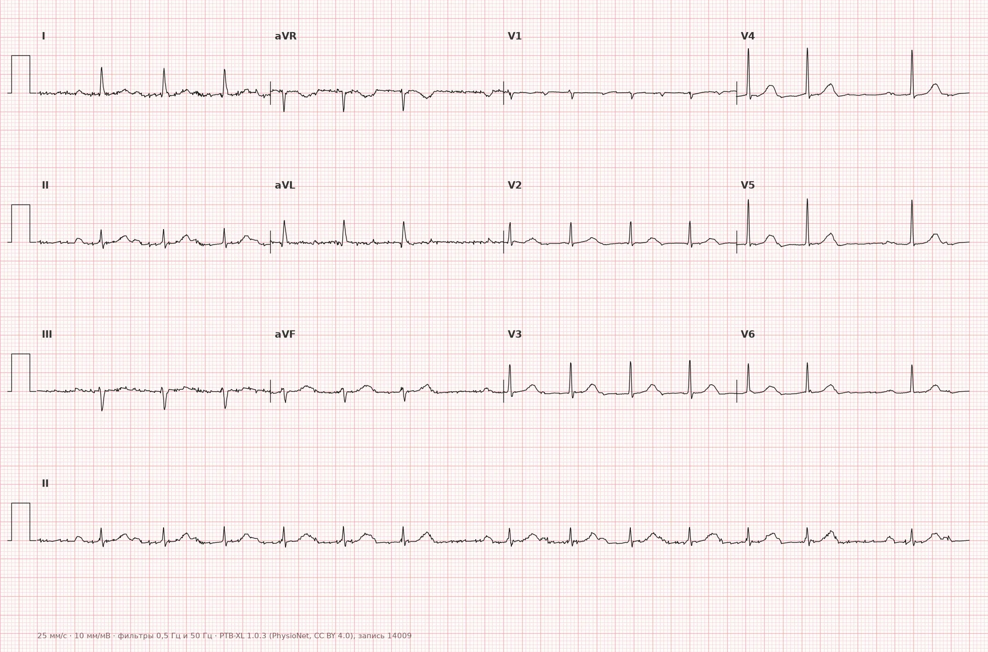
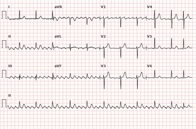
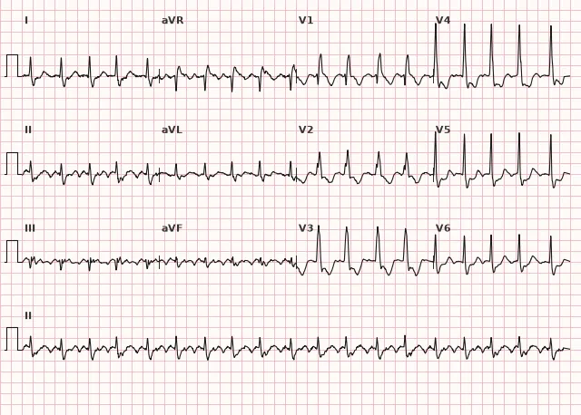
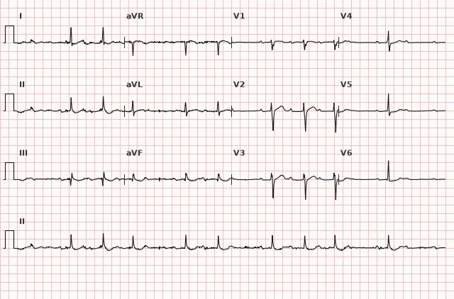
ECG from the open PTB-XL dataset (Wagner P. et al., Scientific Data, 2020; PhysioNet), CC BY 4.0 license. Rendering by WhiteCoat Pro.
Mechanism
In Mobitz I, the block occurs in the AV node due to decremental (progressively worsening) conduction: each successive impulse finds the nodal cells slightly less recovered, the PR interval increases until conduction is completely interrupted. When the block is at the nodal level, the junctional escape rhythm is usually more stable, but its presence and rate are not guaranteed — prognosis is determined not only by the mechanism but also by the level of the block, symptoms, QRS width, and the reversibility of the cause.
In Mobitz II, the block occurs distal to the AV node — in the bundle of His or its branches. Conduction through surviving fibers follows an all-or-none pattern without a gradient of slowing; thus, the PR interval is stable, and the dropped beat is sudden. This indicates organic damage to the conduction system, carrying a higher risk of progression to complete block with a slow, unreliable escape rhythm.
Causes
- Increased vagal tone (sleep, in well-trained athletes) — more commonly causes Mobitz I
- Coronary artery disease, especially inferior myocardial infarction (blood supply to the AV node originates from the right coronary artery) — usually Mobitz I
- Anterior myocardial infarction involving the interventricular septum — more frequently Mobitz II
- Degenerative fibrosis of the conduction system (Lev-Lenègre disease) in the elderly
- Drugs that slow AV conduction: beta-blockers, non-dihydropyridine calcium channel blockers, digoxin, amiodarone
- Myocarditis, infiltrative diseases (sarcoidosis, amyloidosis)
- Post-cardiac surgery or catheter-based valvular interventions
- Electrolyte disturbances (hyperkalemia)
Differential diagnosis
- Sinoatrial block — an entire PQRST cycle drops out rather than just the QRS complex; the interval between the remaining P waves is a multiple of the initial PP interval
- Blocked atrial extrasystole — the dropped complex is associated with a premature, deformed P wave rather than a regular sinus P wave
- High-degree AV block — two or more consecutive P waves fail to conduct (rather than just one), unlike classic second-degree block with single non-conducted P waves
- AV dissociation — P waves and QRS complexes proceed at their own independent rhythms without signs of blocked conduction, lacking the regular pattern of non-conducted impulses
- Respiratory sinus arrhythmia with variable PR — the PR interval varies, but no QRS complexes are dropped
Clinical significance and management
Mobitz I in asymptomatic individuals with a narrow QRS and likely vagal etiology (e.g., during sleep, in athletes) generally does not require treatment — it is a normal variant in some healthy and conditioned individuals.
New or unexplained 2:1 or high-degree AV block with a wide QRS, particularly in the presence of bradycardia, symptoms, or suspected infranodal block, warrants prompt in-person evaluation and monitoring. Acquired Mobitz II and high-grade AV block also require urgent assessment: hospitalization or continuous monitoring, immediate investigation for reversible causes (ischemia, drugs, electrolyte imbalances), and determination of the block level. Known chronic stable block with an AV nodal level (e.g., nodal 2:1 block without QRS widening and without symptoms) may not demand the same urgency, but requires planned follow-up. Once a reversible cause has been excluded, permanent cardiac pacing for Mobitz II, high-grade, or infranodal block is generally indicated regardless of symptoms — a single Holter monitor and delayed consultation are insufficient.
For hemodynamically significant bradycardia or pauses, emergency management is indicated: atropine for a likely nodal block level and early readiness for transcutaneous or transvenous pacing. Temporary pacing is indicated in cases of instability, inefficacy of drug therapy, or suspected infranodal/high-grade block, rather than for every symptomatic Mobitz presentation — invasive temporary pacing carries its own procedural risks.
In acute coronary syndrome complicated by second-degree AV block, ischemia must be treated immediately, reversible causes corrected, and hemodynamics and infarct localization evaluated; in cases of instability, temporary pacing is used as a bridge. Permanent pacemakers are implanted if significant AV block persists after the acute phase, particularly in anterior infarction and infranodal involvement; transient block in inferior infarction typically resolves following reperfusion and generally does not warrant permanent pacing.
Common interpretation errors
- Labeling any non-conducted P wave as 'Mobitz I' without analyzing PR dynamics — one must look specifically for progressive lengthening rather than just the fact that a QRS was dropped
- Attempting to definitively categorize a 2:1 conduction ratio into Mobitz I or II based on a single ECG strip — this is impossible without additional data
- Confusing a blocked atrial extrasystole with second-degree AV block without assessing the morphology and timing of the 'missing' P wave
- Failing to measure QRS width, which provides an indirect clue to the level of the block
- Mistaking sinoatrial block (dropping the entire cycle) for AV block (dropping only QRS)
Mobitz I — PQ lengthens, lengthens, lengthens... and drops (Wenckebach periodicity); Mobitz II — PQ remains constant, and suddenly a drop occurs without warning
1. Which sign is characteristic of second-degree AV block, Mobitz type I?
Progressive prolongation of PQ before a non-conducted P followed by shortening is the classic Samoilov–Wenckebach periodicity characteristic of Mobitz I.
2. Why is it impossible to immediately determine the type (Mobitz I or II) from a single ECG strip in 2:1 AV block?
With a constant 2:1 ratio, every conducted complex is immediately followed by a non-conducted P, so there is no series of lengthening PQ intervals to distinguish Mobitz I from II.
3. What level of conduction system block is typical for Mobitz II?
Mobitz II occurs distally to the AV node — in the bundle of His or its branches, which explains the sudden nature of the dropped beats and the frequent association with a wide QRS.
Frequently asked questions
How to distinguish Mobitz I from Mobitz II on ECG?
In Mobitz I, the PQ interval progressively lengthens before a non-conducted P and shortens after the pause — this is Wenckebach periodicity. In Mobitz II, PQ remains constant across all conducted complexes, and the drop is sudden, without preceding changes.
Is second-degree AV block dangerous?
It depends on the type. Mobitz I with a narrow QRS in healthy individuals (vagal origin) is usually benign, although the reliability of the escape rhythm is not guaranteed. Mobitz II and blocks with a wide QRS are considered prognostically serious due to the risk of progression to complete AV block.
Can the block type be determined in a 2:1 ratio?
A single strip with a constant 2:1 ratio cannot definitively determine whether it is Mobitz I or II, as there is no series of PQ intervals to compare. One must examine the QRS width, the duration of PQ in conducted complexes, segments of the tracing with a different ratio, or prolonged monitoring; when in doubt, an EPS is indicated.
How does second-degree AV block differ from first-degree?
In first-degree block, PQ is prolonged, but every P wave is conducted to the ventricles — there are no non-conducted P waves. In second-degree block, some P waves are not conducted, and the corresponding QRS complexes are dropped.
Is a pacemaker needed for second-degree AV block?
Asymptomatic Mobitz I with a narrow QRS and vagal origin is usually observed without a pacemaker. In confirmed Mobitz II, high-grade, or complete AV block not due to a reversible cause, permanent pacing is generally indicated regardless of symptoms. When QRS is wide and the block type is undetermined, urgent evaluation of the block level is required.
ECG Course
Interpretation from basics to myocardial infarction — learn to read any ECG using a systematic algorithm.
- Video lessons and theory for every topic
- Simulator case studies under every lesson
- Unlimited ECG simulator practice
- Study guide: "All ECGs in 25 minutes"
ECG Strip Simulator
ECG, radiology, histology, pathomorphology, pharmacology — thousands of flashcards with detailed explanations.
- Real ECG strips, not textbook drawings
- Instant answers and explanations
- Streaks and progress tracking in your dashboard
Related terms
More in the «AV Blocks» section
Sources
- Hampton J.R. The ECG Made Easy
- Surawicz B., Knilans T.K. Chou's Electrocardiography in Clinical Practice
- Orlov V.N. Guide to Electrocardiography
- ESC Guidelines on cardiac pacing and cardiac resynchronization therapy (bradyarrhythmias)
Educational material for medical students and physicians. Does not replace clinical guidelines and physical examination; the ECG must always be interpreted in conjunction with the clinical picture.