ECG criteria
- ST-segment depression with a concave (scooped) downward slope, maximal displacement in the middle of the segment, often with a smooth transition into the T wave — leads II, III, aVF, V5–V6; the morphology itself is nonspecific and may mimic ischemic depression
- The QT interval is often shortened due to a decrease in ventricular action potential duration; QTc shortening is an inconstant and nonspecific finding, and its absence does not rule out the drug effect
- Decreased amplitude, flattening, or inversion of the T wave, often with a smooth ST–T transition forming a single continuous curve
- A slight increase in U-wave amplitude may occur, but this is an inconstant finding with no independent diagnostic significance; a prominent U wave requires evaluation for bradycardia, hypokalemia, and other etiologies
- Changes are more pronounced in leads with greater R-wave amplitude; in leads with a small R wave, the changes are usually less noticeable — visibility depends on the actual QRS morphology in the specific lead
- Under normal pharmacologic effect, rhythm and AV conduction are often not significantly altered, but sinus bradycardia and slight PR prolongation may occur due to the vagotonic action of the drug; new, pronounced rhythm or conduction disturbances require evaluation for toxicity rather than being interpreted as an expected effect
- During stable therapy, ST-T changes often persist on serial ECGs, but this stability does not rule out ischemia — evaluate dynamics relative to the baseline ECG, symptoms, troponin, and the clinical context
What it looks like on ECG
The classic sign is ST-segment depression with a characteristic scooped (saucer-like) shape: the segment sags downward in a smooth curve resembling a saddle or moustache (referred to in English literature as the Salvador Dalí moustache sign). The transition from the ST segment to the T wave is often smooth, without a distinct angle; however, this morphology is not absolute and does not reliably differentiate drug effect from ischemia by visual assessment alone.
The effect is best seen in leads with a tall R wave — II, III, aVF, V5–V6, where the magnitude of ST depression correlates with R-wave amplitude. In leads with a small R wave, changes are usually less noticeable; this also applies to aVL, where R-wave amplitude depends on the electrical axis, and a horizontal or left axis deviation may result in a tall R wave in aVL with a noticeable effect.
| Element | Parameter |
|---|---|
| ST | scooped depression |
| T | flattened, decreased, sometimes biphasic |
| QT/QTc | often shortened, but inconstant |
| U | may increase slightly |
| PQ | may be slightly prolonged |
| Rhythm and AV conduction | usually without significant changes |
Heart rate may decrease slightly due to the vagotonic effect of the drug, but this is not a mandatory finding and is not among the primary criteria of the ECG effect. New marked bradycardia, sinus arrest, AV block, or other rhythm disturbances should prompt evaluation for potential toxicity rather than normal drug effect.
Real ECGs: 8 examples
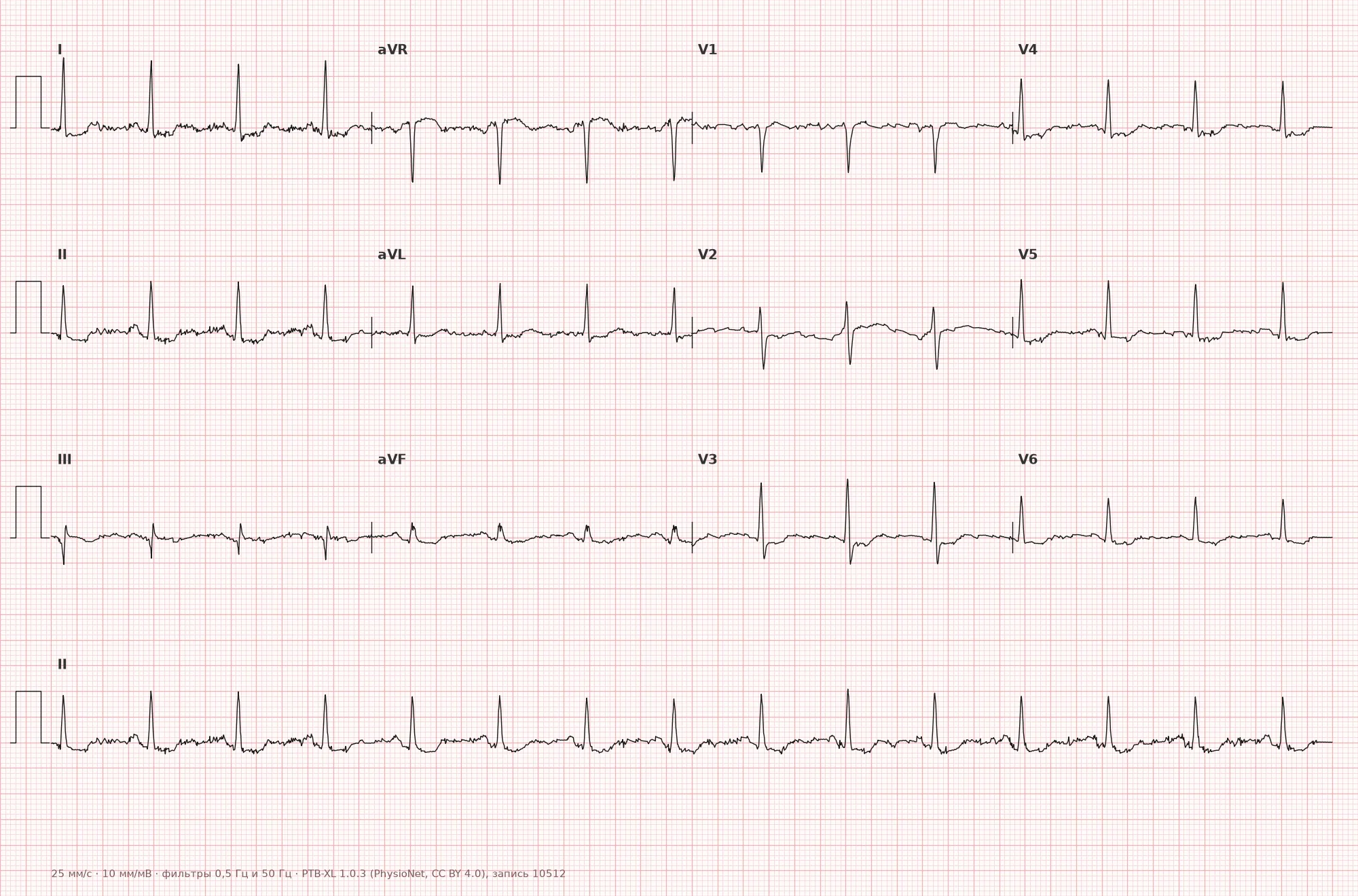
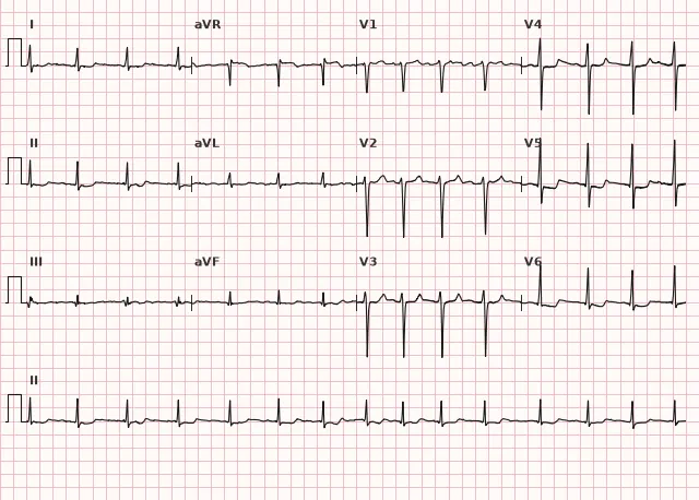
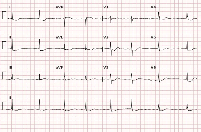
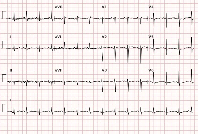
ECG from the open PTB-XL dataset (Wagner P. et al., Scientific Data, 2020; PhysioNet), CC BY 4.0 license. Rendering by WhiteCoat Pro.
Mechanism
Digoxin inhibits cardiomyocyte Na⁺/K⁺-ATPase, which, through a secondary rise in intracellular Ca²⁺, enhances contractility (positive inotropic effect). Concurrently, the drug alters ventricular repolarization and frequently shortens action potential duration and the QT interval. Alteration of the shape and slope of the repolarization phase at the ventricular myocardial level produces the characteristic ST depression and T-wave changes. Additionally, the drug increases vagal tone, slowing AV conduction (slight PR prolongation) and sinus node activity; in cases of excessive effect or toxicity, the same electrophysiology underlies bradyarrhythmias, AV blocks, and — through enhanced automaticity and triggered activity — ventricular and supraventricular arrhythmias.
Causes
- Digoxin therapy (for heart failure or rate control in atrial fibrillation) — ST-T changes can occur at both therapeutic and elevated concentrations
- Other cardiac glycosides (digitoxin) — similar, less studied effect
- The severity of changes may vary with increased drug exposure, but correlates poorly with dosage and serum concentration and is not used to assess toxicity
Differential diagnosis
- Ischemic ST depression: the morphology alone does not reliably distinguish digitalis effect from ischemia — various patterns of depression can occur in ischemia, and ischemia may coexist with digitalis changes and affect the QT interval; a scooped ST segment increases the likelihood of a glycoside effect but does not rule out ischemia — evaluate symptoms, serial ECG changes compared to baseline, troponin, and the clinical context
- Left ventricular hypertrophy with systolic overload: downsloping ST depression with asymmetric T-wave inversion, accompanied by LVH voltage criteria
- Digoxin toxicity: frequently accompanied by arrhythmias and conduction disturbances — ventricular extrasystoles, nonparoxysmal junctional tachycardia, various degrees of AV block, atrial fibrillation with a slow ventricular response, bidirectional ventricular tachycardia; these findings support the diagnosis of toxicity, but their absence does not rule it out — toxicity can manifest without characteristic arrhythmias (gastrointestinal and neurological symptoms, electrolyte disturbances, renal impairment)
- Short QT syndrome: QT is shortened without the characteristic scooped ST depression
- Hypocalcemia/hypercalcemia: affect QT duration, but the ST morphology differs and there is no association with glycoside use
Clinical significance and management
An isolated, stable ECG digoxin effect without clinical and laboratory signs of toxicity generally does not require discontinuation of the drug — it is an expected pharmacodynamic finding; decisions are made based on the totality of data rather than the ST morphology alone. It is important for the clinician to document these changes in the medical record (to avoid misinterpreting them as ischemia on subsequent ECGs) and evaluate the clinical context: symptoms of toxicity (nausea, visual disturbances, confusion), rhythm and conduction, electrolytes, and renal function.
The diagnosis of toxicity is based not on a single ECG, but on the clinical picture, time of the last drug dose (levels are typically drawn no earlier than 6–8 hours post-dose), renal function, and electrolytes. The risk of toxicity, even at normal doses, is increased by hypokalemia, hypomagnesemia, hypercalcemia, as well as advanced age, renal impairment, and drug interactions (which raise digoxin levels or enhance its effects on AV conduction); when toxicity is suspected, electrolytes and other risk factors should be evaluated comprehensively rather than in isolation. If toxicity is suspected — new arrhythmias, AV conduction blocks, marked bradycardia, hypercalcemia, hemodynamic instability — urgent clinical assessment, continuous ECG monitoring, electrolyte correction, and consultation with a toxicologist/intensivist are required; in severe toxicity with life-threatening arrhythmias, the administration of digoxin-specific antibody fragments (anti-digoxin Fab) should be considered based on clinical indications without delaying treatment for serum concentration results.
Exercise stress testing and interpretation of ST depression for angina in patients taking digoxin should be approached with caution — the drug's effect reduces test specificity for ischemia.
Common interpretation errors
- Interpreting scooped ST depression as a sign of ischemia or LV overload without accounting for drug therapy
- Automatically diagnosing digoxin toxicity based solely on ST morphology, or conversely, ruling out toxicity solely due to the absence of arrhythmias
- Relying on QT shortening as a mandatory and specific sign of drug effect
- Interpreting any bradycardia or PR prolongation in a patient on digoxin as a definitively benign effect without evaluating for toxicity
- Performing and interpreting exercise ECG stress testing without accounting for the fact that digoxin distorts the criteria for ischemic ST depression
- Waiting for digoxin level results before initiating urgent treatment in severe clinical toxicity
"Salvador Dalí's mustache" — a form of ST depression associated with digoxin effect
1. Which form of ST depression is characteristic of digoxin effect?
Scoop-like ST depression with a gradual transition into the T wave is typical of cardiac glycoside effect, although the morphology is not entirely specific and does not exclude ischemia.
2. Which of the following is true regarding the QT interval during digoxin therapy?
Digoxin shortens ventricular action potential duration, which is why the QT interval is frequently shortened; however, this is neither a mandatory nor a specific criterion.
3. Which finding supports the diagnosis of digoxin toxicity rather than merely its pharmacological effect?
The appearance of new arrhythmias and AV conduction disturbances supports the diagnosis of toxicity, although their absence does not rule out intoxication — the diagnosis is based on the combination of clinical and laboratory data.
Frequently asked questions
How to distinguish between digoxin effect and toxicity?
Under normal pharmacological action, ST-T changes and potential QT shortening predominate without clinical signs of toxicity. Toxicity is suspected based on a combination of clinical symptoms (nausea, visual and neurological disturbances), new rhythm and conduction abnormalities (ventricular extrasystoles, non-paroxysmal junctional tachycardia, AV blocks), renal function, electrolytes, and accurately measured serum drug concentrations. The absence of arrhythmias does not rule out intoxication.
Are digoxin-induced ECG changes dangerous?
Isolated ST-T changes are not dangerous on their own and do not constitute an indication to discontinue the drug. The risk is associated with the development of toxicity, which is established based on clinical presentation, arrhythmias, and laboratory findings, rather than ST morphology alone.
Can an ECG be used to assess ischemia in a patient taking digoxin?
The interpretation of ST depression during an exercise stress test becomes less specific because the drug itself induces a similar depression. Baseline ECG, dynamic changes, clinical context, and, if necessary, myocardial injury biomarkers must be taken into account.
In which leads are digoxin effects most prominent?
In leads with the highest R wave amplitude — typically II, III, aVF, V5–V6. In leads with a small R wave, the changes are generally less pronounced, although this depends on individual QRS morphology.
Does the digoxin effect disappear after drug discontinuation?
Typically, the effect gradually regresses after drug discontinuation as digoxin is eliminated, but this may take several days or longer in renal failure or overdose due to an increased half-life. Persistent ST-T changes require evaluation for other underlying causes.
ECG Course
Interpretation from basics to myocardial infarction — learn to read any ECG using a systematic algorithm.
- Video lessons and theory for every topic
- Simulator case studies under every lesson
- Unlimited ECG simulator practice
- Study guide: "All ECGs in 25 minutes"
ECG Strip Simulator
ECG, radiology, histology, pathomorphology, pharmacology — thousands of flashcards with detailed explanations.
- Real ECG strips, not textbook drawings
- Instant answers and explanations
- Streaks and progress tracking in your dashboard
Related terms
More in "Pharmacology and Toxicology"
Sources
- Goldberger's Clinical Electrocardiography, 9th ed.
- Surawicz B., Knilans T. Chou's Electrocardiography in Clinical Practice, 6th ed.
- Orlov V.N. Guide to Electrocardiography
- Hampton J.R. The ECG Made Easy
Educational material for medical students and physicians. Does not replace clinical guidelines and physical examination; the ECG must always be interpreted in conjunction with the clinical picture.