ECG criteria
- PQ (PR) interval shorter than 120 ms in adults
- Delta wave — a slurred, gradual initial upstroke of the R wave, assessed across all 12 leads (may not be prominent in all of them)
- Widened QRS complex — in adults, typically ≥ 120 ms due to the delta wave (pseudo-lengthening rather than true conduction delay); width depends on the degree of fusion between pre-excited and normal conduction. Age-specific norms are used in children.
- Secondary ST-segment and T-wave abnormalities are typically discordant with the primary QRS vector; in pre-excitation, they can complicate ischemia assessment.
- QT and QTc intervals may be prolonged due to QRS widening; repolarization assessment in pre-excitation is difficult, sometimes JT/JTc intervals are used.
- The polarity of the delta wave in leads V1, II, III, aVF, and aVL helps suspect the localization of the accessory pathway.
- WPW syndrome = pre-excitation pattern + documented tachyarrhythmia (WPW phenomenon refers to the pattern alone, without arrhythmias)
What it looks like on ECG
The three classic signs of pre-excitation are visible on sinus rhythm ECG:
| Sign | Description |
|---|---|
| Short PQ | < 120 ms |
| Delta wave | initial gradual, slurred QRS phase |
| Wide QRS | typically ≥ 120 ms in adults, morphology varies from lead to lead |
The delta wave must be sought in all 12 leads, but its prominence varies: in some leads it may be minimal or barely visible, especially with intermittent or mild pre-excitation. The absence of a clear delta wave in isolated leads does not rule out pre-excitation if the sign is present in others. The delta wave reflects early ventricular activation via the accessory pathway, bypassing the physiological delay in the AV node; the QRS width depends on the degree of fusion between the pre-excited and normally conducted components, and therefore it can vary in the same patient.
Secondary ST-T changes are typically discordant with the main QRS vector, which is typical for pre-excitation and complicates ischemia evaluation. However, WPW does not rule out ACS: pre-excitation can mask or mimic ischemic changes, so when ACS is clinically suspected, comparison with the patient's baseline ECG, serial troponins, and standard ACS management are required rather than relying solely on the background pattern.
The polarity of the delta wave and QRS in lead V1, as well as in the inferior leads, is used for approximate localization of the accessory pathway (left free wall, septal region, right free wall) — this is covered separately in localization algorithms.
During tachycardia, the picture changes: orthodromic AVRT with a narrow QRS resembles typical SVT without a visible delta wave, whereas antidromic tachycardia produces a wide QRS mimicking VT. In atrial fibrillation with conduction via the Kent bundle, the rhythm is irregularly irregular, QRS complexes are often wide with variable width and morphology due to varying degrees of fusion — relatively narrow complexes may also occur with predominant conduction through the AV node. The ventricular rate can reach 200–300 bpm, a potentially life-threatening situation.
Real ECGs: 8 examples
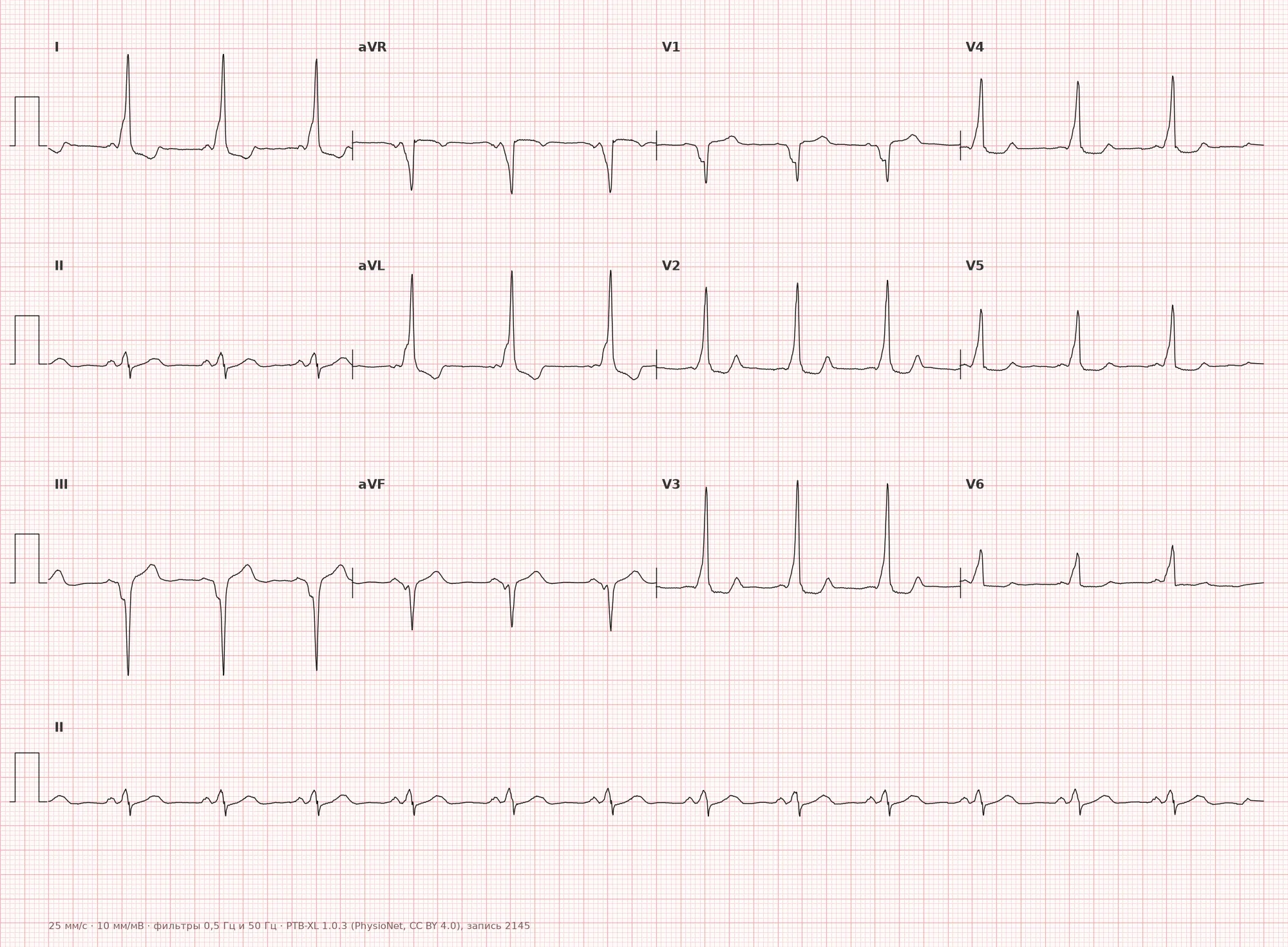
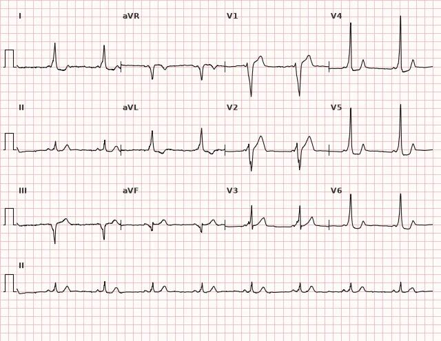
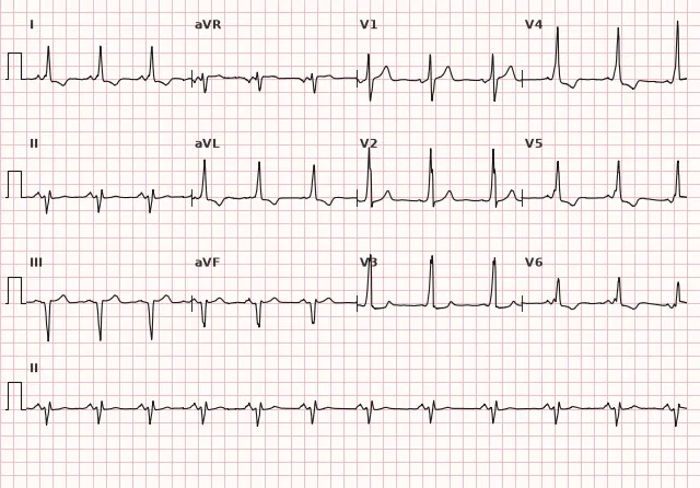
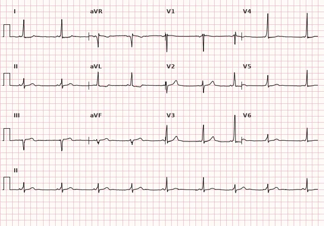
ECG from the open PTB-XL dataset (Wagner P. et al., Scientific Data, 2020; PhysioNet), CC BY 4.0 license. Rendering by WhiteCoat Pro.
Mechanism
Normally, the only conduction pathway from the atria to the ventricles is the AV node with its physiological delay. In WPW, there is an accessory muscular pathway (Kent bundle) connecting the atrium and ventricle directly, bypassing the AV node. The impulse travels faster down this pathway and activates a part of the ventricular myocardium earlier, which produces the delta wave and short PQ interval. The rest of the ventricle is activated normally via the AV node and the His-Purkinje system, so the QRS represents a fusion complex of the pre-excited and normally conducted components. The presence of two conduction pathways (AV node and accessory pathway) creates the anatomical substrate for impulse re-entry and reciprocating tachycardias.
Causes
- Congenital anomaly — persistence of an accessory muscular connection between the atrium and ventricle that failed to undergo resorption during embryogenesis
- Mostly sporadic, in most cases without structural heart disease
- Association with congenital heart defects, primarily Ebstein's anomaly
- Familial forms and rare genetic syndromes associated with pre-excitation have been described
Differential diagnosis
- Short PR with normal QRS (historically Lown-Ganong-Levine / LGL syndrome): PQ is shortened, but QRS is not widened and there is no delta wave. The concept of a separate 'bypass tract' without ventricular pre-excitation is historical and controversial, and proof of an anatomical substrate is usually lacking; today, this pattern is more commonly described as a short PR/accelerated AV conduction rather than a distinct syndrome
- Bundle branch block: QRS is wide, but the PQ interval is normal, there is no delta wave, and QRS morphology is typical of BBB
- Mahaim fibers: typically decremental right-sided atriofasicular pathways (less commonly atrioventricular) that often remain concealed during sinus rhythm without a constant delta wave; during tachycardia, they can produce wide-complex antidromic AVRT. Nodoventricular and fasciculoventricular variants are separate, rarer forms rather than the typical definition of this pathway
- WPW antidromic tachycardia vs ventricular tachycardia: in WPW, there may be a known pre-excitation pattern on baseline ECG, QRS concordance in chest leads, and morphology can help, but definitive differentiation sometimes requires an electrophysiology study
- True myocardial infarction/ischemia: discordant ST-T waves in WPW complicate evaluation but do not rule out ischemia; decisions require comparison with baseline ECG and clinical assessment rather than relying solely on the background pattern
Clinical significance and management
The danger of WPW syndrome is the development of atrial fibrillation with rapid antegrade conduction over the accessory pathway: the short effective refractory period of the pathway allows very rapid and irregular ventricular responses to degenerate into ventricular fibrillation. This is a rare but potentially fatal complication that dictates the indications for invasive management.
Management guidelines at the professional level:
- for a newly detected pre-excitation pattern, management is individualized and depends on age, occupation, pathway characteristics, and patient preferences: referral to an arrhythmologist for risk stratification (non-invasive tests, stress testing, and EPS when indicated). Sudden complete disappearance of pre-excitation during exercise or increased sinus heart rate indicates a relatively low risk, but even this sign does not replace clinical evaluation; gradual reduction of the delta wave, its masking by fusion, or incomplete disappearance does not reliably indicate low risk — EPS is required when in doubt
- during EPS, signs of a high-risk pathway include an effective refractory period of the accessory pathway ≤ 250 ms and/or the shortest pre-excited RR interval ≤ 250 ms during induced AF; the inducibility of AVRT and the presence of multiple accessory pathways are also considered
- for documented tachyarrhythmias (orthodromic/antidromic AVRT, AF with pre-excitation), radiofrequency catheter ablation of the accessory pathway is considered the treatment of choice
- for documented or strongly suspected AF with pre-excitation, AV nodal blocking agents (verapamil, diltiazem, digoxin, beta-blockers) and intravenous amiodarone are contraindicated because they can enhance conduction over the accessory pathway and accelerate the ventricular response; in hemodynamic stability, intravenous procainamide or ibutilide is used according to local protocols, while immediate synchronized cardioversion is used in instability
- for unclear wide-complex tachycardia, manage it as ventricular tachycardia until proven otherwise; the diagnosis of antidromic AVRT should not be established solely based on a known history of WPW, and verapamil should not be used empirically
Common interpretation errors
- Missing a short PQ interval when the delta wave is poorly expressed in some leads — review all 12 leads
- Mistaking discordant ST-T changes for ischemia or hypertrophy without checking them against prior ECGs and the clinical picture
- Confusing a wide QRS in WPW with bundle branch block by failing to check the PQ interval
- Prescribing verapamil or digoxin for wide QRS tachycardia in a patient with WPW without considering antidromic AVRT or AF with pre-excitation
- Confusing WPW pattern (ECG pattern alone without symptoms) with WPW syndrome (pattern + clinically significant tachyarrhythmia)
Short PQ + wide QRS + delta wave — think of WPW
1. Which of the following features is most specific for WPW syndrome on a sinus ECG?
The delta wave is a direct reflection of early ventricular excitation via an accessory pathway and a key sign of pre-excitation.
2. Why is verapamil contraindicated in atrial fibrillation in a patient with WPW?
AV nodal blockade eliminates the competing conduction pathway, causing the entire impulse to travel via the accessory pathway with its short refractory period, which can accelerate the ventricular response up to VF.
3. How does WPW syndrome differ from WPW pattern?
Both conditions present the same ECG pre-excitation pattern, but the syndrome is defined by its association with clinically documented tachyarrhythmias.
Frequently asked questions
How does WPW pattern differ from WPW syndrome?
WPW pattern (phenomenon) refers to ECG signs of pre-excitation (short PQ, delta wave) without documented tachyarrhythmia. WPW syndrome is the same pre-excitation associated with clinically significant tachyarrhythmia or symptoms, usually confirmed by ECG or EPS.
Is WPW syndrome dangerous?
The main danger is the development of atrial fibrillation with very rapid, irregular conduction over the accessory pathway, which can degenerate into ventricular fibrillation. For most patients, the course is benign, but the risk of sudden death exists and is assessed individually.
Why is verapamil contraindicated in wide QRS tachycardia in WPW?
In antidromic tachycardia or AF with conduction over an accessory pathway, AV nodal blocking agents can paradoxically enhance accessory pathway conduction and accelerate the ventricular response, precipitating ventricular fibrillation.
Is it true that LGL syndrome is a distinct disease?
A short PR interval with a normal QRS and no delta wave was long termed Lown-Ganong-Levine (LGL) syndrome, assuming a separate bypass tract. Today, this concept is considered historical and controversial: evidence for an independent anatomical substrate is usually lacking, and this pattern is more commonly described simply as a short PR or accelerated AV conduction.
What treatment is considered for symptomatic WPW?
The method of choice is catheter ablation of the accessory pathway via radiofrequency ablation, which radically eliminates the arrhythmia substrate in most cases.
ECG Course
Interpretation from basics to myocardial infarction — learn to read any ECG using a systematic algorithm.
- Video lessons and theory for every topic
- Simulator case studies under every lesson
- Unlimited ECG simulator practice
- Study guide: "All ECGs in 25 minutes"
ECG Strip Simulator
ECG, radiology, histology, pathomorphology, pharmacology — thousands of flashcards with detailed explanations.
- Real ECG strips, not textbook drawings
- Instant answers and explanations
- Streaks and progress tracking in your dashboard
Related terms
More in the section «Ventricular pre-excitation»
Sources
- Goldberger's Clinical Electrocardiography, latest edition
- Surawicz B., Knilans T. Chou's Electrocardiography in Clinical Practice
- Orlov V.N. Guide to Electrocardiography
- AHA/ACCF/HRS Recommendations for the Standardization of the Electrocardiogram, 2009
Educational material for medical students and physicians. Does not replace clinical guidelines and physical examination; the ECG must always be interpreted in conjunction with the clinical picture.