ECG criteria
- PQ (PR) interval is short — less than 120 ms (3 small squares at 25 mm/s)
- Initial part of the QRS is slurred and gradual — a delta wave, which is an initial smoothing of the upstroke or downstroke of the QRS, typically most prominent in the first 20–40 ms of the complex
- QRS complex is widened — often ≥ 120 ms due to fusion of the delta wave with the normal QRS; with minimal pre-excitation, it may be less than 120 ms
- Secondary ST-T changes may be present, typically discordant with the main direction of the QRS
- The polarity of the delta wave (positive/negative) varies across leads depending on the localization of the accessory pathway
- The feature is non-constant: it may disappear during intermittent pre-excitation, including during exercise
What it looks like on ECG
A delta wave is the initial, more gradual segment of the QRS, most noticeable roughly in the first 20–40 ms of the complex, which "flows into" the main, steeper R or S wave. Visually, this makes the QRS look widened and seemingly "slurred" at the start, unlike the sharp, steep upstroke seen in normal ventricular activation.
Associated features:
| Element | Parameter |
|---|---|
| PQ interval | shortened, less than 120 ms |
| QRS | often ≥ 120 ms; may be shorter with minimal pre-excitation |
| ST-T | secondary changes may be present, typically discordant with the main direction of the QRS |
The delta wave is more prominent in leads with high QRS amplitude and during manifest pre-excitation; it may be barely discernible or absent with a minimal contribution from the accessory pathway, as well as in intermittent WPW, where the same tracing contains complexes with and without a delta wave.
The polarity of the delta wave in various leads is used to roughly localize the accessory pathway (for details, see the separate topic «ECG Localization of Accessory Pathways»): a negative delta wave in the inferior leads II, III, and aVF can resemble a pathological Q wave and mimic an inferior myocardial infarction; a negative delta wave in V1–V2 can mimic left bundle branch block-like morphology and pathological QS/Q waves in anteroseptal leads.
Real ECGs: 8 examples
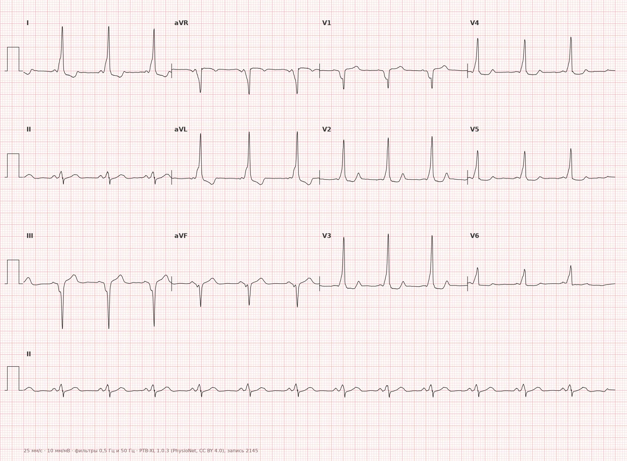
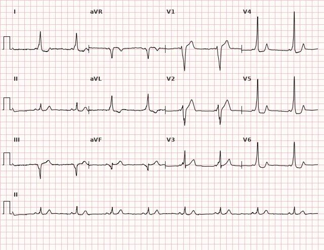
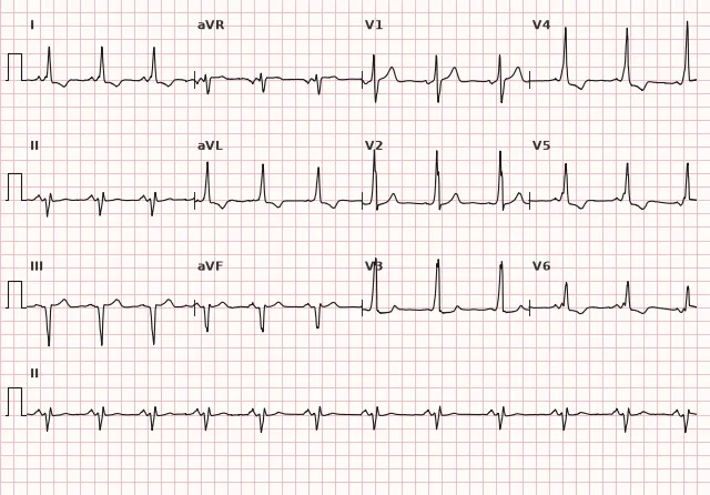
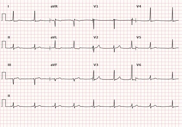
ECG from the open PTB-XL dataset (Wagner P. et al., Scientific Data, 2020; PhysioNet), CC BY 4.0 license. Rendering by WhiteCoat Pro.
Mechanism
Normally, the only pathway for impulse conduction from the atria to the ventricles is the AV node and the bundle of His, which physiologically delays the impulse. In pre-excitation syndrome, there is an accessory pathway (such as the bundle of Kent) that connects the atrial myocardium directly to the ventricular myocardium, bypassing the AV nodal delay.
The impulse travels simultaneously via two pathways: faster through the accessory pathway (without delay) and slower through the AV node and the His-Purkinje system. The ventricle begins to activate earlier than usual via the myocardium at the insertion site of the accessory pathway — this slow propagation of excitation directly through the working ventricular myocardium forms the gradual delta wave. Then, when the impulse via the normal pathway reaches the His-Purkinje system, activation rapidly captures the rest of the ventricle, forming the normal, steep part of the QRS. The QRS complex thus represents a fusion of two activation waves.
Causes
- WPW syndrome (manifest pre-excitation) — presence of an accessory AV pathway (bundle of Kent)
- Mahaim atriofascicular fibers — can cause a specific variant of pre-excitation, often with a normal PR and a left bundle branch block-like QRS; this is not the typical AV pathway of WPW
- Multiple accessory pathways — variable delta wave morphology is possible
- Intermittent pre-excitation — the delta wave appears and disappears on the same tracing
Differential diagnosis
- Inferior myocardial infarction — a negative delta wave in II, III, and aVF can mimic a pathological Q wave; WPW can produce pseudo-pathological Q waves and ST-T changes regardless of the PQ interval (pre-excitation and infarction can coexist), so a short PQ by itself does not rule out infarction — clinical suspicion of ACS requires serial ECGs and high-sensitivity troponin
- Right or left bundle branch block — QRS widening without PQ shortening in isolated bundle branch block; in WPW, the PQ is short and a delta wave is present
- Ventricular hypertrophy — high QRS voltage without PQ shortening and without a gradual initial component
- Extrasystole or fusion complexes — the delta wave is consistently present in sinus complexes, rather than appearing episodically as in ventricular extrasystoles
- Lown–Ganong–Levine (LGL) syndrome — a historical designation for a short PR without a delta wave; the independence of LGL syndrome has not been proven, so the diagnosis is not made based on this feature
Clinical significance and management
The delta wave is an ECG marker of the anatomical substrate for re-entrant tachyarrhythmias (orthodromic and antidromic AV re-entrant tachycardia) and, more dangerously, for rapid conduction to the ventricles during atrial fibrillation in a patient with WPW — this combination can lead to ventricular fibrillation.
At the ECG interpretation level, a physician must: identify the presence of a delta wave, a short PQ, and a widened QRS; describe the approximate localization of the accessory pathway based on the delta wave polarity across different leads; and refer the patient to an arrhythmologist/cardiologist to assess risk (symptoms, family history of sudden cardiac death) and consider an electrophysiological study and catheter ablation of the accessory pathway.
In irregular wide-complex tachycardia suspected to be pre-excited atrial fibrillation, management depends on the patient's stability. In hemodynamic instability, immediate synchronized electrical cardioversion is indicated. In a stable patient, intravenous procainamide or ibutilide is used if indications and monitoring conditions are met — these drugs slow conduction through the accessory pathway and can restore sinus rhythm. AV nodal blockers (adenosine, verapamil/diltiazem, β-blockers, digoxin) must not be used — they block the AV node but not the accessory pathway and can paradoxically accelerate ventricular response through it. Intravenous amiodarone is also not considered a safe alternative in pre-excited AF: it can slow AV conduction and promote preferential impulse conduction via the accessory pathway, so it is generally avoided in this setting. In regular wide-QRS tachycardia, the diagnosis should not be made solely on a history of WPW, and it should be managed as potential ventricular tachycardia until proven otherwise.
Common interpretation errors
- Mistaking a negative delta wave in the inferior or anterior leads for a pathological Q wave and erroneously diagnosing a prior myocardial infarction, failing to recognize that WPW does not exclude concurrent ischemia
- Failing to notice a subtle delta wave in minimal pre-excitation and missing WPW
- Confusing QRS widening with a bundle branch block without paying attention to the shortened PQ
- Failing to recognize intermittent pre-excitation if both normal and "delta" complexes are present on the tracing
- In irregular wide-complex tachycardia, erroneously prescribing an AV nodal blocker or amiodarone without ruling out pre-excited AF
1. Which interval is typically shortened in the presence of a delta wave?
The accessory pathway conducts the impulse to the ventricles without the delay of the AV node, which is why the PQ interval is shortened (usually < 120 ms).
2. How does a delta wave differ diagnostically from a pathological Q wave in myocardial infarction?
A short PQ interval does not rule out infarction: pre-excitation and ischemic injury can coexist, and WPW can mimic a pathological Q wave. Therefore, suspected ACS requires serial ECGs and troponin testing, rather than relying solely on the morphology of the initial QRS deflection.
3. What complication is most dangerous in a patient with a delta wave (WPW) who develops atrial fibrillation?
A typical AV accessory pathway usually lacks protective decremental conduction, so atrial fibrillation can lead to extremely high ventricular response rates, which may trigger ventricular fibrillation.
Frequently asked questions
How does a delta wave differ from a pathological Q wave?
The delta wave is a slurred, prolonged initial QRS deflection, usually combined with a shortened PR interval and signs of pre-excitation. Pathological Q/QS waves in myocardial infarction are evaluated by duration, depth, and distribution across adjacent leads, as well as by serial ECG dynamics, clinical presentation, and troponin levels—because in WPW these criteria can produce a pseudopathological pattern, and pre-excitation can coexist with myocardial infarction in the same patient.
Is the delta wave dangerous on its own?
The delta wave itself is an ECG sign, not a life-threatening condition. The danger is associated with the risk of tachyarrhythmias in a patient with an accessory pathway, especially when combined with atrial fibrillation, where rapid ventricular conduction can occur, up to and including ventricular fibrillation.
Can a delta wave disappear on an ECG?
Yes, with intermittent pre-excitation, the delta wave can appear and disappear from beat to beat on the same recording, including during exercise. The mechanism is related to changes in the relative conduction velocity and refractoriness of the accessory and normal pathways, rather than strictly being driven by sympathetic tone.
Does a delta wave always mean WPW syndrome?
The delta wave indicates manifest ventricular pre-excitation, most commonly via an atrioventricular accessory pathway (bundle of Kent), although other types of accessory pathways are possible. In an asymptomatic patient, this is referred to as the "WPW pattern" (asymptomatic pre-excitation); WPW syndrome is diagnosed when pre-excitation is combined with clinical symptoms—namely, paroxysms of tachycardia.
Can the location of the accessory pathway be determined from the delta wave?
Yes, approximately: the polarity of the delta wave in various leads (especially precordial and inferior leads) is used in algorithms for localizing the accessory pathway, although precise localization is confirmed during an electrophysiological study.
ECG Course
Interpretation from basics to myocardial infarction — learn to read any ECG using a systematic algorithm.
- Video lessons and theory for every topic
- Simulator case studies under every lesson
- Unlimited ECG simulator practice
- Study guide: "All ECGs in 25 minutes"
ECG Strip Simulator
ECG, radiology, histology, pathomorphology, pharmacology — thousands of flashcards with detailed explanations.
- Real ECG strips, not textbook drawings
- Instant answers and explanations
- Streaks and progress tracking in your dashboard
Related terms
More in the section «Ventricular pre-excitation»
Sources
- Goldberger A.L. Clinical Electrocardiography: A Simplified Approach
- Surawicz B., Knilans T. Chou's Electrocardiography in Clinical Practice
- Orlov V.N. Guide to Electrocardiography
- AHA/ACCF/HRS Recommendations for the Standardization and Interpretation of the Electrocardiogram, 2009
Educational material for medical students and physicians. Does not replace clinical guidelines and physical examination; the ECG must always be interpreted in conjunction with the clinical picture.