ECG criteria
- Measure QT from the onset of the QRS complex to the end of the T wave (return to the isoelectric line); evaluate several leads and select the most reliably measurable one, often the longest QT, avoiding leads with a prominent U wave or T–U fusion
- Measure RR in the same cycle (in case of sinus arrhythmia, average over several cycles)
- Calculate QTc using Bazett's formula: QTc = QT / √RR (RR in seconds); in tachycardia or bradycardia, Fridericia's or Framingham's formula is preferable, but they also have limitations
- QTc ≥480 ms on repeat ECGs or a Schwartz score ≥3.5 makes the diagnosis of LQTS highly probable in the absence of secondary causes
- QTc of 460–479 ms on repeat ECGs yields 2 points on the Schwartz scale; the final score is calculated taking into account the circumstances of syncope (unexplained — 1 point, during exertion/stress — 2 points) and other signs (T-wave morphology, family history, bradycardia) — only the total score determines the probability of the diagnosis
- QTc >450 ms in men and >460 ms (up to 470 ms according to some guidelines) in women represents the upper limit of normal / borderline value and is not a diagnostic criterion; it requires repeat measurement and exclusion of acquired causes
- Evaluate T-wave morphology: broad, notched, or biphasic T wave in V2–V4 (typical for LQT2), delayed onset of the T wave (LQT3)
- Exclude secondary causes (electrolytes, medications, bradycardia, hypothermia) before diagnosing congenital LQTS; note that in bradycardia, QTc may be falsely low
What it looks like on ECG
The main sign is prolongation of the QT interval relative to heart rate, i.e., QTc prolongation. Absolute QT values must not be measured in isolation from RR: during tachycardia, QT is physiologically shorter; during bradycardia, it is longer.
QT is measured from the beginning of the Q wave (or R wave if Q is absent) to the end of the T wave — the point where the T-wave curve returns to the isoelectric line. QT is assessed in multiple leads: most commonly leads II or V5–V6 are used, but the lead with the clearest and generally longest measurable QT is chosen for calculation, avoiding leads with a prominent U wave or T–U fusion, as differences between leads can mask the maximum prolongation. If a prominent U wave fused with the T wave is present, the end of the T wave is determined by the tangent to the steepest slope of the latter part of the T wave until it intersects the isoelectric line.
T-wave morphology often points to the genetic type:
| LQTS type | Typical T-wave morphology | Trigger of events |
|---|---|---|
| LQT1 | broad, smooth T wave | physical exertion, swimming |
| LQT2 | low-amplitude, biphasic, notched T wave (especially in V2–V4) | sudden noise, emotion, postpartum period |
| LQT3 | delayed onset of the T wave following a long isoelectric ST segment | sleep, rest, bradycardia |
Prominent U-wave amplitude, electrical T-wave alternans (at high arrhythmia risk), and age-relative sinus bradycardia may be present. QRS is usually normal. During an episode, polymorphic ventricular tachycardia (torsades de pointes) occurs with characteristic "twisting" of the QRS axis around the isoelectric line. Pause-dependent initiation (starting after a pause, e.g., following an extrasystole) is typical for acquired QT prolongation but also occurs in congenital LQTS, particularly in LQT2 and LQT3, and therefore does not reliably differentiate between acquired and congenital forms.
Real ECGs: 8 examples
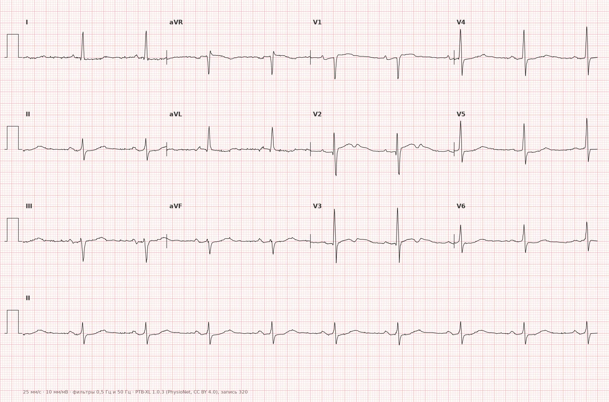
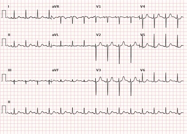
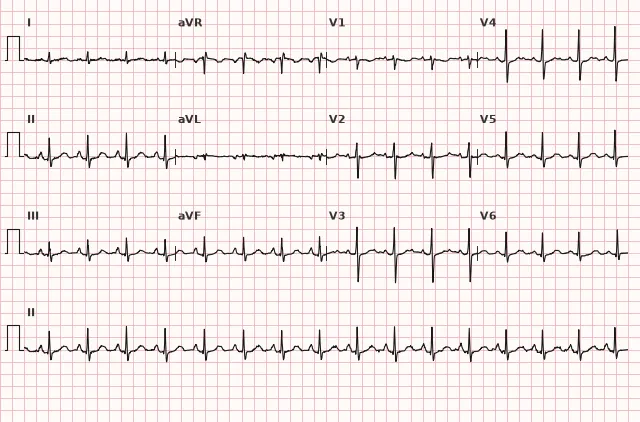
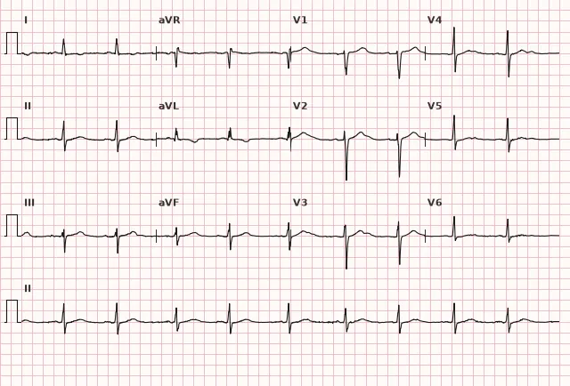
ECG from the open PTB-XL dataset (Wagner P. et al., Scientific Data, 2020; PhysioNet), CC BY 4.0 license. Rendering by WhiteCoat Pro.
Mechanism
Congenital LQTS is based on mutations in cardiomyocyte ion channel genes that disrupt the balance of inward and outward currents during phases 2–3 of the action potential. In LQT1 and LQT2, the function of potassium channels (IKs and IKr, respectively) is reduced, and repolarization is slowed. In LQT3, a mutation in the SCN5A gene leads to an excessive late inward sodium current, which prolongs the plateau phase.
Action potential prolongation promotes the development of early afterdepolarizations and triggered ventricular activity. Spatial heterogeneity (dispersion) of repolarization between myocardial layers and adjacent cells increases electrical vulnerability of the ventricles and can sustain polymorphic ventricular tachycardia (torsades de pointes), which may spontaneously terminate or degenerate into ventricular fibrillation.
Causes
- Congenital (channelopathies): Romano–Ward syndrome (autosomal dominant, isolated); Jervell and Lange-Nielsen syndrome (autosomal recessive, with congenital deafness); genes LQT1–LQT3 and rarer forms are known
- Acquired: certain QT-prolonging drugs (class IA and III antiarrhythmics, certain antipsychotics, macrolides, certain antidepressants, and other agents — the risk depends on the specific drug, dose, and interactions; it is advisable to check against the current list of QT-risk medications)
- Electrolyte disturbances: hypokalemia, hypomagnesemia, hypocalcemia
- Marked bradycardia, high-degree AV block
- Hypothyroidism
- Acute ischemia and myocardial infarction, myocarditis
- Intracranial pathology (subarachnoid hemorrhage)
- Malnutrition, anorexia, electrolyte depletion due to diets and alcohol abuse
Acquired factors (medications, electrolyte disturbances, bradycardia) can not only cause independent QT prolongation but also unmask a latent congenital channelopathy, so their presence does not rule out genetic predisposition.
Differential diagnosis
- Short QT syndrome — the opposite deviation; QTc ≤330 ms is diagnostically convincing, 331–360 ms is suspicious or supports the diagnosis only in the presence of additional signs (symptoms, family history, genetics)
- Normal U wave without T-wave prolongation — including the U wave in the QT measurement falsely lengthens the interval
- Bradycardia and QT correction — absolute QT during bradycardia is physiologically long, but Bazett's formula at low heart rates usually underestimates QTc, and a formally "normal" QTc does not exclude congenital LQTS; several consecutive complexes must be evaluated, Fridericia's or Framingham's formulas should be applied with caution, and clinical presentation, T-wave morphology, and family history must be considered
- Acquired QT prolongation (medications, electrolytes, bradycardia) — differentiated by history and reversibility after removing the cause, but the absence of a family history does not rule out the congenital form, and an acquired trigger may merely reveal hidden genetic predisposition
- Brugada syndrome — a different mechanism and ST-segment morphology in V1–V3, not directly related to QT duration, but both belong to channelopathies and can coexist
Clinical significance and management
LQTS is dangerous due to the development of torsades de pointes progressing to ventricular fibrillation, manifesting as syncope, seizures (often misdiagnosed as epilepsy), or sudden cardiac death, frequently during physical or emotional stress (LQT1, LQT2) or during sleep (LQT3).
For the physician at the ECG stage: confirm QTc prolongation on repeat tracings, evaluate T-wave morphology, calculate the Schwartz risk score taking into account history (syncope, family history of sudden death, deafness), obtain a medication history, and check electrolytes. If a congenital form is suspected, refer for genetic testing and arrhythmologist consultation, and screen first-degree relatives; a negative genetic result does not rule out a clinical diagnosis.
In developed torsades de pointes: immediately discontinue QT-prolonging drugs, administer intravenous magnesium regardless of baseline levels, actively raise potassium to a high-normal level (usually around 4.5–5.0 mmol/L) rather than merely correcting marked hypokalemia. In ongoing sustained polymorphic ventricular tachycardia where synchronization is impossible, perform immediate unsynchronized electrical therapy without waiting for overt hemodynamic instability; upon progression to ventricular fibrillation, perform defibrillation. In the recurrent form, predominantly acquired, consider heart rate acceleration (temporary cardiac pacing or an isoproterenol infusion to increase heart rate) to shorten the QT interval in addition to magnesium and discontinuation of causative drugs. Further risk management for the congenital form (beta-blockers, implantable cardioverter-defibrillator, left sympathetic denervation in selected cases) is a matter for specialized guidelines on ventricular arrhythmias and sudden death prevention.
Common interpretation errors
- Measurement of absolute QT without heart rate correction — leads to under- or overdiagnosis
- Inclusion of the U wave in the QT measurement, especially during bradycardia
- Using Bazett's formula in severe tachycardia or bradycardia without accounting for its known limitations (overestimation in tachycardia, underestimation in bradycardia)
- Diagnosing LQTS based on a single tracing without repeat measurements and without considering reversible causes
- Ignoring T-wave morphology, which helps suspect the genetic type and the triggering factor
1. Which lead is preferred for measuring the QT interval?
QT is assessed in multiple leads, and the one where the T wave is clearly visible and measurement is reliable is chosen (often II or V5–V6), avoiding fusion with the U wave.
2. What is the underlying mechanism of LQT3?
In LQT3, the SCN5A mutation leads to an excessive late inward sodium current, which prolongs the plateau phase of the action potential.
3. Which arrhythmia is most characteristic of long QT syndrome?
Repolarization prolongation and early afterdepolarizations create the substrate specifically for polymorphic ventricular tachycardia of the torsades de pointes type.
Frequently asked questions
What QTc is considered definitively pathological?
A QTc ≥480 ms on repeat tracings or a Schwartz score ≥3.5 supports the diagnosis of LQTS in the absence of secondary causes. Values of 460–479 ms alone only contribute partial points to the score—the diagnosis depends on the sum with other features (circumstances of syncope, T-wave morphology, history). Values of 450–459 ms in men and 450–469 ms in women are borderline and require the exclusion of acquired causes rather than serving as an independent diagnostic criterion.
How do QTc thresholds differ between men and women?
In most guidelines, the upper limit of normal QTc is 450 ms in men and 460 ms in women, with some sources citing up to 470 ms for women. These are reference values, not diagnostic thresholds: exceeding them is a reason for repeat measurement and searching for the cause, not a diagnosis of LQTS per se.
Is a prolonged QT dangerous if there are no symptoms?
Yes, the risk of torsades de pointes and sudden death exists even in the absence of symptoms, especially with marked QTc prolongation and a positive family history. Asymptomatic mutation carriership does not rule out future events, so such patients require follow-up with an arrhythmologist.
How to distinguish congenital LQTS from acquired QT prolongation?
Acquired prolongation is usually associated with a specific medication, electrolyte imbalance, or bradycardia and is reversible once the cause is eliminated. The congenital form typically presents with persistent QTc prolongation, a family history of syncope or sudden death, and specific triggers (exercise, acoustic stimuli, sleep), though the absence of a family history does not exclude it. Genetic testing can confirm the etiology and allows for cascade screening of relatives, but a negative result does not rule out a clinical diagnosis of LQTS.
Which QT correction formula is more accurate—Bazett's or Fridericia's?
Bazett's formula (QT/√RR) is the most common, but it overestimates QTc in tachycardia and underestimates it in bradycardia, especially when severe. Fridericia's formula (cube root of RR) and the Framingham formula are considered more accurate in these heart rate ranges, but they also have limitations; therefore, in bradycardia, a normal QTc does not rule out congenital LQTS and requires the evaluation of multiple complexes and clinical data.
ECG Course
Interpretation from basics to myocardial infarction — learn to read any ECG using a systematic algorithm.
- Video lessons and theory for every topic
- Simulator case studies under every lesson
- Unlimited ECG simulator practice
- Study guide: "All ECGs in 25 minutes"
ECG Strip Simulator
ECG, radiology, histology, pathomorphology, pharmacology — thousands of flashcards with detailed explanations.
- Real ECG strips, not textbook drawings
- Instant answers and explanations
- Streaks and progress tracking in your dashboard
Related terms
More in the «Channelopathies and Cardiomyopathies» section
Sources
- Surawicz B., Knilans T. Chou's Electrocardiography in Clinical Practice
- Goldberger A. Clinical Electrocardiography: A Simplified Approach
- Orlov V.N. Guide to Electrocardiography
- AHA/ACCF/HRS Recommendations for the Standardization and Interpretation of the Electrocardiogram, 2009
Educational material for medical students and physicians. Does not replace clinical guidelines and physical examination; the ECG must always be interpreted in conjunction with the clinical picture.