ECG criteria
- The pacing artifact (spike) is very short, approximately 0.1–2 ms; it appears simultaneously in all leads, but its visibility varies in each lead — the absence of a spike in a single lead does not prove its absence
- In atrial pacing: a spike precedes the P wave, the P wave morphology differs from the native sinus P wave and depends on the lead placement site
- In right ventricular myocardial pacing: a spike precedes a wide QRS complex (often ≥ 120 ms); during conduction system pacing, the QRS width may be closer to normal
- Typical morphology of a paced ventricular complex from the RV apex is left bundle branch block (LBBB) pattern with left axis deviation
- In biventricular (resynchronization) pacing, the QRS morphology is often different: frequently a dominant R wave in V1 (resembling RBBB) and a narrower QRS than in pure right ventricular pacing, though this is not a mandatory finding
- In dual-chamber pacing, two spikes (atrial and ventricular) are possible with a programmed AV delay between them, but during AS-VP or AP-VS, only one spike is visible or pacing is absent due to preserved native activity; the mode is confirmed by pacemaker interrogation
- Assess capture for every output pulse intended for pacing, keeping in mind that the response may be hidden, fused with a native complex (fusion/pseudofusion), or fall within the refractory period — the clear absence of a P wave/QRS complex after a spike across several complexes indicates failure to capture
- Assess whether there are native (non-paced) complexes and how they relate to the spikes — this serves as a guide for presumed sensing function, which is ultimately confirmed by the programmer
What it looks like on ECG
The pacing artifact is a very short "spike" lasting approximately 0.1–2 ms, generated by the pacemaker pulse. Electrically, it occurs simultaneously in all leads, but its visibility varies: in some leads it is distinct, while in others it is barely visible — this depends on the vector projection of the impulse and the electrocardiograph filters, rather than device malfunction. The absence of a visible spike in a single lead does not prove its absence; all 12 leads must be checked.
In atrial pacing (lead usually in the right atrial appendage), the spike precedes the P wave. With right atrial appendage pacing, the P wave generally remains positive in the inferior leads (II, III, aVF) because the overall vector of depolarization is directed inferiorly, but the P wave's shape and axis differ from sinus rhythm and depend on fusion with native activation. A negative or biphasic P wave in the inferior leads is more characteristic of low atrial or lower septal lead placement rather than typical right atrial appendage pacing.
In right ventricular myocardial pacing (lead in the apex — the most common variant), the spike precedes a wide, deformed QRS complex, often ≥ 120 ms. The morphology resembles left bundle branch block: a wide S wave in V1, a wide R wave in I, aVL; the electrical axis is often deviated to the left or indeterminate because depolarization proceeds from the apex to the base and from right to left — slowly through the myocardium, bypassing the His-Purkinje system. In conduction system pacing (His bundle, left bundle branch area), the QRS may remain narrow or moderately widened because activation partially or entirely utilizes physiological conduction pathways.
In dual-chamber (DDD) pacing, two spikes are possible — atrial and ventricular — with a programmed AV delay between them. However, in this mode, one of the channels is often inhibited by native activity (AS-VP, AP-VS variants), so only a single spike may be visible on the tracing, or pacing may be absent altogether; the AV delay following a paced versus a native atrial event may differ programmatically. Two consecutive spikes are a common, but not absolute, finding in DDD; the mode and parameters are confirmed by device interrogation.
In biventricular pacing (cardiac resynchronization therapy), a lead is added in the coronary sinus to pace the left ventricle; the resulting QRS is frequently, but not necessarily, narrower than with isolated right ventricular pacing and often features a dominant R wave in V1. The morphology strongly depends on the LV lead position, pacing sequence, and fusion with native conduction, so the CRT type cannot be reliably determined from a single ECG without device data.
In fully ventricular-paced complexes, secondary repolarization changes are expected — the ST segment and T wave are usually, but not necessarily in every lead, discordant with the QRS (directed oppositely). Concordant or excessively pronounced discordant ST-T changes require evaluation for ischemia using modified Sgarbossa criteria and the clinical context, rather than being automatically dismissed as normal.
Real ECGs: 8 examples
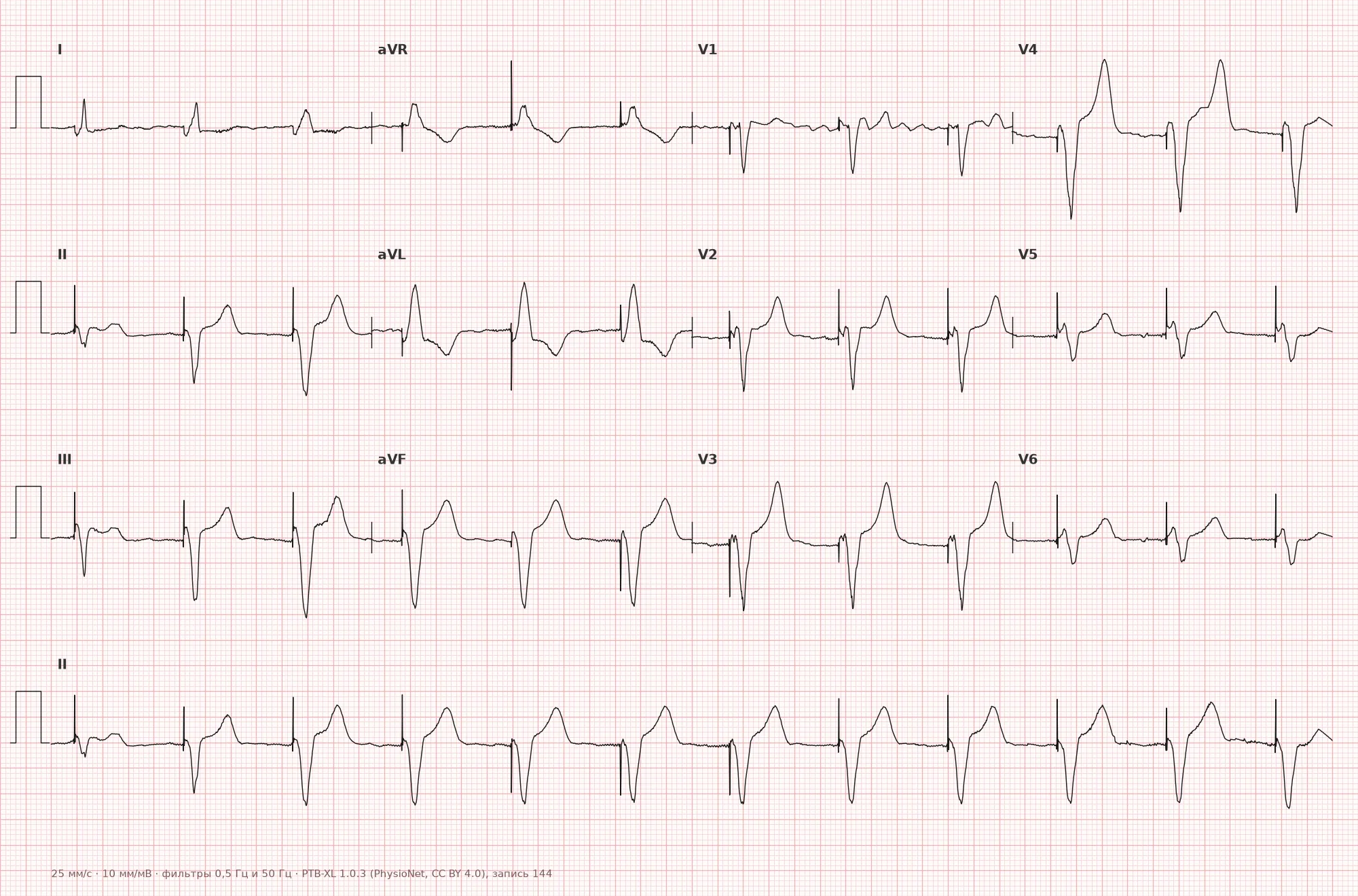
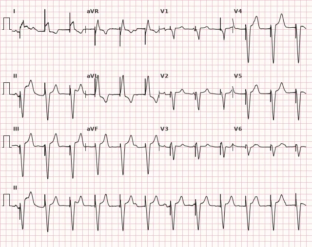
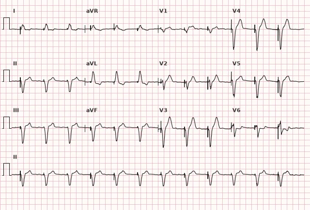
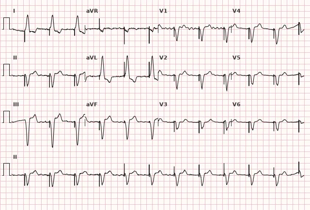
ECG from the open PTB-XL dataset (Wagner P. et al., Scientific Data, 2020; PhysioNet), CC BY 4.0 license. Rendering by WhiteCoat Pro.
Mechanism
The cardiac pacemaker delivers an electrical impulse via a lead placed in the atrium and/or ventricle. The impulse depolarizes the myocardium at the contact point of the electrode, and the wave of excitation spreads from this point rather than from the physiological pacemaker (sinus node or His-Purkinje system). Therefore, the direction and speed of depolarization are altered: the paced atrial complex has a different P wave morphology. In conventional right ventricular myocardial pacing, the QRS is typically wide because excitation travels slowly through the myocardium, bypassing the fast conduction system; during pacing of the conduction system itself (His bundle, left bundle branch), the QRS may remain narrow or moderately widened since the physiological conduction pathway is utilized. Modern pacemakers operate on demand: an impulse is delivered only if no native activity is detected within a set time (sensing); if a native rhythm is present, the stimulus is inhibited.
Causes
- Sick sinus syndrome, bradyarrhythmias
- Complete and high-grade AV block
- Acquired Mobitz II block and symptomatic second-degree AV block are standard indications for permanent pacing; with 2:1 block, a single ECG cannot reliably distinguish Mobitz I from Mobitz II, requiring determination of the block level (QRS width, provocative maneuvers, EPS) and clinical significance, especially in the presence of symptoms or signs of infra-nodal block
- Heart failure with reduced ejection fraction and wide QRS is a distinct indication for cardiac resynchronization therapy (CRT) according to guideline criteria (ejection fraction, QRS width and morphology, clinical status)
- Post-AV node ablation
- Prevention of sudden cardiac death in high-risk patients with life-threatening ventricular arrhythmias (including specific channelopathies and cardiomyopathies) — implantable cardioverter-defibrillator (ICD) with pacing capabilities, rather than a standard pacemaker
Differential diagnosis
- Native rhythm with LBBB — distinction: no pacing spike precedes the QRS.
- Ventricular extrasystole — morphology resembles a paced complex, but there is no preceding spike and no fixed coupling interval to an atrial event.
- Myocardial infarction in a paced patient — evaluation using modified criteria (modified Sgarbossa criteria), since the baseline morphology is already wide and discordant.
- Mains interference artifact — unlike a pacing spike, it is not time-locked to the complexes and has no fixed interval preceding the QRS.
- Pseudofusion and fusion with intrinsic QRS — the complex is partly formed by intrinsic excitation and partly by the stimulus, with a morphology intermediate between paced and intrinsic; not to be confused with loss of capture.
Clinical significance and management
The mere presence of a paced rhythm is not a pathology, but rather a functioning device. ECG is used to assess signs of pacing, presumed capture, and presumed sensing, but final evaluation of parameters, battery, leads, and programmed rate requires a programmer interrogation of the pacemaker: the rate depends on mode, hysteresis, rate response, intrinsic AV conduction, and fusion, and cannot be reliably determined from a single tracing.
The absence of a P or QRS wave following an individual output pulse intended for pacing is a reason to consider loss of capture (e.g., due to lead dislocation, battery depletion, perforation, increased pacing threshold), but one must account for the possibility of a concealed response, fusion/pseudofusion, and the impulse falling within the refractory period; when in doubt, check other leads, a long rhythm strip, and device data.
Spikes occurring against the background of an intrinsic rhythm earlier than expected indicate undersensing of intrinsic activity or programmed asynchronous/other pacing; oversensing, conversely, more often causes suppression of expected spikes and pauses rather than their appearance. Suspected malfunction of any kind, especially in a pacemaker-dependent patient, requires urgent programmer check; additionally — referral to an arrhythmologist and chest radiography to assess lead position. Modified criteria (analogous to Sgarbossa criteria) are used to assess ischemia during ventricular pacing, as standard ST-T criteria are inapplicable to a wide discordant paced QRS.
Common interpretation errors
- Mistaking the absence of a visible spike in some leads for its total absence — check all 12 leads.
- Interpreting a wide paced QRS as a sign of ischemia or bundle branch block without considering the pacing context.
- Failing to evaluate the interval between the atrial and ventricular spike during dual-chamber pacing and forgetting that in DDD, often only a single spike is visible.
- Confusing a fusion beat with loss of capture.
- Automatically equating 2:1 block with Mobitz II and forgetting that discordant ST-T waves in paced complexes are an expected norm, but concordant changes require evaluation for ischemia.
1. Which ECG sign indicates right ventricular apex pacing?
Right ventricular apex pacing creates excitation spreading atypically through the myocardium, forming a wide QRS with LBBB morphology and left axis deviation.
2. What indicates loss of capture during cardiac pacing?
Capture means the pacemaker impulse successfully depolarized the myocardium; a consistent absence of a response P or QRS after an impulse, not explained by fusion or the refractory period, indicates loss of capture.
3. Why can standard ST criteria not be used to assess ischemia during paced ventricular rhythm?
The wide discordant paced complex operates by a mechanism analogous to LBBB, so modified (adapted) criteria rather than standard ST elevation thresholds are used to assess ischemia.
Frequently asked questions
How to distinguish paced rhythm from intrinsic left bundle branch block?
The main sign is the presence of a pacing spike immediately preceding a wide QRS complex. In intrinsic LBBB, there is no spike, and the QRS onset is smooth, without a sharp vertical artifact.
Is a wide QRS dangerous during ventricular pacing?
A wide complex during conventional RV myocardial pacing is an expected consequence of single-site pacing and is not dangerous in itself. Attention should be paid not to the width, but to the presence of capture after every output pulse and signs of adequate sensing.
Can myocardial infarction be seen against the background of paced rhythm?
Yes, but standard ST elevation criteria do not work due to the baseline wide discordant complex. Modified criteria analogous to Sgarbossa criteria, adapted for paced rhythm, are used.
Why is the spike not visible in all leads?
A spike occurs electrically simultaneously in all leads, but its amplitude and visibility on the tracing depend on the projection of the pacemaker impulse vector onto the axis of a specific lead, as well as on electrocardiograph filters. This is a variant of normal and does not indicate device malfunction.
How does dual-chamber pacing differ from single-chamber pacing on ECG?
In dual-chamber pacing in AP-VP mode, two consecutive spikes are visible — atrial and ventricular — with a programmed AV delay between them. However, with preserved intrinsic activity, one of the spikes is often absent (AS-VP, AP-VS), whereas in single-chamber pacing, fundamentally only a single spike may be present — before P or before QRS.
ECG Course
Interpretation from basics to myocardial infarction — learn to read any ECG using a systematic algorithm.
- Video lessons and theory for every topic
- Simulator case studies under every lesson
- Unlimited ECG simulator practice
- Study guide: "All ECGs in 25 minutes"
ECG Strip Simulator
ECG, radiology, histology, pathomorphology, pharmacology — thousands of flashcards with detailed explanations.
- Real ECG strips, not textbook drawings
- Instant answers and explanations
- Streaks and progress tracking in your dashboard
Related terms
More in the section "Cardiac Pacing"
Sources
- Goldberger A. Clinical Electrocardiography: A Simplified Approach
- Surawicz B., Knilans T. Chou's Electrocardiography in Clinical Practice
- AHA/ACCF/HRS Recommendations for the Standardization and Interpretation of the Electrocardiogram, 2009
- Orlov V.N. Guide to Electrocardiography
Educational material for medical students and physicians. Does not replace clinical guidelines and physical examination; the ECG must always be interpreted in conjunction with the clinical picture.